Download presentation
Presentation is loading. Please wait.

1
Inhaled Anesthetic Delivery Systems
พญ.เพชรรัตน์ วิสุทธิเมธีกร พ.บ., ป. ชั้นสูงสาขาวิสัญญีวิทยา, วว.(วิสัญญี) ภาควิชาวิสัญญีวิทยา วิทยาลัยแพทยศาสตร์กรุงเทพมหานคร และวชิรพยาบาล
 ภาควิชาวิสัญญีวิทยา. วิทยาลัยแพทยศาสตร์กรุงเทพมหานคร. และวชิรพยาบาล.")
2
Inhaled Anesthetic Delivery Systems
Anesthesia machine Vaporizers Anesthetic breathing circuit Ventilator Scavenging system

3
Anesthesia Machine เครื่องดมยาสลบ

4
Vaporizers

5
ANESTHESIA MACHINES

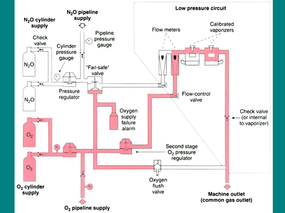
6
Generic Anesthetic Machine
The pressures within the anesthesia machine can be divided into three circuits High-pressure Intermediate-pressure Low-pressure circuit

8
Gas supply แหล่งจ่ายก๊าซ
Pipeline Cylinder

9
Pipeline supply แหล่งจ่ายก๊าซหลัก
primary gas source for the anesthesia machine oxygen, nitrous oxide, and air "normal working pressure" 50 psi DISS (diameter index safety system)
")
10
Pipeline

11
Nuts Nipples Body Adaptors Nut and Nipple Combinations

12
Cylinder supply แหล่งจ่ายก๊าซสำรอง
reserve E cylinders Color-coded Pin Index Safety System (PISS) high-pressure cylinder source pressure regulator oxygen 2200 psig to 45 psig nitrous oxide 745 psig to 45 psig
 high-pressure cylinder source. pressure regulator. oxygen 2200 psig to 45 psig. nitrous oxide 745 psig to 45 psig.")
13
ขนาดและความจุของท่อออกซิเจน ที่ความดัน 2200 Psig
เส้นผ่าศูนย์กลาง x สูง (นิ้ว) ความจุ N2O เต็มที่ 745 Psig (L) ความจุ O2 เต็มที่ 2200 Psig (L) ค่าคงที่ D E F G H 4.5 X 17 4.5 X 26 5.5 X 36 8.5 X 51 9 X 51 940 1590 - 13800 15800 360 622 1273 5259 6905 0.28 3.14
 ความจุ N2O เต็มที่ 745 Psig (L) ความจุ O2 เต็มที่ 2200 Psig (L) ค่าคงที่ D. E. F. G. H. 4.5 X X X X X")
14
Pin-Indexed Yoke Assemblies
Cylinder Valve Connections

15
Safety Devices for Oxygen Supply Pressure Failure
Oxygen Supply Failure Alarm oxygen supply pressure decreases to 30 psig activated within 5 seconds Second-Stage Pressure Regulator for Oxygen set at between 12 and 19 psig supplies a constant pressure to the oxygen flow control valve

16
Safety Devices for Oxygen Supply Pressure Failure
Fail-Safe Valves Pressure sensor's shut-off valve Oxygen failure protection device (OFPD)
")
17
Pressure sensor's shut-off valve
A, The valve is open because the oxygen supply pressure is greater than the threshold value of 20 psig. B, The valve is closed because of inadequate oxygen pressure

18
The oxygen failure protection device (OFPD)
OFPD responds proportionally to changes in oxygen supply pressure

19
Flow Meter Assemblies

20
Physical Principles of Conventional Flow Meters
The clearance between the head of the float and the flow tube is known as the annular space. It can be considered an equivalent to a circular channel of the same cross-sectional area.

21
Physical Principles of Conventional Flow Meters
density (turbulent flow ) viscosity (laminar flow)
 viscosity (laminar flow)")
22
Flow Meter Assemblies Flow Control Valve Flow Meter Subassembly
FLOW TUBES fine flow tube mL/min to 1 L/min coarse flow tube – 1 L/min to between 10 and 12 L/min INDICATOR FLOATS AND FLOAT STOPS

23
The flow meter sequence is a potential cause of hypoxia
A and B, In the event of a flow meter leak, a potentially dangerous arrangement exists when nitrous oxide is located in the downstream position. C and D, The safest configuration exists when oxygen is located in the downstream position

24
An oxygen leak from the flow tube can produce a hypoxic mixture, regardless of the arrangement of the flow tubes

25
Proportioning Systems
Prevent delivery of a hypoxic mixture N2O and O2 are interfaced mechanically or pneumatically Minimum O2 concentration at the common gas outlet is between 23% and 25%

26
N2O and O2 flow control valves are identical
N2O and O2 flow control valves are identical. A 14-tooth sprocket is attached to the N2O flow control valve, and a 28-tooth sprocket is attached to the O2 flow control valve. A chain links the sprockets. The combination of the mechanical and pneumatic aspects of the system yields the final oxygen concentration. The Datex-Ohmeda Link-25 proportioning system can be thought of as a system that increases oxygen flow when necessary to prevent delivery of a fresh gas mixture with an oxygen concentration of less than 25%

27
North American Dräger Oxygen Ratio Monitor Controller (ORMC)
The ORMC is composed of an O2 chamber, a N2O chamber, and a N2Oslave control valve, all of which are interconnected by a mobile horizontal shaft. The pneumatic input into the device is from the O2 and the N2O flow meters. These flow meters have resistors located downstream from the flow control valves that create backpressures directed to the O2 and N2O chambers. The value of the O2 flow tube's resistor is three to four times that of the N2O flow tube's resistor, and the relative value of these resistors determines the value of the controlled fresh gas concentration of O2. The backpressure in the O2 and the N2O chambers pushes against rubber diaphragms attached to the mobile horizontal shaft. Movement of the shaft regulates the N2O slave control valve, which feeds the N2Oflow control valve.

28
Oxygen Flush Valve

29
Oxygen Flush Valve Direct communication between the oxygen high-pressure circuit and the low-pressure circuit Delivers 100% oxygen at a rate of 35 to 75 L/min to the breathing circuit High pressure of 50 psig

30
Oxygen Flush Valve Several hazards Barotrauma Awareness
dilutes the inhaled anesthetic

31
VAPORIZERS Vapor Pressure Latent Heat of Vaporization Specific Heat
calories required to change 1 g of liquid into vapor without a temperature change Specific Heat calories required to increase the temperature of 1 g of a substance by 1°C. Thermal Conductivity

32
Vapor pressure versus temperature curves for desflurane, isoflurane, halothane, enflurane, and sevoflurane The vapor pressure curve for desflurane is steeper and shifted to higher vapor pressures compared with the curves for other contemporary inhaled anesthetics.

33
Variable-bypass vaporizer

34
Ohmeda Tec-type vaporizer
Ohmeda Tec-type vaporizer. At high temperatures, the vapor pressure inside the vaporizing chamber is high. To compensate for the increased vapor pressure, the bimetallic strip of the temperature-compensating valve leans to the right, allowing more flow through the bypass chamber and less flow through the vaporizing chamber. The net effect is a constant vaporizer output. In a cold operating room environment, the vapor pressure inside the vaporizing chamber decreases. To compensate for the decreased vapor pressure, the bimetallic strip swings to the left, causing more flow through the vaporizing chamber and less through the bypass chamber. The net effect is a constant vaporizer output

35
North American Dräger Vapor 19. 1 vaporizer
North American Dräger Vapor 19.1 vaporizer. Automatic temperature-compensating mechanisms in bypass chambers maintain a constant vaporizer output with varying temperatures. An expansion element directs a greater proportion of gas flow through the bypass chamber as temperature increases.

36
Tec 6 desflurane vaporizer
Tec 6 desflurane vaporizer. The vaporizer has two independent gas circuits arranged in parallel. The fresh gas circuit is shown in red, and the vapor circuit is shown in white. The fresh gas from the flow meters enters at the fresh gas inlet, passes through a fixed restrictor (R1), and exits at the vaporizer gas outlet. The vapor circuit originates at the desflurane sump, which is electrically heated and thermostatically controlled to 39°C, a temperature well above desflurane's boiling point. The heated sump assembly serves as a reservoir of desflurane vapor. Downstream from the sump is the shut-off valve. After the vaporizer warms up, the shut-off valve fully opens when the concentration control valve is turned to the on position. A pressure-regulating valve located downstream from the shut-off valve downregulates the pressure. The operator controls desflurane output by adjusting the concentration control valve (R2), which is a variable restrictor
, and exits at the vaporizer gas outlet. The vapor circuit originates at the desflurane sump, which is electrically heated and thermostatically controlled to 39°C, a temperature well above desflurane s boiling point. The heated sump assembly serves as a reservoir of desflurane vapor. Downstream from the sump is the shut-off valve. After the vaporizer warms up, the shut-off valve fully opens when the concentration control valve is turned to the on position. A pressure-regulating valve located downstream from the shut-off valve downregulates the pressure. The operator controls desflurane output by adjusting the concentration control valve (R2), which is a variable restrictor.")
37
ANESTHETIC CIRCUITS Deliver oxygen and anesthetic gases to the patient
Eliminate carbon dioxide adequate inflow of fresh gas carbon dioxide absorbent Semiclosed rebreathing circuits and the circle system.

38
Mapleson Systems

39
Mapleson Systems Factors influence carbon dioxide rebreathing
the fresh gas inflow rate the minute ventilation the mode of ventilation (spontaneous or controlled), the tidal volume the respiratory rate the inspiratory to expiratory ratio the duration of the expiratory pause the peak inspiratory flow rate the volume of the reservoir tube the volume of the breathing bag ventilation by mask ventilation through an endotracheal tube the carbon dioxide sampling site.
, the tidal volume. the respiratory rate. the inspiratory to expiratory ratio. the duration of the expiratory pause. the peak inspiratory flow rate. the volume of the reservoir tube. the volume of the breathing bag. ventilation by mask. ventilation through an endotracheal tube. the carbon dioxide sampling site.")
40
Mapleson Systems Prevention of rebreathing, during spontaneous ventilation: A > DFE > CB. During controlled ventilation, DFE > BC > A A, B, and C systems are rarely used today

41
The Bain circuit a modification of the Mapleson D system
spontaneous and controlled ventilation.

42
The Bain circuit Exhaled gases in the outer reservoir tubing add warmth to inspired fresh gases unrecognized disconnection or kinking of the inner fresh gas hose The fresh gas inflow rate necessary to prevent rebreathing is 2.5 times the minute ventilation

43
Components of the Circle system
APL, adjustable pressure limiting; B, reservoir bag; V, ventilator

44
Circle Breathing System
A circle system can be semiopen, semiclosed, or closed, depending on the amount of fresh gas inflow Semiopen system has no rebreathing and requires a very high flow of fresh gas Semiclosed system is associated with rebreathing of gases Closed system is one in which the inflow gas exactly matches that being consumed by the patient

45
Circle Breathing System
Components of The circle system (1) a fresh gas inflow source (2) inspiratory and expiratory unidirectional valves (3) inspiratory and expiratory corrugated tubes (4) a Y-piece connector (5) an overflow or pop-off valve, referred to as the APL valve (6) a reservoir bag (7) a canister containing a carbon dioxide absorbent
 a fresh gas inflow source. (2) inspiratory and expiratory unidirectional valves. (3) inspiratory and expiratory corrugated tubes (4) a Y-piece connector. (5) an overflow or pop-off valve, referred to as the APL valve. (6) a reservoir bag. (7) a canister containing a carbon dioxide absorbent.")
46
Circle Breathing System
Rules to prevent rebreathing of carbon dioxide in a traditional circle system Unidirectional valves must be located between the patient and the reservoir bag on the inspiratory and expiratory limbs of the circuit. The fresh gas inflow cannot enter the circuit between the expiratory valve and the patient. The overflow (pop-off) valve cannot be located between the patient and the inspiratory valve.
 valve cannot be located between the patient and the inspiratory valve.")
47
Circle Breathing System
Advantages stability of inspired gas concentrations, conservation of respiratory moisture and heat, prevention of operating room pollution Disadvantage complex design

48
ABSORPTION Lack of toxicity with common anesthetics, low resistance to airflow, low cost, ease of handling, and efficiency 3 formulations soda lime Baralyme calcium hydroxide lime (Amsorb)
")
49
ABSORPTION Soda lime (most commonly used ) The equations
80% calcium hydroxide, 15% water, 4% sodium hydroxide, and 1% potassium hydroxide (an activator) silica The equations 1) CO2 + H2 O ⇔ H2 CO3 2) H2 CO3 + 2NaOH (KOH) ⇔ Na2 CO3 (K2 CO3 ) + 2H2 O + Heat 3) Na2 CO3 (K2 CO3 ) + Ca(OH)2 ⇔ CaCO3 + 2NaOH (KOH)
 silica. The equations. 1) CO2 + H2 O ⇔ H2 CO3. 2) H2 CO3 + 2NaOH (KOH) ⇔ Na2 CO3 (K2 CO3 ) + 2H2 O + Heat. 3) Na2 CO3 (K2 CO3 ) + Ca(OH)2 ⇔ CaCO3 + 2NaOH (KOH)")
50
ABSORPTION Baralyme Calcium hydroxide lime
20% barium hydroxide and 80% calcium hydroxide Calcium hydroxide lime lack of sodium and potassium hydroxides carbon monoxide and the nephrotoxic substance known as compound A

51
ABSORPTION Absorptive Capacity size of the absorptive granules
soda lime is 26 L of carbon dioxide per 100 g of absorbent calcium hydroxide lime has been reported at 10.2 L per 100 g of absorbent size of the absorptive granules surface area air flow resistance

52
ABSORPTION Indicators
Ethyl violet :pH indicator added to soda lime and Baralyme from colorless to violet when the pH of the absorbent decreases as a result of carbon dioxide absorption Fluorescent lights can deactivate the dye

53
ABSORPTION Sevoflurane interaction with carbon dioxide absorbents
Compound A fluoromethyl-2,2-difluoro-1-(trifluoromethyl)vinyl ether Factors low-flow or closed-circuit concentrations of sevoflurane higher absorbent temperatures fresh absorbent Baralyme dehydration increases the concentration of compound A, and soda lime dehydration decreases the concentration of compound A
vinyl ether. Factors. low-flow or closed-circuit. concentrations of sevoflurane. higher absorbent temperatures. fresh absorbent. Baralyme dehydration increases the concentration of compound A, and soda lime dehydration decreases the concentration of compound A.")
54
ABSORPTION Desiccated soda lime and Baralyme carbon monoxide
after disuse of an absorber for at least 2 days, especially over a weekend

55
ABSORPTION Several factors appear to increase the production of CO and carboxyhemoglobin: Anesthetic agents (desflurane ≥ enflurane > isoflurane ≥ halothane = sevoflurane) The absorbent dryness (completely dry absorbent produces more carbon monoxide than hydrated absorbent) The type of absorbent (at a given water content, Baralyme produces more carbon monoxide than does soda lime)
 The absorbent dryness (completely dry absorbent produces more carbon monoxide than hydrated absorbent) The type of absorbent (at a given water content, Baralyme produces more carbon monoxide than does soda lime)")
56
ABSORPTION Several factors appear to increase the production of CO and carboxyhemoglobin: The temperature (a higher temperature increases carbon monoxide production) The anesthetic concentration (more carbon monoxide is produced from higher anesthetic concentrations) Low fresh gas flow rates Reduced animal size per 100 g of absorbent
 The anesthetic concentration (more carbon monoxide is produced from higher anesthetic concentrations) Low fresh gas flow rates. Reduced animal size per 100 g of absorbent.")
57
ABSORPTION Interventions have been suggested to reduce the incidence of carbon monoxide exposure Educating anesthesia personnel regarding the cause of carbon monoxide production Turning off the anesthesia machine at the conclusion of the last case of the day to eliminate fresh gas flow, which dries the absorbent Changing carbon dioxide absorbent if fresh gas was found flowing during the morning machine check

58
ABSORPTION Interventions have been suggested to reduce the incidence of carbon monoxide exposure Rehydrating desiccated absorbent by adding water to the absorbent Changing the chemical composition of soda lime (e.g., Dragersorb 800 plus, Sofnolime, Spherasorb) to reduce or eliminate potassium hydroxide Using absorbent materials such as calcium hydroxide lime that are free of sodium and potassium hydroxides
 to reduce or eliminate potassium hydroxide. Using absorbent materials such as calcium hydroxide lime that are free of sodium and potassium hydroxides.")
59
Inspiratory (A) and expiratory (B) phases of gas flow in a traditional circle system with an ascending bellows anesthesia ventilator. The bellows physically separates the driving-gas circuit from the patient's gas circuit. The driving-gas circuit is located outside the bellows, and the patient's gas circuit is inside the bellows. During the inspiratory phase (A), the driving gas enters the bellows chamber, causing the pressure within it to increase. This causes the ventilator's relief valve to close, preventing anesthetic gas from escaping into the scavenging system, and the bellows to compress, delivering the anesthetic gas within the bellows to the patient's lungs. During the expiratory phase (B), the driving gas exits the bellows chamber. The pressure within the bellows chamber and the pilot line declines to zero, causing the mushroom portion of the ventilator's relief valve to open. Gas exhaled by the patient fills the bellows before any scavenging occurs because a weighted ball is incorporated into the base of the ventilator's relief valve. Scavenging happens only during the expiratory phase, because the ventilator's relief valve is open only during expiration
, the driving gas enters the bellows chamber, causing the pressure within it to increase. This causes the ventilator s relief valve to close, preventing anesthetic gas from escaping into the scavenging system, and the bellows to compress, delivering the anesthetic gas within the bellows to the patient s lungs. During the expiratory phase (B), the driving gas exits the bellows chamber. The pressure within the bellows chamber and the pilot line declines to zero, causing the mushroom portion of the ventilator s relief valve to open. Gas exhaled by the patient fills the bellows before any scavenging occurs because a weighted ball is incorporated into the base of the ventilator s relief valve. Scavenging happens only during the expiratory phase, because the ventilator s relief valve is open only during expiration.")
60
Inspiratory (A) and expiratory (B) phases of gas flow in a Dräger-type circle system with a piston ventilator and fresh gas decoupling. NPR valve, negative-pressure relief valve.

61
SCAVENGING SYSTEMS The collection and the subsequent removal of vented gases from the operating room Components (1) the gas-collecting assembly (2) the transfer means (3) the scavenging interface (4) the gas-disposal assembly tubing (5) an active or passive gas-disposal assembly
 the gas-collecting assembly. (2) the transfer means. (3) the scavenging interface. (4) the gas-disposal assembly tubing. (5) an active or passive gas-disposal assembly.")
62
Components of a scavenging system
Components of a scavenging system. APL valve, adjustable pressure limiting valve

63
Each of the two open scavenging interfaces requires an active disposal system. An open canister provides reservoir capacity. Gas enters the system at the top of the canister and travels through a narrow inner tube to the canister base. Gases are stored in the reservoir between breaths. Relief of positive and negative pressure is provided by holes in the top of the canister. A and B, The open interface shown in A differs somewhat from the one shown in B. The operator can regulate the vacuum by adjusting the vacuum control valve shown in B. APL, adjustable pressure limiting valve

64
Closed scavenging interfaces
Closed scavenging interfaces. Interface used with a passive disposal system (left). Interface used with an active system (right)
. Interface used with an active system (right)")
65
Anesthesia Apparatus Checkout Recommendations

66
EMERGENCY VENTILATION EQUIPMENT
1. Verify Backup Ventilation Equipment Is Available and Functioning

67
HIGH-PRESSURE SYSTEM 2. Check Oxygen Cylinder Supply
Open O2 cylinder and verify that it is at least half full (about 1000 psi). Close cylinder. 3. Check Central Pipeline Supplies Check that hoses are connected and that pipeline gauges read about 50 psi
. Close cylinder. 3. Check Central Pipeline Supplies. Check that hoses are connected and that pipeline gauges read about 50 psi.")
68
LOW-PRESSURE SYSTEM 4. Check Initial Status of the Low-Pressure System
Close flow control valves, and turn vaporizers off. Check the fill level, and tighten the vaporizers' filler caps

69
LOW-PRESSURE SYSTEM 5. Perform a Leak Check of the Machine's Low-Pressure System Verify that the machine master switch and flow control valves are OFF. Attach a suction bulb to the common (fresh) gas outlet. Squeeze the bulb repeatedly until fully collapsed. Verify bulb stays fully collapsed for at least 10 seconds. Open one vaporizer at a time, and repeat steps c and d above. Remove the suction bulb, and reconnect the frésh gas hose.
 gas outlet. Squeeze the bulb repeatedly until fully collapsed. Verify bulb stays fully collapsed for at least 10 seconds. Open one vaporizer at a time, and repeat steps c and d above. Remove the suction bulb, and reconnect the frésh gas hose.")
70
LOW-PRESSURE SYSTEM 6. Turn on the Machine's Master Switch and All Other Necessary Electrical Equipment. 7. Test Flow Meters Adjust flow of all gases through their full range, checking for smooth operation of floats and undamaged flow tubes. Attempt to create a hypoxic O2 /N2 O mixture, and verify correct changes in the flow and/or alarms.

71
SCAVENGING SYSTEM 8. Adjust and Check the Scavenging System
Ensure proper connections between the scavenging system and both the adjustable pressure limiting (APL) (pop-off) valve and the ventilator's relief valve. Adjust the waste gas vacuum (if possible). Fully open the APL valve and occlude the Y-piece. With minimum O2 flow, allow the scavenger reservoir bag to collapse completely, and verify that the absorber pressure gauge reads about zero. With the O2 flush activated, allow the scavenger reservoir bag to distend fully, and then verify that absorber pressure gauge reads <10 cm H2 O.
 (pop-off) valve and the ventilator s relief valve. Adjust the waste gas vacuum (if possible). Fully open the APL valve and occlude the Y-piece. With minimum O2 flow, allow the scavenger reservoir bag to collapse completely, and verify that the absorber pressure gauge reads about zero. With the O2 flush activated, allow the scavenger reservoir bag to distend fully, and then verify that absorber pressure gauge reads <10 cm H2 O.")
72
BREATHING SYSTEM 9. Calibrate the O2 Monitor
Ensure the monitor reads 21% in room air. Verify that the low O2 alarm is enabled and functioning. Reinstall the sensor in the circuit, and flush the breathing system with O2 . Verify that monitor now reads greater than 90%

73
BREATHING SYSTEM 10. Check Initial Status of Breathing System
Set the selector switch to Bag mode. Check that the breathing circuit is complete, undamaged, and unobstructed. Verify that the carbon dioxide absorbent is adequate. Install the breathing circuit accessory equipment (e.g., humidifier, PEEP valve) to be used during the case.
 to be used during the case.")
74
BREATHING SYSTEM 11. Perform a Leak Check of the Breathing System
Set all gas flows to zero (or minimum). Close the APL (pop-off) valve, and occlude the Y-piece. Pressurize the breathing system to about 30 cm H2 O with an O2 flush. Ensure that pressure remains fixed for at least 10 seconds. Open the APL (pop-off) valve, and ensure that the pressure decreases.
. Close the APL (pop-off) valve, and occlude the Y-piece. Pressurize the breathing system to about 30 cm H2 O with an O2 flush. Ensure that pressure remains fixed for at least 10 seconds. Open the APL (pop-off) valve, and ensure that the pressure decreases.")
75
MANUAL AND AUTOMATIC VENTILATION SYSTEMS
12. Test the Ventilation Systems and Unidirectional Valves Place a second breathing bag on the Y-piece. Set appropriate ventilator parameters for the next patient. Switch to automatic ventilation mode (i.e., Ventilator). Turn the ventilator ON, and fill the bellows and breathing bag with an O2 flush. Set the O2 flow to minimum and other gas flows to zero. Verify that the bellows deliver an appropriate tidal volume during inspiration and that the bellows fill completely during expiration.
. Turn the ventilator ON, and fill the bellows and breathing bag with an O2 flush. Set the O2 flow to minimum and other gas flows to zero. Verify that the bellows deliver an appropriate tidal volume during inspiration and that the bellows fill completely during expiration.")
76
MANUAL AND AUTOMATIC VENTILATION SYSTEMS
12. Test the Ventilation Systems and Unidirectional Valves Set the fresh gas flow to about 5 L/min. Verify that the ventilator's bellows and simulated lungs fill and empty appropriately without sustained pressure at end expiration. Check for proper action of unidirectional valves. Exercise breathing circuit accessories to ensure proper function. Turn the ventilator off, and switch to manual ventilation mode (i.e., Bag/APL). Ventilate manually, and ensure inflation and deflation of artificial lungs and appropriate feel of system resistance and compliance. Remove second breathing bag from the Y-piece.
. Ventilate manually, and ensure inflation and deflation of artificial lungs and appropriate feel of system resistance and compliance. Remove second breathing bag from the Y-piece.")
77
MONITORS 13. Check, Calibrate, and/or Set Alarm Limits of all Monitors
Capnometer Oxygen analyzer Pressure monitor with alarms for high and low airway pressure Pulse oximeter Respiratory volume monitor (i.e., spirometer)
")
Similar presentations