Download presentation
Presentation is loading. Please wait.

1
By DR MARYAM FARGHADANI RADIOLOGIST

2
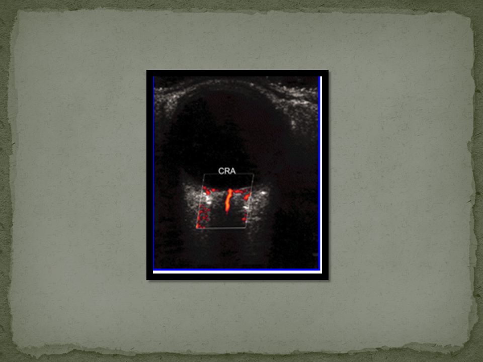
1 Opacity of light-conducting media, making direct vision by ophthalmoscopy difficult 2 Suspected intraocular tumour-solid lesions are readily diagnosed, sited and measured by ultrasound 3 Differentiation of serous and solid retinal detachment; a detachment may conceal a tumour-the subretinal area is clearly demonstrated by ultrasound 4 Examination of the vitreous 5 Localisation of foreign bodies 6 Ocular measurements (biometry by calibrated A-scan) 7 Proptosis (CT and MRI are usually more helpful) 8 Doppler investigation of orbital vascular disease and tumours.
 7 Proptosis (CT and MRI are usually more helpful) 8 Doppler investigation of orbital vascular disease and tumours.")
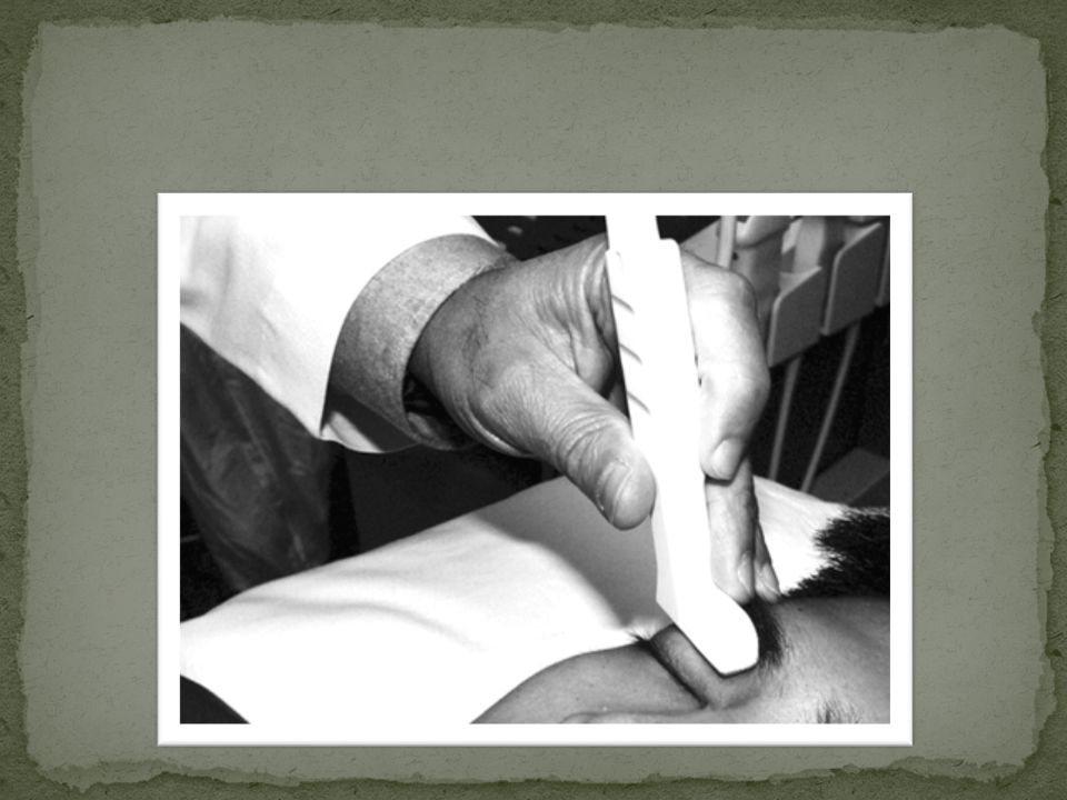
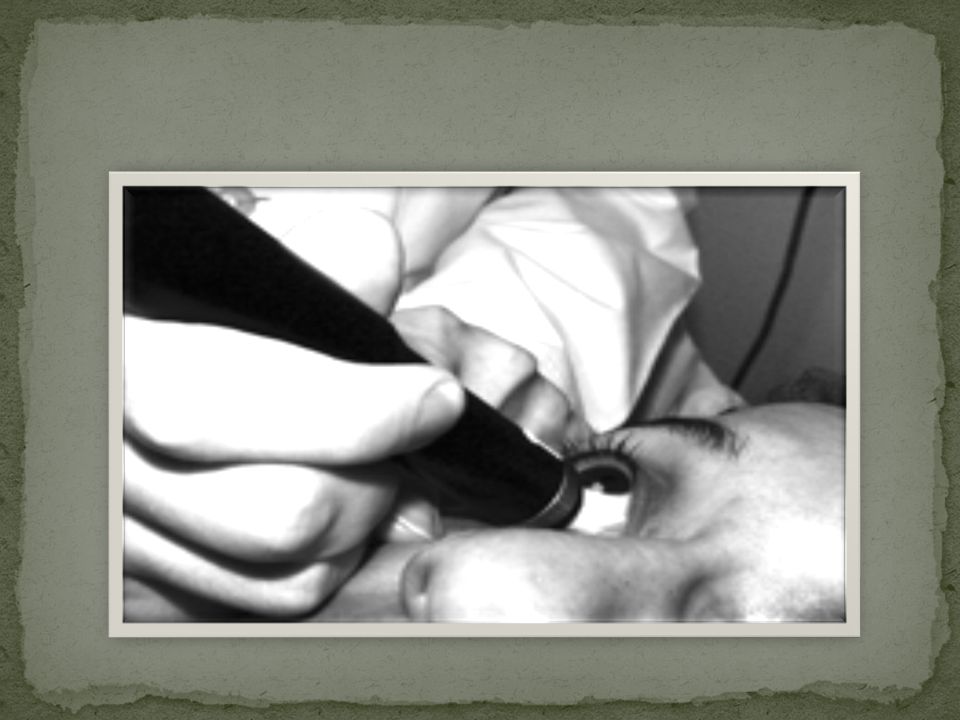
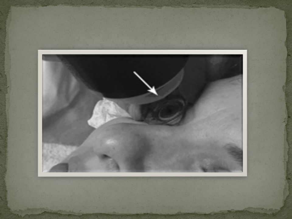
3
Patients with opaque light-conducting media form the majority of referrals, especially those with cataracts and haemorrhages. It is not necessary to scan every patient with a cataract, but if other symptoms develop, for example inflammation, pain, rapidly worsening vision or the development of glaucoma, then a scan must be performed to determine any coexistent pathology. When vitreoretinal surgery is contemplated, ultrasound assessment of the globe is mandatory. The information required includes: The state of the vitreous The position and extent of any intraocular lesion visible by ultrasound The condition of the retina, and particularly the macula The mobility of the contents of the globe, which has a direct influence on operability The relation between the vitreous and retina, mapping out any vitreoretinal adhesions.

12
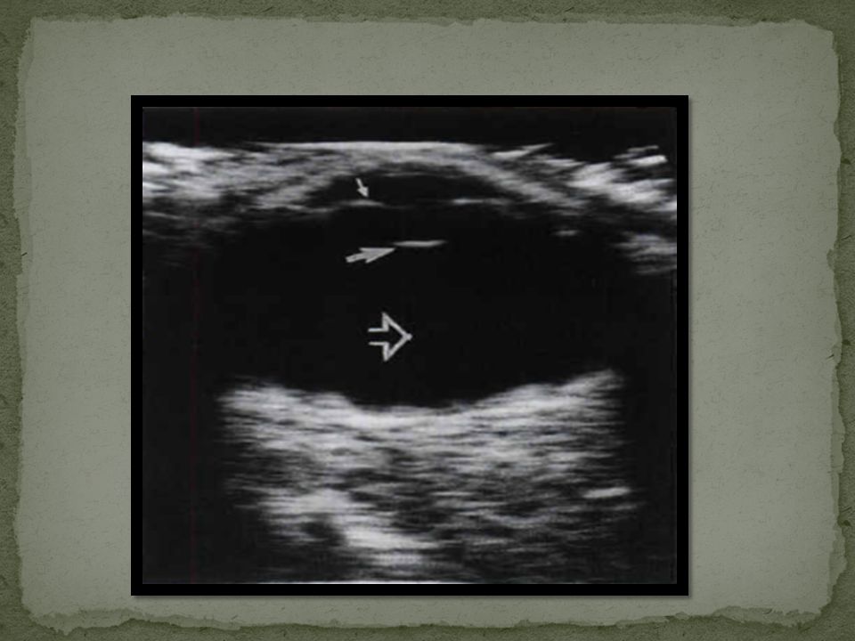
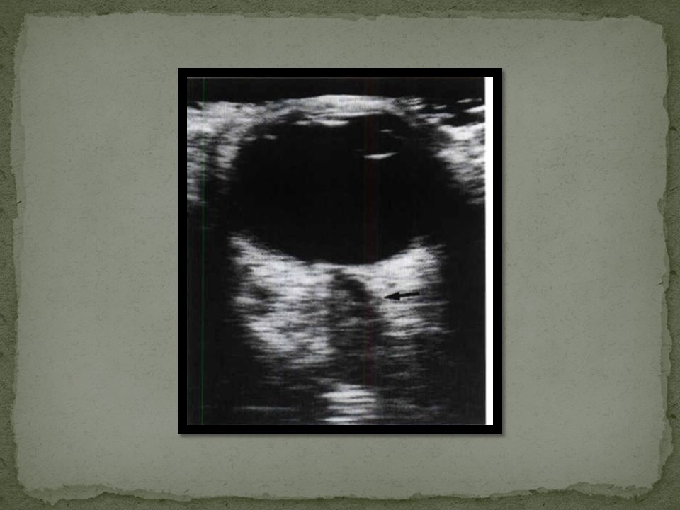
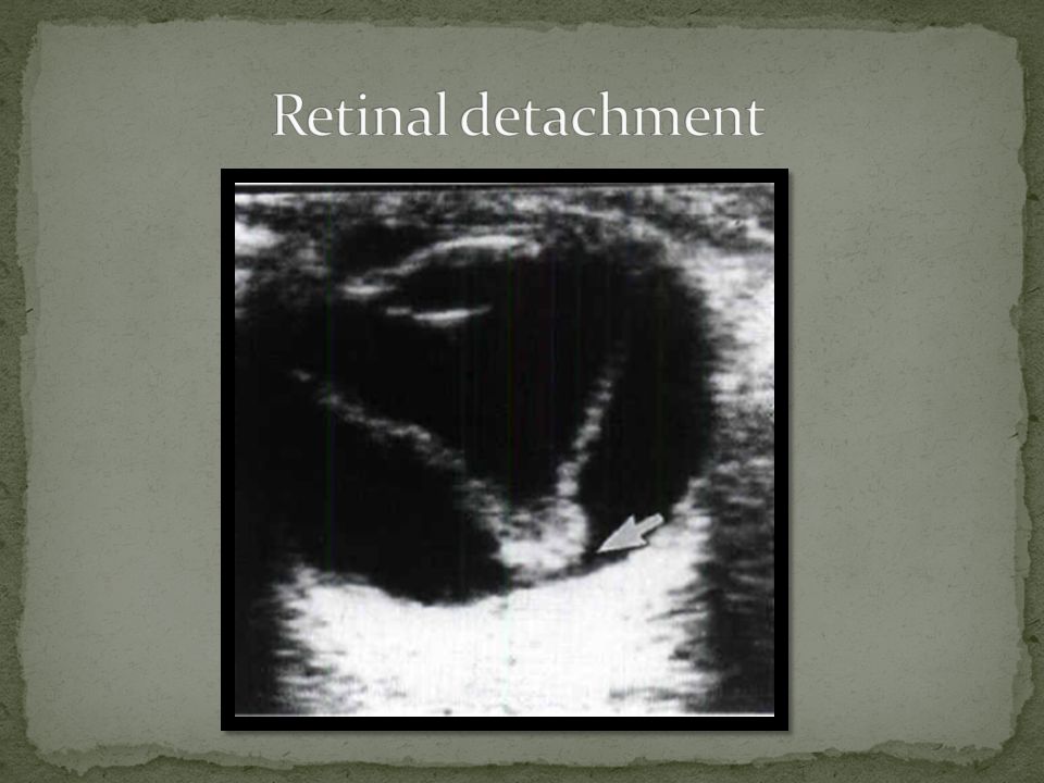
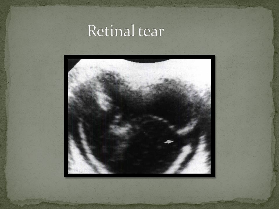
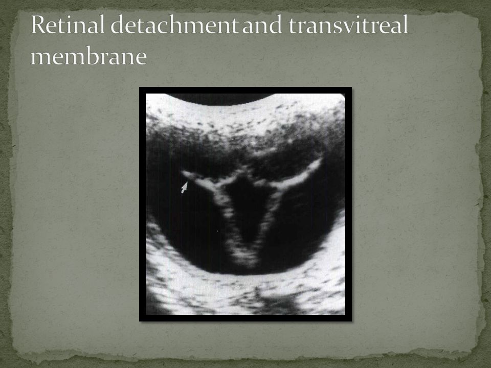
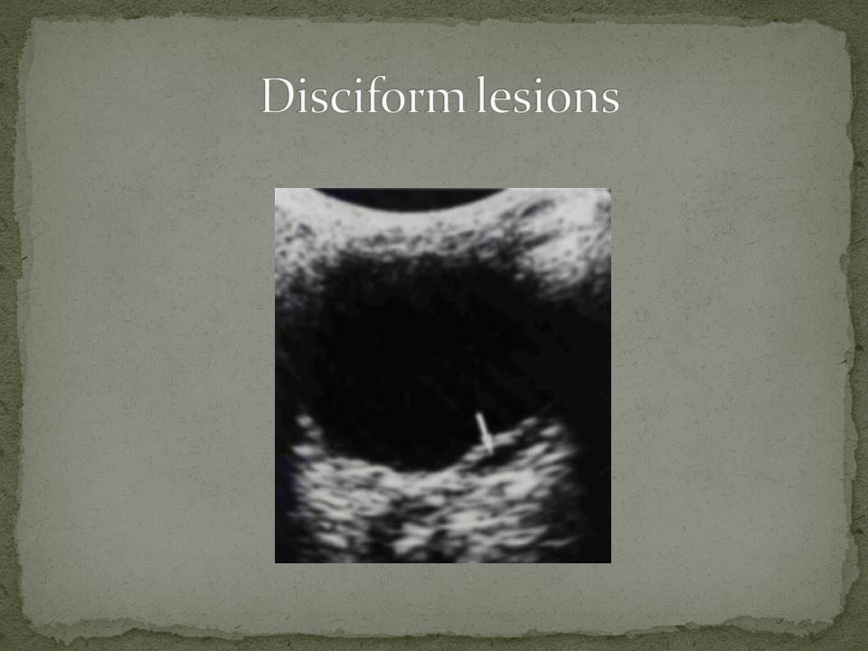
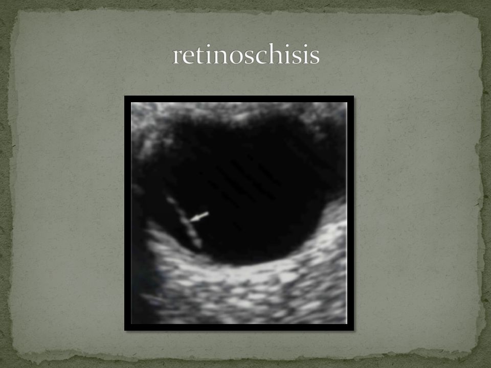
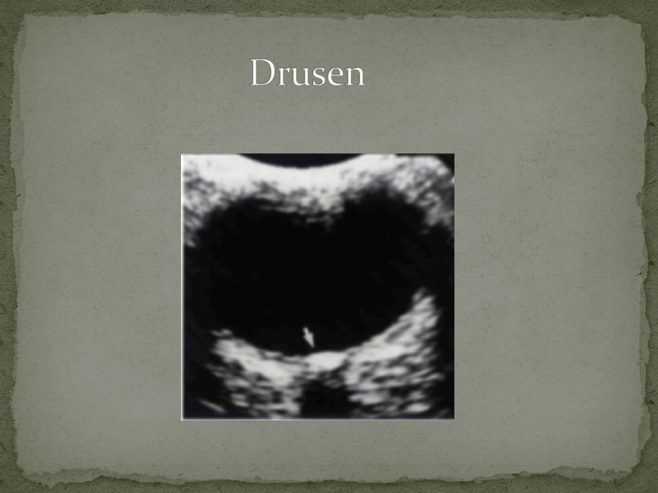
Retinal detachment Acquired retinoschisis Disciform lesions Drusen(hyalin bodies)
")
21
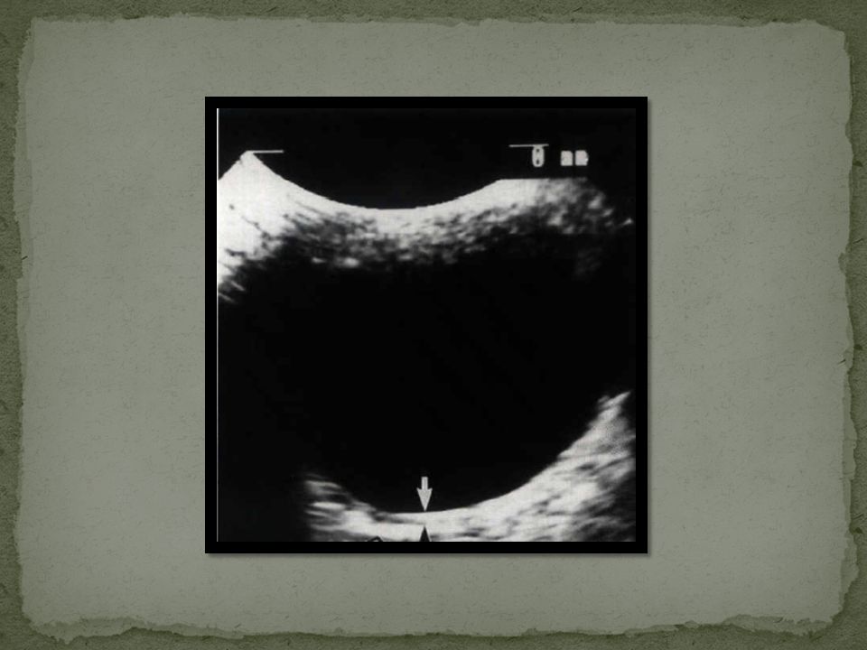
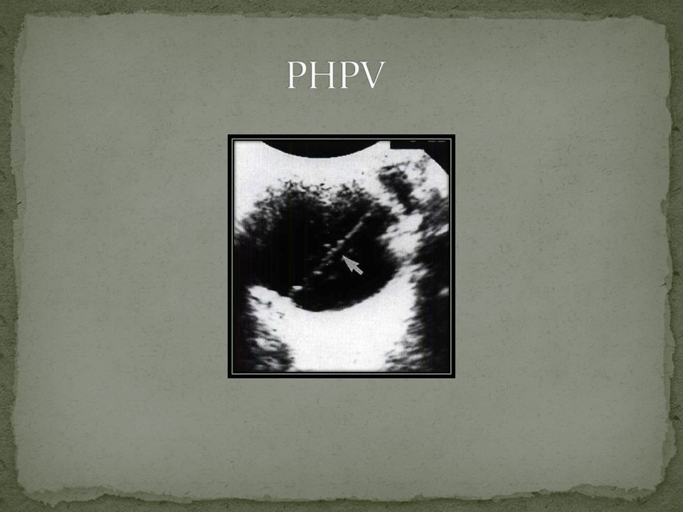
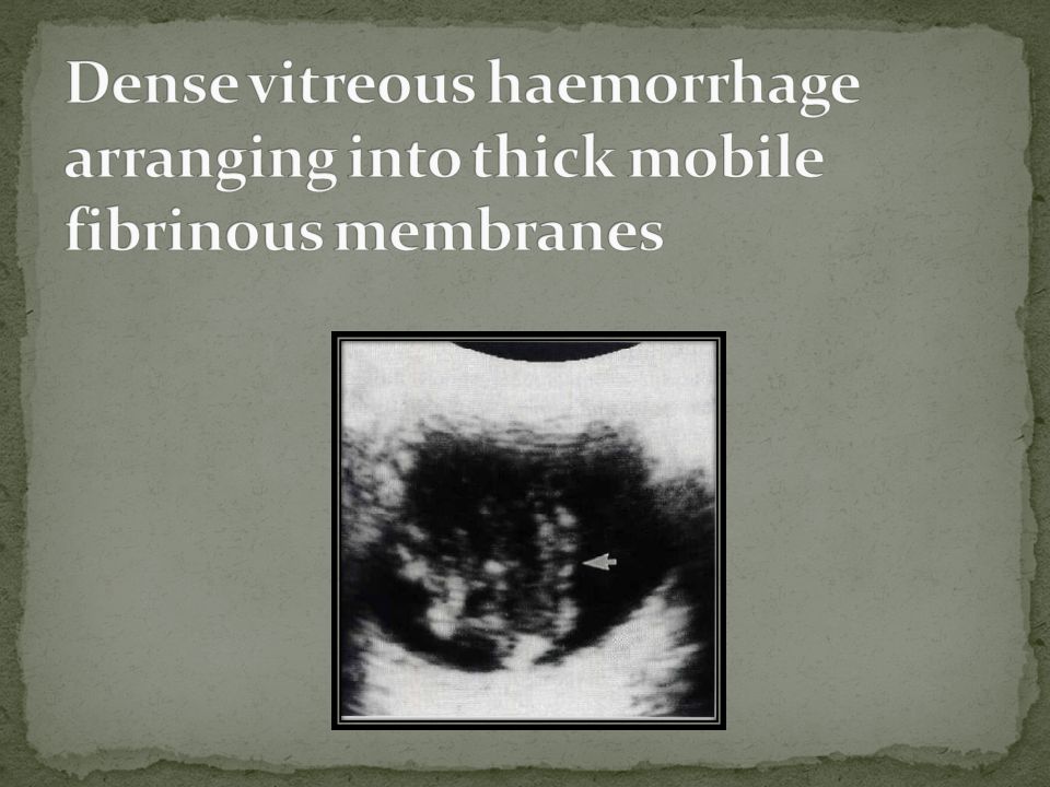
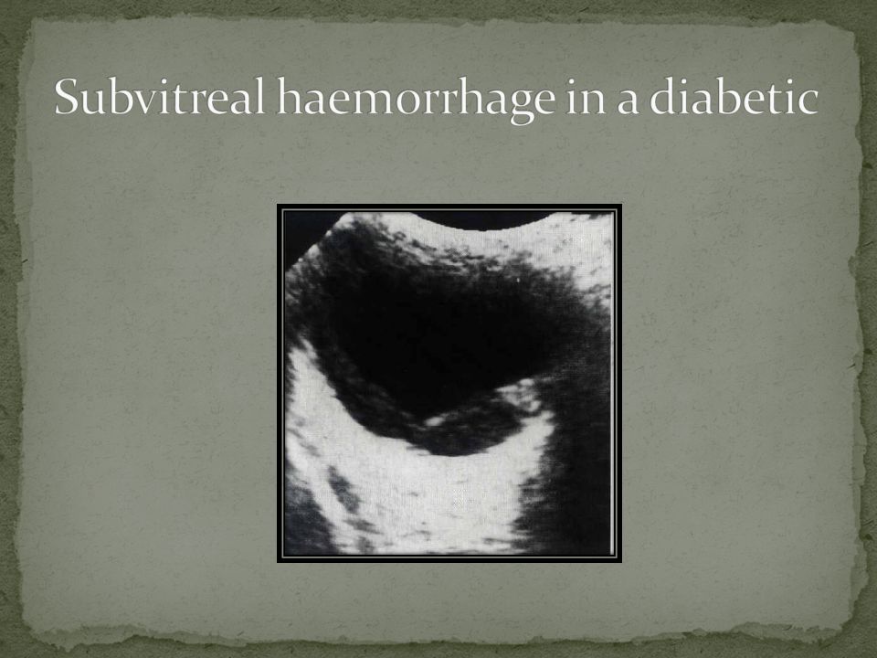
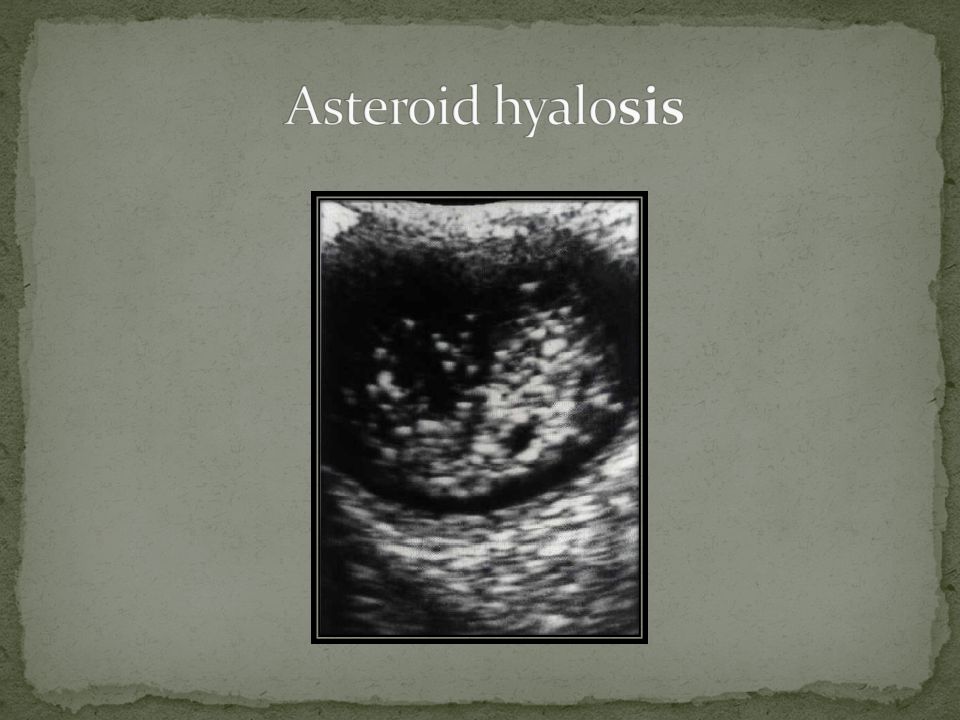
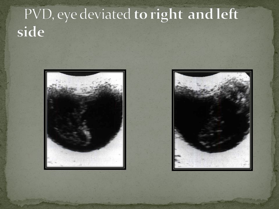
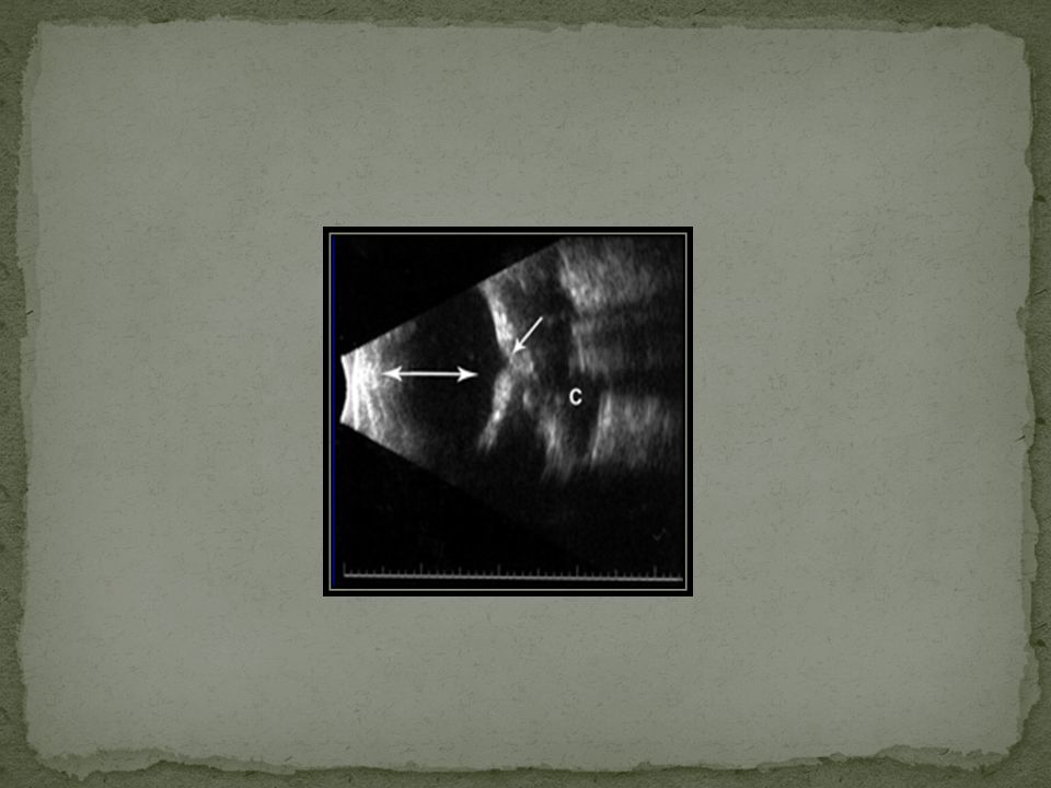
Persistant hyperplastic primary vitreos Vitreous hemorrhage Asteroid hyalosis Posterior vitreous detachment

36
Sonography of the eye shows a variety of diseases with remarkable clarity. The technique is more cost- efficient than other diagnostic techniques and is well tolerated by the patient. We have experienced no limitations and have received no complaints from patients. We do not advocate the routine use of sonography in the asymptomatic eye, but it may serve as a useful extension of the initial investigation of the symptomatic patient.

Similar presentations


1 RETINOBLASTOMA. 2 RETINOBLASTOMA It is the most common primary ocular malignancy of childhood. It formed 15% of all childhood cancers.>")


