Download presentation
Presentation is loading. Please wait.

1
Dr.Amal Al-Abdulkareem
Breast disease Dr.Amal Al-Abdulkareem

2
Breast Modified Sebaceous Glands
Upper border - Collar bone. Lower border th or 7th rib. Inner Border Edge of sternum. Outer border - Mid-axillary line.

3
Breast Divisions 5 Segments
Four Quadrants - By horizontal and vertical lines. Tail of Spence Majority of benign or malignant tumors in the Upper Outer Quadrant

4
External Anatomy of the Breast
Nipple -Pigmented, Cylindrical -4th inter-costal space * at age 18 Areola -Pigmented area surrounding nipple Glands of Montgomery -Sebaceous glands within the areola -Lubricate nipple during lactation

5
Montgomery’s Tubercles
Blocked Montgomery Tubercle

6
Terminal Lobular Unit and Branching Systems of Ducts

7
Anatomy Axillary lymph nodes defined by pectoralis minor muscle:
- Level 1 – lateral - Level 2 – posterior - Level 3 – medial Long Thoracic Nerve - Serratus anterior Thoracodorsal Nerve - Latissimus Dorsi Intercostalbrachial Nerve - Lateral cutaneous - Sensory to medial arm & axilla

8
Tissue Types Glandular Tissue - Milk producing tissue Fibrous Tissue
Fatty Tissue

9
Internal Anatomy of the Breast

10
Fibrous Tissue Cooper’s Ligaments -Suspensor ligaments
- Extending through the breast to underlying muscle Benign or malignant lesions may affect these ligament Skin retraction or dimpling

11
Fatty Tissue Subcutaneous and retro-mammary fat Bulk of breast.
No fat beneath areola and nipple

12
Chest Muscles Pectoralis Major/Minor Serratus Anterior
Latissimus Dorsi Subscapularis External Oblique Rectus Abdominus

13
Lymph Nodes Most drain towards axilla.
Superficial lymphatic nodes drain skin . Deep lymphatic nodes drain mammary lobules

14
Lymph Drainage of Breast

15
Levels of Axillary Nodes

16
Lymph Nodes Palpate ALL nodes - From distal arm to under arm with deep palpation Axillary Supraclavicular Infra-clavicular Nodes deep in the chest or abdomen Infra-mammary ridge - Shelf in the lower curve of each breast

17
Normal Variations of Breast
Accessory breast tissue. Supernumerary nipples. Hair Lifelong Asymmetry

18
Milk Lines Sites of Accessory Nipples and Breasts

19
Accessory Beast Tissue
Accessory Tissue Biopsy

20
Accessory Nipple

21
Accessory Nipple and Bilateral Accessory Breasts

22
Breast with Two Nipples

23
Breast Hair

24
Physiology of Breast Puberty - Need estrogen and progesterone
Estrogen - Growth and appearance - Milk-producing system Progesterone - Lobes and alveoli - Alveolar cells become secretory Asymmetry is common.

25
Breast Asymmetry

26
Breast Asymmetry

27
Physiology of Breast Pregnancy and lactation
Glandular tissue displaces connective tissue Increase in size Nipples prominent and darker Mammary vascularization increases Colostrum present Attain Tanner Stage V with birth

28
Physiology of Breast Aging Perimenopause Flatten, elongate, pendulous
- Decrease in glandular tissue - Loss of lobular and alveolar tissue Flatten, elongate, pendulous Infra-mammary ridge thickens Suspensory ligaments relax Nipples flatten Tissue feels “grainy”

29
Clinical Breast Exam

30
Clinical Exam Inspection Palpable Skin Symmetry Masses Gland
Axilla, Supraclavicular spaces Nipple-areola complex

31
Inspect Both Breasts

32
Skin Dimpling and Change in Contour
Dimpling due to Carcinoma Change in contour due to carcinoma

33
Skin Dimpling Both Breasts Involution Due to Aging

34
Skin Dimpling Breast Infection

35
Skin Dimpling Previous Breast Surgery

36
Inverted Nipple Since Puberty

37
Palpate Axilla and Clavicular Nodes

38
Breast Palpation

39
Common Benign Breast Disorders

40
Common Benign Breast Disorders
Fibrocystic changes Fibroadenoma Intraductal papilloma Mammary duct ectasia Mastitis Fat necrosis Phylloides tumor Male gynecomastia

41
Fibrocystic Changes Lumpy, bumpy breasts
50-80% of all menstruating women Age % in women less than 21 Caused by hormonal changes prior to menses Relationship to breast cancer doubtful

42
Fibrocystic Disease Histology Adenosis Apocrine metaplasia Fibrosis
Duct ectasia Mild duct ectasia

43
Signs and Symptoms Mobile cysts with well-defined margins
Singular or multiple May be symmetrical Upper outer quadrant or lower breast border

44
Signs and Symptoms Pain and tenderness
Cysts may appear quickly and decrease in size Lasts half of a menstrual cycle Subside after menopause -If no HRT

45
Breast Mass Breast Cysts Fluid-filled 1 out of every 14 women
50% multiple and recurrent Hormonally influenced Needle aspirated

46
Breast Mass

47
Treatment Aspirate cyst fluid Imaging for questionable cysts
Treatment based on symptoms Reassure ‘’Atypical Hyperplasia’’ on pathology report indicates increased risk of breast cancer

48
Breast Pain Cyclical pain – hormonal Non-cyclical pain
Dull, diffuse and bilateral Luteal phase Treatment: Reassurance, NSAIDS, evening primrose oil Non-cyclical pain Non-breast vs breast Imaging

49
Fibroadenoma Second most common breast condition
Most common in black women Late teens to early adulthood Rare after menopause

50
Fibroadenoma

51
Signs and Symptoms Firm, rubbery, round, mobile mass
Painless, non-tender Solitary % are multiple Well circumscribed Upper-outer quadrant 1-5 cm or larger

52
Mammogram Multiple Calcified Fibroadenomas

53
Intraductal Papilloma
Slow-growing Overgrowth of ductal epithelial tissue Usually not palpable Cauliflower-like lesion Length of involved duct Most common of bloody nipple discharge 40-50 years of age

54
Signs and Symptoms Watery, serous, serosanguinous, or bloody discharge
Spontaneous discharge Usually unilateral Often from single duct - Pressure elicits discharge from single duct 50% no mass palpated

55
Bloody Breast Discharge

56
Treatment Test for occult blood Ductogram Biopsy
Excision of involved duct

57
Intraductal Papilloma

58
Galactorrhea

59
Mammary Duct Ectasia Inflammation and dilation of sub-areolar ducts
behind nipples May result in palpable mass because of ductal rupture Greatest incidence after menopause Etiology Unclear - Ducts become distended with cellular debris causing obstruction

60
Mammary Duct Ectasia versus Breast Cancer
Left breast – slit-like nipple characteristic of mammary duct eclasia Right breast – nipple retraction from carcinoma

61
Signs and symptoms Multi-colored discharge - Thick, pasty (like toothpaste) - White, green, greenish-brown or serosanguinous Intermittent, no pattern Bilaterally from multiple ducts Nipple itching Drawing or pulling (burning) sensation
 sensation.")
62
Dried Secretions from Mammary Duct Ectasia

63
Yellow Breast Discharge Duct Ectasia

64
Multi-colored Breast Discharge

65
Treatment Test for occult blood Imaging - Mammogram - Sonogram
Biopsy - Excision of ducts if mass present Antibiotics Close follow-up

66
Mastitis Breast infection when bacteria enter the breast via the nipple Ducts infected Fluid stagnates in lobules Usually during lactation Penicillin resistant staphylococcus common cause

67
Mastitis Treatment Antibiotics Continue breast feeding Close follow-up

68
Puerperal Mastitis

69
Puerperal Mastitis Left Breast

70
Inflammatory Carcinoma
Erythema and peau d’orange

71
Signs and Symptoms of Mastitis
Pain Nipple discharge - Pus - Serum - Blood Localized induration Fever Breast Abscess

72
Breast Abscess

73
Non-Lactating Breast Abscess
Arrow points to inverted nipple

74
Breast Abscess Treatment Antibiotics Needle aspiration
Incision and drainage

75
Draining Breast Abscess

76
Abscess Drained under Local Anesthesia

77
Puerperal Breast Abscess
Before treatment Local anesthetic After treatment Abscess occurred during lactation

78
Peripheral Breast Abscess
Left – before management Right – after recurrent aspiration and antibiotics

79
Fat Necrosis Cause - Trauma to breast - Surgery
Necrosis of adipose tissue Pain or mass - Usually non-mobile mass - Resolves over time without treatment may be excised

80
Fat Necrosis Seat Belt Trauma

81
Breast Hematoma

82
Phylloides Tumor Giant fibroadenoma with rapid growth
Malignant potential Often occurs in women aged 40+ Treatment - Excision

83
Giant Fibroadenoma Before Surgery After Surgery

84
Cross Section of Giant Fibroadenoma

85
Malignant Phylloides Tumor

86
Left-Sided Gynecomastia

87
Treatment If pre-puberty - Wait to see if it resolves
Change medication Treat underlying illness Occurs in families with genetic mutation - Colon, prostate cancer

88
Differential Diagnosis of Nipple Discharge
Common causes in non-pregnant women Carcinoma Intraductal papilloma Fibrocystic changes Duct ectasia Hypothyroid Pituitary adenoma

90
Clinical Characteristic
Physiologic - Usually bilateral - Multiple ducts - Non-spontaneous - Screen for phenothiazine use and stimulation

91
Physiological Breast Discharge

92
Clinical Characteristic
Pathologic discharge - Spontaneous - Unilateral - Single duct - Discolored discharge Bloody discharge

93
Bloody Nipple Discharge

94
Mammography Screening tool - Age of 40
Estimated reduction in mortality 15 – 25% 10% false positive rate Densities and calcification

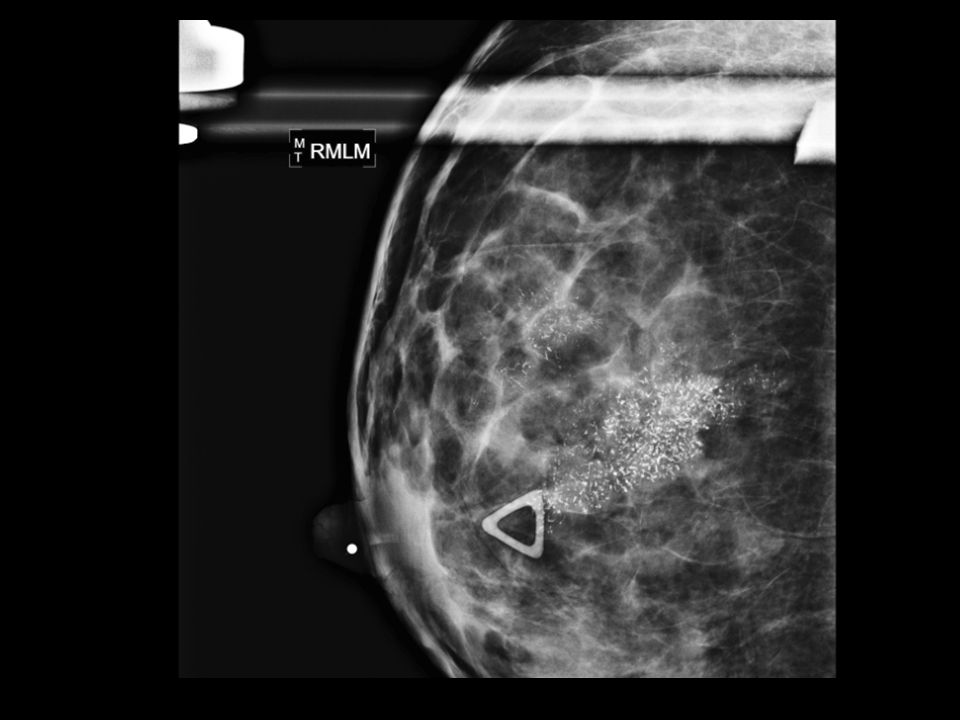
95
Calcification Macrocalcifications - Large white dots - Almost always non-cancerous and require no further follow-up Microcalcifications - Very fine white specks - Usually non-cancerous but can sometimes be a sign of cancer - Size, shape and pattern

99
Features BI-RADS Classification Need additional imaging 1
Need additional imaging 1 Negative – routine in 1 year 2 Benign finding – routine in 1 year 3 Probably benign – 6 month follow-up 4 Suspicious abnormality – biopsy recommended 5 Highly suggestive of malignancy – appropriate action must be taken

100
Ultrasound Benign Malignant Pure hyperechoic Hypoechoic, spiculated
Elliptical shape (wider than tall) Taller than wide Lobulated Duct extension Complete tine capsule Microlobulation
 Taller than wide. Lobulated. Duct extension. Complete tine capsule. Microlobulation.")
101
Ultrasound

102
MRI High risk patients - History of breast cancer - LCIS, atypia - 1st degree relative with breast cancer - Very dense breast High sensitivity - 10 – 20% will have a biopsy

103
Diagnosis Fine needle aspiration - Cytology Core biopsy
- Image guided - Stereotactic Excisional biopsy - Needle localization

104
Fine Needle Aspiration
Fast, inexpensive 96% accuracy Institution dependent Unable to differentiate between in-situ vs CA

105
Core Needle Biopsy 14 – 18 gauge spring loaded needle Tissue Multiple

107
Large Core Biopsy 6 – 14 gauge core Large Samples Single insertion

108
Core Biopsy Vacuum Assisted

109
Stereotactic Biopsy Suspicious mammographic abnormalities
Patients lay prone

112
Excisional Biopsy Atypical lesions LCIS Radial scar
Atypical papillary lesions Radiologic-pathologic discordance Phyllodes Inadequate tissue harvesting

113
Screening Prior breast cancer or atypia - Annual mammography month CBE Family Hx - 10 years younger than relative’s diagnosis - 6 month CBE BRCA y.o, annual mammography month CBE

114
Genetics Early age of onset 2 breast primaries or breast & ovarian CA
Clustering of breast CA with: - Male breast CA - Thyroid CA - Sarcoma - Adrenocortical CA - Pancreatic CA - Leukemia/Lymphoma on same side of family Family member with BRCA gene Male breast CA Ovarian CA

115
BRCA Account for 25% of early-onset breast cancers
% lifetime risk of breast cancer 16 – 60% lifetime risk of ovarian cancer

116
BRCA Management Monthly BSE – 18 y.o
6 month CBE & annual mammo – 25 y.o Discuss risk reducing options Prophylactic Mastectomies Salpingo-oophorectomy upon completion of child bearing 6 month transvaginal US & CA125 – 35. y.o

117
Any Questions?

Similar presentations