Download presentation
Presentation is loading. Please wait.

1
CHAPTER II: CARDIAC MECHANICS Asst. Prof. Dr. Emre Hamurtekin EMU Faculty of Pharmacy

2
1.CARDIAC CYCLE 2.CARDIAC OUTPUT 3.DETERMINANTS of CARDIAC OUTPUT 4.CARDIAC WORK

3
encyclopedia.lubopitko-bg.com

4
medical-dictionary.thefreedictionary.com

5
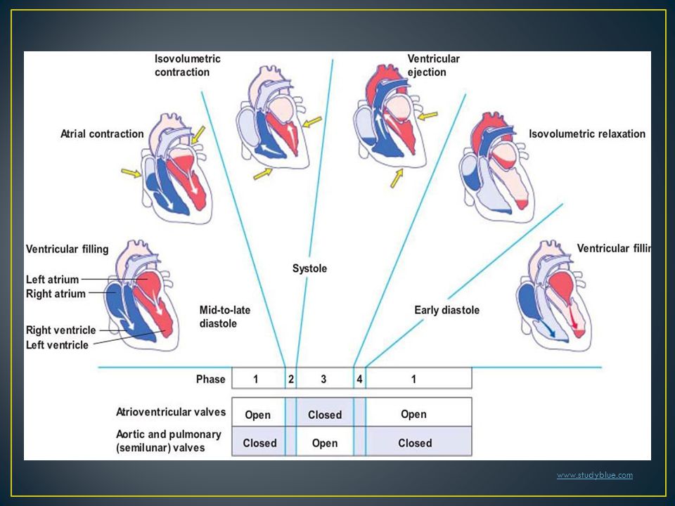
Cardiac cycle can be divided into seven phases: 1.Atrial systole 2.Isovolumic ventricular contraction 3.Rapid ventricular ejection 4.Reduced ventricular ejection 5.Isovolumic ventricular relaxation 6.Rapid ventricular filling 7.Reduced ventricular filling

6
Atrial systole is initiated by …………………... Atrial systole follows the crest of P wave on the ECG. Atrial contraction forces a small additional blood into the venticular chamber (atrial kick). isovolumic ventricular contraction Ventricular systole begins with isovolumic ventricular contraction. In isovolumic ventricular contraction, when intraventricular pressure rises, mitral valve closes. In isovolumic ventricular contraction, aortic valve is still held closed by higher aortic pressure. atrial excitation
. isovolumic ventricular contraction Ventricular systole begins with isovolumic ventricular contraction. In isovolumic ventricular contraction, when intraventricular pressure rises, mitral valve closes. In isovolumic ventricular contraction, aortic valve is still held closed by higher aortic pressure. atrial excitation.")
7
www.studyblue.com

8
hypocaffeinic.pbworks.com

9
Rapid Ventricular Ejection In Rapid Ventricular Ejection phase, aortic valve finally opens and blood exits the ventricle. In this phase, atrium relaxes and the blood starts to fill the atrium. Reduced Ventricular Ejection In Reduced Ventricular Ejection phase, ejection velocity decreases (reduced ejection). At the end of this phase, aortic valve is finally closed. Isovolumic Ventricular Relaxation Once the aortic valve is closed, Isovolumic Ventricular Relaxation period starts. In this period, LV volume is lowest.
. At the end of this phase, aortic valve is finally closed. Isovolumic Ventricular Relaxation Once the aortic valve is closed, Isovolumic Ventricular Relaxation period starts. In this period, LV volume is lowest..")
10
www.studyblue.com
11
hypocaffeinic.pbworks.com

12
Once intraventricular pressure drops below atrial pressure, mitral valve opens. Rapid Ventricular Filling Blood in the atrium starts to move into the ventricle in Rapid Ventricular Filling phase. Reduced Ventricular Filling (diastasis) In Reduced Ventricular Filling (diastasis) phase, atrium and ventricle are both fully relaxed. Arterial pressure continues to fall as blood flows into capillary beds. This phase typically disappears when HR increases.
 In Reduced Ventricular Filling (diastasis) phase, atrium and ventricle are both fully relaxed. Arterial pressure continues to fall as blood flows into capillary beds. This phase typically disappears when HR increases..")
13
www.studyblue.com

14
hypocaffeinic.pbworks.com

15
LV does not empty completely during systole. ESV is around 50 ml. EDV - ESV = SV (stroke volume). EDV - ESV = SV (stroke volume). SV is the amount of blood transferred from LV to the arterial system during systole. In healty person SV should be > 60 ml. EF (ejection fraction) = SV \ EDV EF (ejection fraction) = SV \ EDV (normally about 55% - 75%. EF is an important measurement of cardiac efficiency. EF is used clinically to assess cardiac status in patients with heart failure.
. EDV - ESV = SV (stroke volume). SV is the amount of blood transferred from LV to the arterial system during systole. In healty person SV should be > 60 ml. EF (ejection fraction) = SV \ EDV EF (ejection fraction) = SV \ EDV (normally about 55% - 75%. EF is an important measurement of cardiac efficiency. EF is used clinically to assess cardiac status in patients with heart failure..")
16
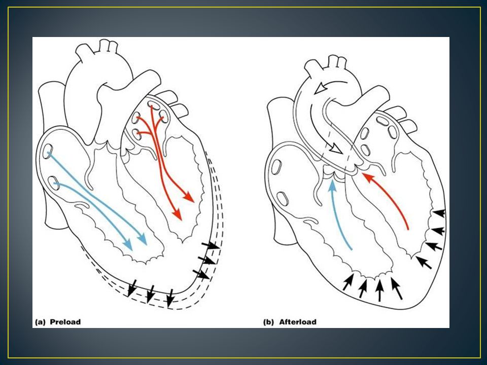
CO (L/min) = HR x SV CO (L/min) = HR x SV HR is established on the SA node and is controlled by ANS. SV is dependent on SV is dependent on, LV preload LV afterload Contractility Preload Preload: Muscle length before contraction begins. EDV Preload is related with the volume of blood entering the chamber (EDV) Afterload Afterload: The load against which a myocyte must shorten. The principal component of afterload is arterial pressure. Contractility Contractility: measure of a muscle’s ability to shorten against a afterload. Contractility equates with the cytoplasmic free Ca concentration.
 Afterload Afterload: The load against which a myocyte must shorten. The principal component of afterload is arterial pressure. Contractility Contractility: measure of a muscle’s ability to shorten against a afterload. Contractility equates with the cytoplasmic free Ca concentration..")
18
Ability of a muscle cell to develop force (contractility) (+) inotropic agents -Epinephrine -Norepineprine -Digoxin (-) inotropic agents - β - blockers -Ca channel blockers Ca Contractility equates with intracellular free Ca concentration SNS norepinephrine β -1 receptors cAMP PKA activation L - type Ca channels, Ca release channels, SERCA
 (+) inotropic agents -Epinephrine -Norepineprine -Digoxin (-) inotropic agents - β - blockers -Ca channel blockers Ca Contractility equates with intracellular free Ca concentration SNS norepinephrine β -1 receptors cAMP PKA activation L - type Ca channels, Ca release channels, SERCA")
19
Heart performs two kinds of work: I.Internal work II.External work Internal work Internal work: Expended in «isovolumic contraction» The force necessary to open the aortic and pulmonary valves Accounts for ˃ 90% of total cardiac workload. External work (pressure-volume work) External work (pressure-volume work) : Expended in transferring blood to the arterial system against a resistance. Accounts for ˂ 10% of total cardiac workload.
 External work (pressure-volume work) : Expended in transferring blood to the arterial system against a resistance. Accounts for ˂ 10% of total cardiac workload..")
Similar presentations