Download presentation
Presentation is loading. Please wait.

1
Jeroen Hermans, Frederik Maes, Dirk Vandermeulen, Paul Suetens
Unified Framework for Automatic Segmentation, Probabilistic Atlas Construction, Registration and Clustering of Brain MR Images Annemie Ribbens Jeroen Hermans, Frederik Maes, Dirk Vandermeulen, Paul Suetens

2
Introduction Computer–aided diagnosis

3
Introduction Segmentation

4
Introduction Atlas & Atlas-to-image registration Φ

5
Introduction Population Specific Atlases

6
Introduction Atlas Construction Images I Registrations
Atlas previous iteration Registrations

7
Images I Deformed images New Atlas Averaging

8
Introduction Computer aided-diagnosis Registration Segmentation
Prob. Atlases Registration

9
Framework Aspects: Advantages: Segmentation
Clustering (i.e. computer-aided diagnosis) (+ Localization of cluster specific morphological differences) Groupwise registration (nonrigid probabilistic atlases per cluster) Atlas-to-image registration Advantages: Less prior information necessary Cooperation Statistical framework convergence
 (+ Localization of cluster specific morphological differences) Groupwise registration (nonrigid probabilistic atlases per cluster) Atlas-to-image registration. Advantages: Less prior information necessary. Cooperation. Statistical framework convergence.")
10
Framework Segmentation Atlas-to-image registration Atlas formation &
Clustering

11
Framework: model K = tissue classes number of Gaussians
Y = intensities Image i

12
Framework: model Atlas t (Gray matter map) Image i (Gray matter map)
 Image i (Gray matter map)")
13
Framework: model Uniform prior for all voxels in an image

14
Framework: model G1 G2 Deformations

15
Framework: MAP MAP: Jensen’s inequality
Expectation maximization framework

16
Framework: EM algorithm: E-step
i = images j = voxels k = tissue classes t = clusters Per cluster: atlas deformed towards image Gaussian prior on the deformations of each cluster Uniform prior on the cluster memberships Gaussian mixture model

17
Framework: EM Posterior
Posterior = (clustering) * (segmentation using the atlas of the same cluster) Clustering = probability that voxel j of image i belongs to cluster t = sum over all tissue classes of the posterior = (prior of clustering) * (atlas is sharp & close to intensity model) * (subject specific registration close to groupwise) Segmentation = probability that a voxel belongs to a certain tissue class = sum over all clusters of the posterior = weighted sum of the segmentations using a specific atlas
 * (segmentation using the atlas of the same cluster) Clustering = probability that voxel j of image i belongs to cluster t. = sum over all tissue classes of the posterior. = (prior of clustering) * (atlas is sharp & close to intensity model) * (subject specific registration close to groupwise) Segmentation = probability that a voxel belongs to a certain tissue class. = sum over all clusters of the posterior. = weighted sum of the segmentations using a specific atlas.")
18
Framework: EM algorithm: M-step
Maximum likelihood Q-function parameters All solutions close form (except registration) Solutions (e.g. atlas) ~ literature
 Solutions (e.g. atlas) ~ literature.")
19
Framework: EM algorithm: M-step
Gaussian mixture parameters: Atlas Prior cluster memberships Groupwise registration Atlas-to-image registration No closed form solution Spatial regularization Viscous fluid model on derivative Weighting terms per voxel

20
w1 w8

21
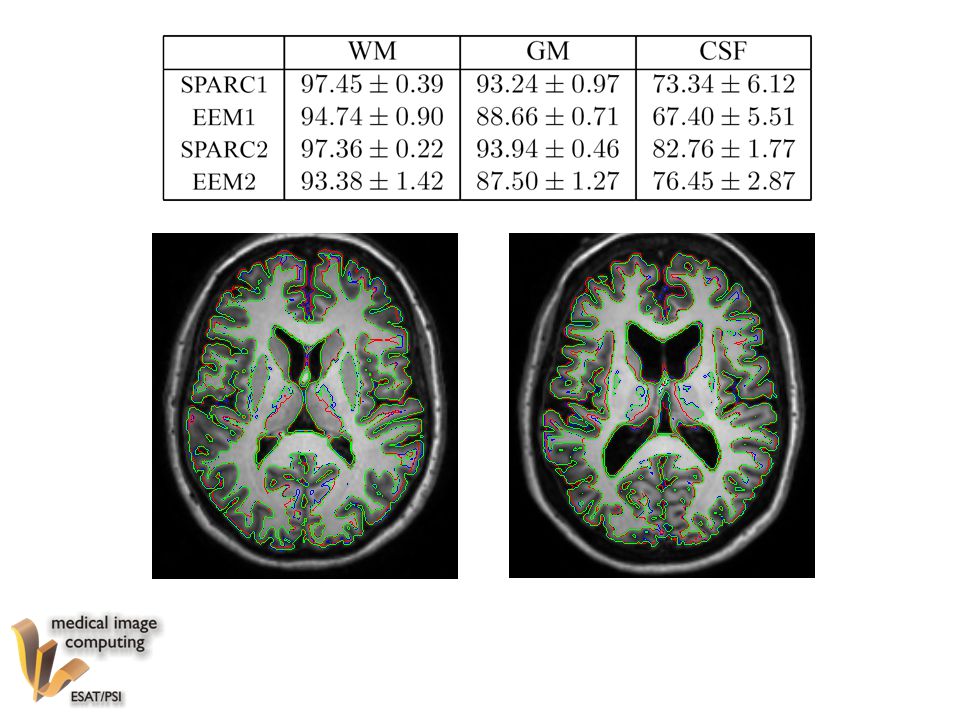
Experiments Brainweb data: 20 simulated normal images One cluster
Segmentation & Atlas: Dice =

22
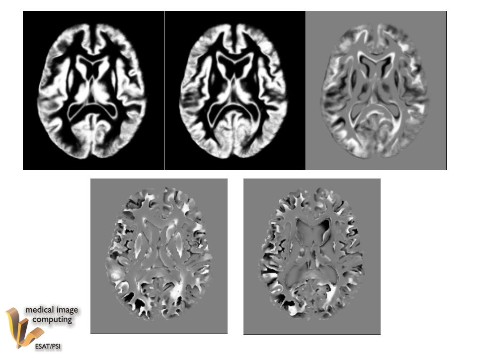
Experiments 8 brain MR images of healthy persons (normals)
8 brain MR images of Huntington disease patients (HD) Cluster memberships: all correctly classified
 Cluster memberships: all correctly classified.")
25
Conclusion Statistical framework combining: Segmentation Clustering
Atlas construction per cluster (weighted) Registration Convergence & cooperation & less prior information needed Validation promising Cluster specific morphological differences are found Easily extendable to incorporate clinical/spatial prior knowledge
 Registration. Convergence & cooperation & less prior information needed. Validation promising. Cluster specific morphological differences are found. Easily extendable to incorporate clinical/spatial prior knowledge.")
Similar presentations


. Goal : assume the.>")

>")


, Expectation Maximization (EM)>")

