Download presentation
Presentation is loading. Please wait.

1
Crystallography and Diffraction Techniques Myoglobin

2
Types of diffraction - X-ray diffraction - Electron diffraction - Neutron diffraction Enhanced visibility of hydrogen atoms by neutron crystallography on fully deuterated myoglobin Myoglobin diffraction pattern 1962 Nobel Prize by Max Perutz and Sir John Cowdery KendrewMax PerutzSir John Cowdery Kendrew

3
X-ray Diffraction

4
Water

5
Light

6
Electron

7
Constructive

8
Destructive

9
Diffraction from atoms

10
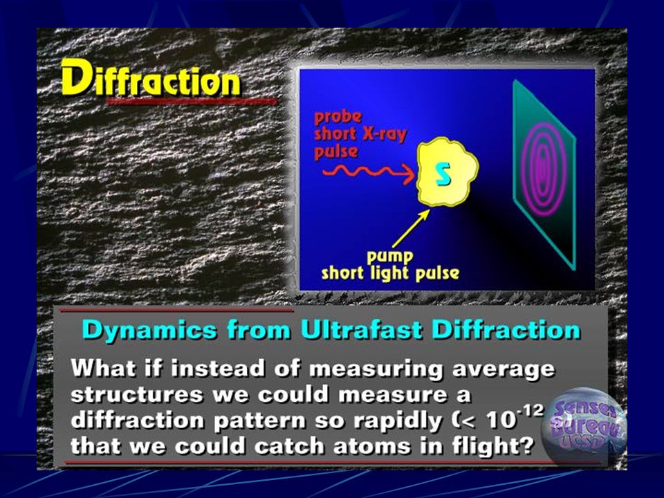
Continue

11
1 A About 1 Å

12
Wave of mater

13
Wave of electrons The electrons are accelerated in an electric potential U to the desired velocity:

14
Crystal diffraction

15
Gas, liquid, powder diffraction

17
Surface diffraction

18
Diffraction by diffractometer

19
Example of spots by diffractometer

20
X-ray Crystallography

21
Electron density

22
Deformation Electron Density

23
Macromolecule X-ray Crystallography

24
Generation of X-rays

25
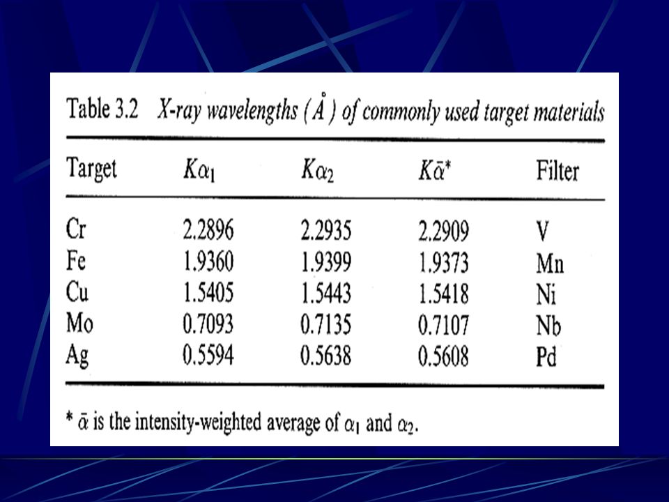
What is K and K (for Cu) ? K : 2p 1s K : 3p 1s
 K : 2p 1s K : 3p 1s")
26
X-ray tube

28
An optical grating and diffraction of light

29
Lattice planes

30
Lattice planes => reflection

31
Lattice planes review

32
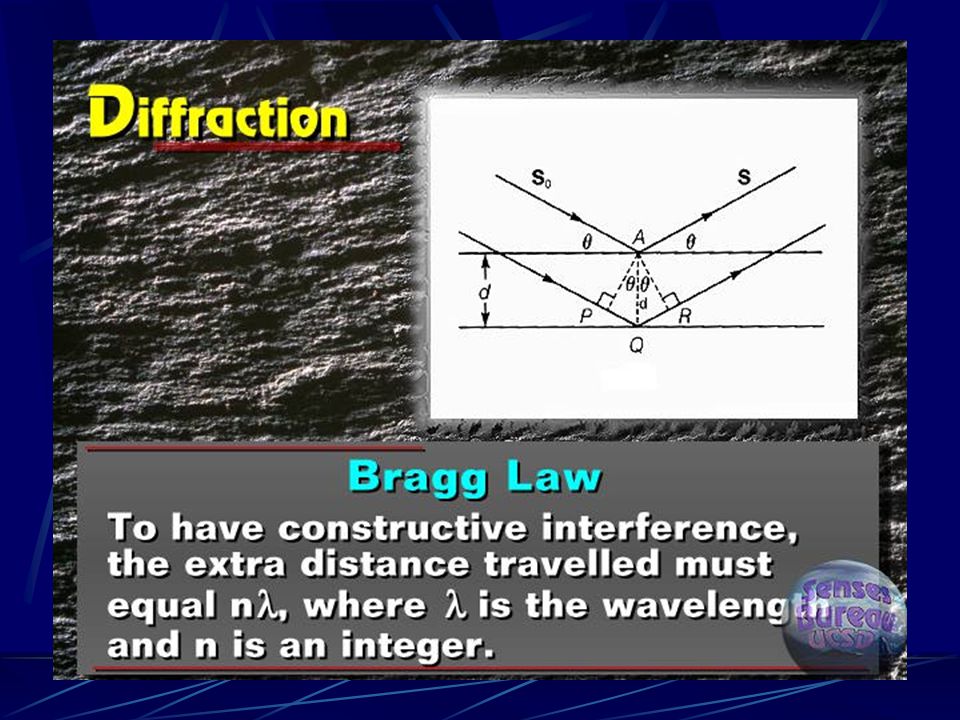
Bragg ’ s Law

35
2dsin(theta)=n lumda
=n lumda")
36
Bragg ’ s Law

37
Atomic scattering factor

39
intensity

40
Phase and intensity

41
Electron density

42
Diffraction of one hole

43
Diffraction of two holes

44
Diffraction of 5 holes

45
2D four holes

46
From real lattice to reciprocal lattice Real holesReflection pattern Crystal lattice is a real lattice, while its reflection pattern is its corresponding reciprocal lattice.

47
TEM image of Si? or Diamond? Real lattice viewed from (110) direction. Si Diamond
 direction. Si Diamond")
48
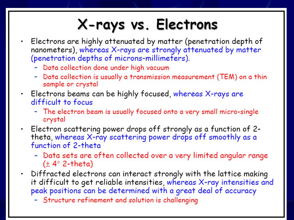
Electron Diffraction

49
Conversion of Real Lattice to Reciprocal Lattice PPP PPP PPP PPP PPP PPP PPP PPP PPP PPP

50
Ewald Sphere and Diffraction Pattern The Ewald sphere is a geometric construct used in X-ray crystallography which neatly demonstrates the relationship between: the wavelength of the incident and diffracted x-ray beams, the diffraction angle for a given reflection, the reciprocal lattice of the crystal Paul Peter Ewald (1888~1985)
")
51
Ewald Sphere

52
A vector of reciprocal lattice represents a set of parallel planes in a crystal lattice 2d sin = n (1/d hkl )/(2/ ) = sin (hkl)
/(2/ ) = sin (hkl) ")
53
Reciprocal Lattice and Ewald Sphere

54
Detector, Reciprocal Lattice and Ewald Sphere

55
3D View of Ewald Sphere and Reciprocal Sphere

56
Techniques of X-ray diffraction Single Crystal and Powder X-ray Diffractions many many many very small single crystals

57
Diffractometers for Single Crystal and Powder X-ray Diffractions

58
Single Crystal and Powder X- ray Diffraction Patterns

59
The powder XRD method

60
Formation of a cone of diffracted radiation

61
XRPD on film electron diffraction of powder sample

62
Finger Print Identification Finger Print Identification for Known Compounds by comparing experimental XRPD to those in PDF database

63
Some peaks may not be observed due to preferred orientation For example, layered structure such as graphite.

64
X-ray powder diffraction patterns of crystalline and amorphous sample

65
Scherrer Formula t = thickness of crystal in Å B = width in radians, at an intensity equal to half the maximum intensity However, this type of peak broadening is negligible when the crystallite size is larger than 200 nm. B is often calculated relative to a reference solid (with crystallite size >500 nm) added to the sample: B 2 =Bs 2 -Br 2.
 added to the sample: B 2 =Bs 2 -Br 2..")
66
2d sin = Some equations to calculate cell parameters (d-spacings)
")
67
X-ray powder diffraction patterns for potassium halides

68
Structure Factor, Intensity and Electron Density R 1 = ||F o | - |F c ||/ |F o | F calc F obs

69
Electron density maps by X-ray diffraction

70
Scattering of X-rays by a crystal-systematic absences

71
Systematic Absences

72
Systematic absence for C-center: (x,y,z) ≣ (x+1/2, y+1/2, z) F hkl = (1/V) f j exp[2 i(hx j +ky j +lz j )] = (1/V) f j [cos2 (hx j +ky j +lz j )+isin2 (hx j +ky j +lz j )] = (1/V) f j {cos2 (hx j +ky j +lz j )+cos2 [h(x j +1/2) +k(y j +1/2)+lz j )]}+i{sin2 (hx j +ky j +lz j ) +sin2 [h(x j +1/2)+k(y j +1/2)+lz j )]} j=1 N N/2
![Systematic absence for C-center: (x,y,z) ≣ (x+1/2, y+1/2, z) F hkl = (1/V) f j exp[2 i(hx j +ky j +lz j )] = (1/V) f j [cos2 (hx j +ky j +lz j )+isin2 (hx j +ky j +lz j )] = (1/V) f j {cos2 (hx j +ky j +lz j )+cos2 [h(x j +1/2) +k(y j +1/2)+lz j )]}+i{sin2 (hx j +ky j +lz j ) +sin2 [h(x j +1/2)+k(y j +1/2)+lz j )]} j=1 N N/2](http://images.slideplayer.com/14/4400731/slides/slide_72.jpg "Systematic absence for C-center: (x,y,z) ≣ (x+1/2, y+1/2, z) F hkl = (1/V) f j exp[2 i(hx j +ky j +lz j )] = (1/V) f j [cos2 (hx j +ky j +lz j )+isin2 (hx j +ky j +lz j )] = (1/V) f j {cos2 (hx j +ky j +lz j )+cos2 [h(x j +1/2) +k(y j +1/2)+lz j )]}+i{sin2 (hx j +ky j +lz j ) +sin2 [h(x j +1/2)+k(y j +1/2)+lz j )]} j=1 N N/2")
73
let 2 (hx j +ky j +lz j )= j cos(A+B)=cosAcosB-sinAsinB sin(A+B)=sinAcosB+cosAsinB (1/V) f j cos2 (hx j +ky j +lz j )+cos2 h(x j +1/2)+k(y j +1/2)+lz j )]} +i sin2 (hx j +ky j +lz j )+sin2 h(x j +1/2)+k(y j +1/2)+lz j )]} =(1/V) f j cos j +cos j + h+k))+i[sin j +sin j + h+k))]} =(1/V) f j cos j +cos j cos h+k)]+i sin j +sin j cos h+k)]} ={[cos h+k) + 1]}/V f j cos j + isin j ] So when cos h+k) = -1 that is when h+k = 2n+1, F hkl = 0 Condition for systematic absences caused by C-center: For all (hkl), when h+k = 2n+1, I hkl = 0
![let 2 (hx j +ky j +lz j )= j cos(A+B)=cosAcosB-sinAsinB sin(A+B)=sinAcosB+cosAsinB (1/V) f j cos2 (hx j +ky j +lz j )+cos2 h(x j +1/2)+k(y j +1/2)+lz j )]} +i sin2 (hx j +ky j +lz j )+sin2 h(x j +1/2)+k(y j +1/2)+lz j )]} =(1/V) f j cos j +cos j + h+k))+i[sin j +sin j + h+k))]} =(1/V) f j cos j +cos j cos h+k)]+i sin j +sin j cos h+k)]} ={[cos h+k) + 1]}/V f j cos j + isin j ] So when cos h+k) = -1 that is when h+k = 2n+1, F hkl = 0 Condition for systematic absences caused by C-center: For all (hkl), when h+k = 2n+1, I hkl = 0](http://images.slideplayer.com/14/4400731/slides/slide_73.jpg "let 2 (hx j +ky j +lz j )= j cos(A+B)=cosAcosB-sinAsinB sin(A+B)=sinAcosB+cosAsinB (1/V) f j cos2 (hx j +ky j +lz j )+cos2 h(x j +1/2)+k(y j +1/2)+lz j )]} +i sin2 (hx j +ky j +lz j )+sin2 h(x j +1/2)+k(y j +1/2)+lz j )]} =(1/V) f j cos j +cos j + h+k))+i[sin j +sin j + h+k))]} =(1/V) f j cos j +cos j cos h+k)]+i sin j +sin j cos h+k)]} ={[cos h+k) + 1]}/V f j cos j + isin j ] So when cos h+k) = -1 that is when h+k = 2n+1, F hkl = 0 Condition for systematic absences caused by C-center: For all (hkl), when h+k = 2n+1, I hkl = 0")
74
F hkl =(1/V) f j cos2 (hx j +ky j +lz j )+isin2 (hx j +ky j +lz j )] =(1/V) f j { cos2 (hx j +ky j +lz j )+cos2 (-hx j +k(y j +1/2)-lz j )] +i sin2 (hx j +ky j +lz j )+ sin2 (-hx j +k(y j +1/2)-lz j )]} For reflections at (0 k 0) F hkl = (1/V) f j {[cos(2 ky j )+ cos(2 ky j )cos(k )] + i[sin(2 ky j )+ sin(2 ky j )cos(k )]} =[(cos(k )+1)/v] f j [cos(2 ky j )+ i[sin(2 ky j )] Systematic absences for 2 1 //b where (x,y,z) ≣ (-x,y+1/2,-z) So the conditions for 2 1 //b screw axis: For all reflections at (0 k 0), when k = 2n+1, I hkl =0
![F hkl =(1/V) f j cos2 (hx j +ky j +lz j )+isin2 (hx j +ky j +lz j )] =(1/V) f j { cos2 (hx j +ky j +lz j )+cos2 (-hx j +k(y j +1/2)-lz j )] +i sin2 (hx j +ky j +lz j )+ sin2 (-hx j +k(y j +1/2)-lz j )]} For reflections at (0 k 0) F hkl = (1/V) f j {[cos(2 ky j )+ cos(2 ky j )cos(k )] + i[sin(2 ky j )+ sin(2 ky j )cos(k )]} =[(cos(k )+1)/v] f j [cos(2 ky j )+ i[sin(2 ky j )] Systematic absences for 2 1 //b where (x,y,z) ≣ (-x,y+1/2,-z) So the conditions for 2 1 //b screw axis: For all reflections at (0 k 0), when k = 2n+1, I hkl =0](http://images.slideplayer.com/14/4400731/slides/slide_74.jpg "F hkl =(1/V) f j cos2 (hx j +ky j +lz j )+isin2 (hx j +ky j +lz j )] =(1/V) f j { cos2 (hx j +ky j +lz j )+cos2 (-hx j +k(y j +1/2)-lz j )] +i sin2 (hx j +ky j +lz j )+ sin2 (-hx j +k(y j +1/2)-lz j )]} For reflections at (0 k 0) F hkl = (1/V) f j {[cos(2 ky j )+ cos(2 ky j )cos(k )] + i[sin(2 ky j )+ sin(2 ky j )cos(k )]} =[(cos(k )+1)/v] f j [cos(2 ky j )+ i[sin(2 ky j )] Systematic absences for 2 1 //b where (x,y,z) ≣ (-x,y+1/2,-z) So the conditions for 2 1 //b screw axis: For all reflections at (0 k 0), when k = 2n+1, I hkl =0")
75
Conditions of Systematic Absences I-center: for all (hkl), h+k+l = 2n+1, I hkl = 0 F-center: for all (hkl), h+k = 2n+1, h+l = 2n+1 k+l = 2n+1, I hkl = 0 (or h, k, l not all even or all odd) c-glide (b-axis), for all (h0l), l = 2n+1, I hkl = 0 n-glide (b-axis), for all (h0l), h+l = 2n+1, I hkl = 0 d-glide (b-axis), for all (h0l), h+l = 4n+1, 2 or 3, I hkl = 0 3 1 //b screw axis, for all (0k0), k = 3n+1, 3n+2, I hkl = 0 其他類推
, h+k+l = 2n+1, I hkl = 0 F-center: for all (hkl), h+k = 2n+1, h+l = 2n+1 k+l = 2n+1, I hkl = 0 (or h, k, l not all even or all odd) c-glide (b-axis), for all (h0l), l = 2n+1, I hkl = 0 n-glide (b-axis), for all (h0l), h+l = 2n+1, I hkl = 0 d-glide (b-axis), for all (h0l), h+l = 4n+1, 2 or 3, I hkl = //b screw axis, for all (0k0), k = 3n+1, 3n+2, I hkl = 0 其他類推")
76
Setup of Conventional Single Crystal X-ray Diffractometer

77
Electron diffraction Electron diffraction e - 0.04 Å Can see crystal structure of very small area Associated with TEM f much larger than that of X-ray: can see superlattice Ni–Mo alloy (18 % Mo) with fcc structure. Weak spots result from superlattice of Mo arrangement.

78
Secondary diffraction of electron diffraction Extra reflections may appear in the diffraction pattern The intensities of diffracted beam are unreliable

80
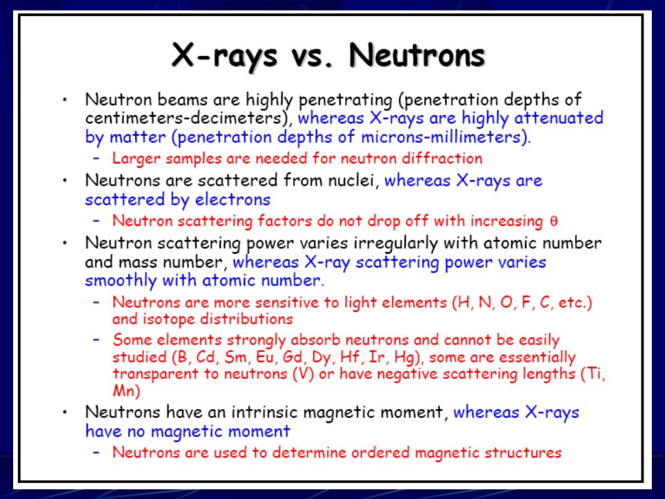
Neutron diffraction

82
Antiferromagnetic superstructure in MnO, FeO and NiO MnO Fe 3 O 4 The most famous anti-ferromagnetic, manganese oxide (MnO) helped earn the Nobel prize for C. Shull, who showed how such magnetic structures could be obtained by neutron diffraction (but not with the more common X-ray diffraction).
..")
83
Schematic neutron and X-ray diffraction patterns for MnO

Similar presentations


>")


>")
 Pure metal target (Cu) Electrons remover inner-shell electrons from target. Other electrons “fall”>")


>")

 Patterns in a TEM MATERIALS SCIENCE &ENGINEERING Anandh Subramaniam & Kantesh Balani.>")
 We also looked at internal ordering of atoms in 3-D structure (230 space.>")


