Download presentation
Presentation is loading. Please wait.

1
PROF DR. ÇİĞDEM ÖZKARA BRAIN STEM ANATOMY & CLINICAL PRESENTATIONS

2
In vertebrate anatomy the brainstem (or brain stem) is the posterior part of the brain, adjoining and structurally continuous with the spinal cord.vertebrateanatomybrainspinal cord The brain stem provides the main motor and sensory innervation to the face and neck via the cranial nerves. cranial nerves Though small, this is an extremely important part of the brain as the nerve connections of the motor and sensory systems from the main part of the brain to the rest of the body pass through the brain stem.

3
This includes the corticospinal tract (motor), the posterior column-medial lemniscus pathway (fine touch, vibration sensation and proprioception) and the spinothalamic tract (pain, temperature, itch and crude touch).corticospinal tract posterior column-medial lemniscus pathwayproprioceptionspinothalamic tract The brain stem also plays an important role in the regulation of cardiac and respiratory function. It also regulates the central nervous system, and is pivotal in maintaining consciousness and regulating the sleep cycle.sleep cycle

4
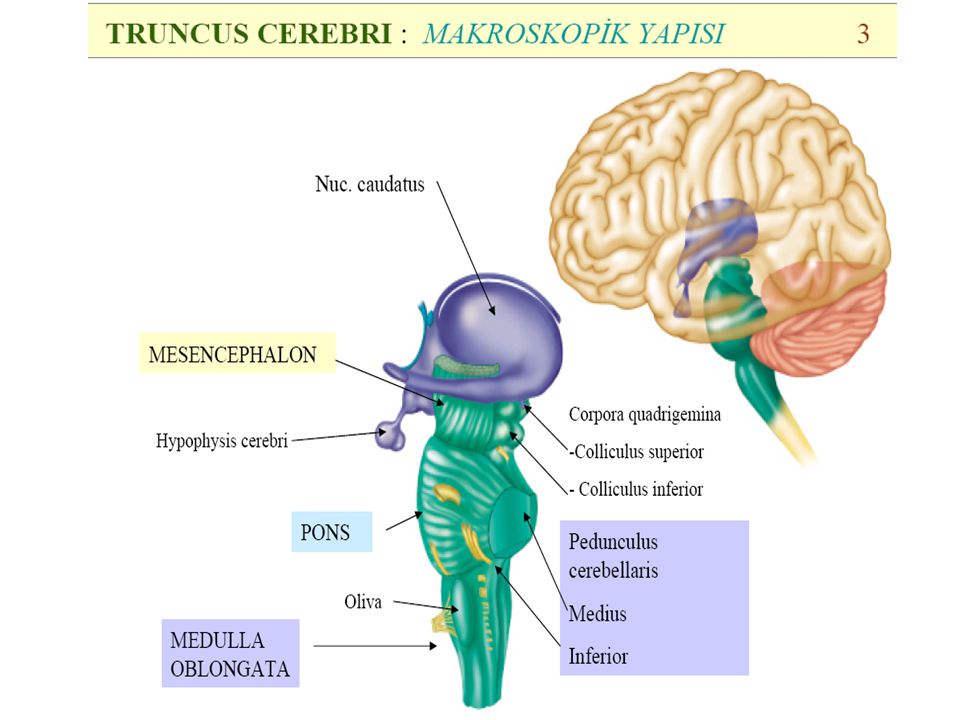
BRAIN STEM Includes: medulla oblongata (myelencephalon), medulla oblongata pons (part of metencephalon), pons midbrain (mesencephalon). midbrain Less frequently, parts of the diencephalon are included.diencephalon

5
1.Cerebrum 2.Thalamus 3.Mesencephalon - Midbrain 4.Pons 5.Medulla oblongata 6.Medulla spinalis - Spinal cord

9
The midbrain is divided into three parts. The first is the tectum, which is "roof" in Latin.tectum The tectum includes the superior and inferior colliculi and is the dorsal covering of the cerebral aqueduct. The inferior colliculus, involved in the sense of hearing sends its inferior brachium to the medial geniculate body of the diencephalon.inferior colliculusmedial geniculate bodydiencephalon Superior to the inferior colliculus, the superior colliculus marks the rostral midbrain. It is involved in the special sense of vision and sends its superior brachium to the lateral geniculate body of the diencephalon.superior colliculuslateral geniculate body The second part is the tegmentum and is ventral to the cerebral aqueduct.tegmentumcerebral aqueduct Several nuclei, tracts and the reticular formation are contained here. Last, the ventral side is composed of paired cerebral peduncles. These transmit axons of upper motor neurons. cerebral peduncles Midbrain (mesencephalon)
.")
10
Midbrain Periaqueductal grayPeriaqueductal gray: The area around the cerebral aqueduct, which contains various neurons involved in the pain desensitization pathway. Neurons synapse here and, when stimulated, cause activation of neurons in the nucleus raphe magnus, which then project down into the dorsal horn of the spinal cord and prevent pain sensation transmission. Occulomotor nerve nucleus: This is the nucleus of CN III. Trochlear nerve nucleus: This is the nucleus of CN IV. Red Nucleus: This is a motor nucleus that sends a descending tract to the lower motor neurons. Substantia nigra: This is a concentration of neurons in the ventral portion of the midbrain that uses dopamine as its neurotransmitter and is involved in both motor function and emotion. Its dysfunction is implicated in Parkinson's Disease. Reticular formation: This is a large area in the midbrain that is involved in various important functions of the midbrain. It contains lower motor neurons, is involved in the pain desensitization pathway, is involved in the arousal and consciousness systems, and contains the locus ceruleus, which is involved in intensive alertness modulation and in autonomic reflexes. Central tegmental tract: Directly anterior to the floor of the 4th ventricle, this is a pathway by which many tracts project up to the cortex and down to the spinal cord.pain nucleus raphe magnus Occulomotor nerve Trochlear nerve Red Nucleus Substantia nigradopamine Parkinson's Disease Reticular formationmidbrainautonomic Central tegmental tract

11
BEYİN SAPI Mezensefalon beyinsapının en üst bölümünü oluşturur. Üçüncü (N. Oculomotorius) ve IV. (N. Trochlearis) kranyal sinirlerin nukleusları buradadır
 ve IV. (N. Trochlearis) kranyal sinirlerin nukleusları buradadır.")
12
Fibria Corticospinalis Substania Nigra

13
N.oculomotorius(CN3): has 2 nuclei: Nüc nervi oculomotorii motor nucleus. At collikulus superior : All extraoculer mucles except for M.obliquus superior & m.rectus lateralis & m.levator palpebra superioris i were innervated. Upward and internal gaze nüc visseralis (edinger westphal): Parasympathetic nucleus. Innervates M. sphincter pupillae & M. ciliaris. Lesion: ptosis, mydriazis, eye down and out deviation, vertical diplopi, light reflex & accomodation loss. N.Oculomotorius III
: Parasympathetic nucleus. Innervates M. sphincter pupillae & M. ciliaris. Lesion: ptosis, mydriazis, eye down and out deviation, vertical diplopi, light reflex & accomodation loss. N.Oculomotorius III.")
14
Only somatomotor. M.obliquus superioru innervation. Unique CN leaves brain stem from posterior Lesion: cannot look down and out, vertikal diplopi Complains when coming down the stairs. If nucleus n. troclearisin is damaged: contrlateral m.obliquus superior is effected (exception for other CN) N. trochlearis(CN4)
 N. trochlearis(CN4).")
15
Weber sendromu: (superior alternating hemiplegia) is a form of stroke characterized by the presence of an oculomotor nerve palsy and contralateral hemiparesis or hemiplegia. It is caused by midbrain infarction as a result of occlusion of the paramedian branches of the posterior cerebral artery or of basilar bifurcation perforating arteries. [1]strokeoculomotor nerve palsyhemiparesismidbraininfarctionparamedian branchesposterior cerebral artery [1] 1. Substantia nigra, akinesia (parkinsonism) 2. Corticospinal fibers, contralateral spastic hemiplegia 3. Corticonuclear fibers, contralateral lower facial and hypoglossal paralysis, supranuclear 4. Corticopontine tract, contralateral dystaxia 5. Root fibers of oculomotor nerve, ipsilateral oculomotor paralysis with wide fixed pupil
 2. Corticospinal fibers, contralateral spastic hemiplegia 3. Corticonuclear fibers, contralateral lower facial and hypoglossal paralysis, supranuclear 4. Corticopontine tract, contralateral dystaxia 5. Root fibers of oculomotor nerve, ipsilateral oculomotor paralysis with wide fixed pupil.")
16
1. Medial lemniscus, contralateral decrease in sensations of touch, position, and vibration 2. Red nucleus, contralateral hyperkinesia (chorea, athetosis) 3. Substantia nigra, contralateral akinesia (parkinsonism) 4. Root fibers of oculomotor nerve, ipsilateral oculomotor paralysis, wide fixed pupil BENEDIKT SYNDROME: Caused by a lesion ( infarction, hemorrhage, tumor, or tuberculosis) in the tegmentum of the midbrain and cerebellum. It can result from occlusion of the posterior cerebral arterylesioninfarctionhemorrhage tuberculosistegmentumposterior cerebral artery Characterized by the presence of an CN III oculomotor nerve palsy and contralateral hemiparesis (weakness) and cerebellar ataxia including tremor.oculomotor nervepalsycontralateralataxia NeuroanatomicalNeuroanatomical structures affected include CNIII nucleus, Red nucleus, corticospinal tracts, brachium conjunctivum, and cerebellum.
 3. Substantia nigra, contralateral akinesia (parkinsonism) 4. Root fibers of oculomotor nerve, ipsilateral oculomotor paralysis, wide fixed pupil BENEDIKT SYNDROME: Caused by a lesion ( infarction, hemorrhage, tumor, or tuberculosis) in the tegmentum of the midbrain and cerebellum. It can result from occlusion of the posterior cerebral arterylesioninfarctionhemorrhage tuberculosistegmentumposterior cerebral artery Characterized by the presence of an CN III oculomotor nerve palsy and contralateral hemiparesis (weakness) and cerebellar ataxia including tremor.oculomotor nervepalsycontralateralataxia NeuroanatomicalNeuroanatomical structures affected include CNIII nucleus, Red nucleus, corticospinal tracts, brachium conjunctivum, and cerebellum..")
17
Parinaud's Syndrome : results from injury, either direct or compressive, to the dorsal midbrain. Specifically, compression or ischemic damage of the mesencephalic tectum, including the superior colliculus adjacent oculomotor (origin of cranial nerve III) and Edinger-Westphal nuclei, causing dysfunction to the motor function of the eye.midbrainmesencephalicsuperior colliculus oculomotorcranial nerveEdinger-Westphal 1. Cerebral aqueduct, stenosis with occlusive hydrocephalus 2. Superior colliculi, conjugated upwards gaze paralysis 3. Oculomotor nucleus, eventual oculomotor paralysis and ptosis (trochlear paralysis) 4. Medial longitudinal fasciculus, nystagmus A. Pinealoma compressing superior colliculi and aqueduct. B. Nuclei of III (IV) and medial longitudinal tracts are within range of deformation.
 and Edinger-Westphal nuclei, causing dysfunction to the motor function of the eye.midbrainmesencephalicsuperior colliculus oculomotorcranial nerveEdinger-Westphal 1. Cerebral aqueduct, stenosis with occlusive hydrocephalus 2. Superior colliculi, conjugated upwards gaze paralysis 3. Oculomotor nucleus, eventual oculomotor paralysis and ptosis (trochlear paralysis) 4. Medial longitudinal fasciculus, nystagmus A. Pinealoma compressing superior colliculi and aqueduct. B. Nuclei of III (IV) and medial longitudinal tracts are within range of deformation..")
18
PONS Named after the Latin word for "bridge" or the 16th-century ], It is superior to (up from) the medulla oblongata, inferior to (down from) the midbrain, and ventral to (in front of) the cerebellum.superior medulla oblongatainferior midbrainventral cerebellum In humans and other bipeds this means it is above the medulla, below the midbrain, and anterior to the cerebellum. This white matter includes tracts that conduct signals from the cerebrum down to the cerebellum and medulla, and tracts that carry the sensory signals up into the thalamus.humansbipedsanteriorwhite mattercerebrum thalamus Posteriorly, it consists mainly of two pairs of thick stalks called cerebellar peduncles. They connect the cerebellum to the pons and midbrain.cerebellar peduncles The pons contains nuclei that relay signals from the forebrain to the cerebellum, along with nuclei that deal primarily with sleep, respiration, swallowing, bladder control, hearing, equilibrium, taste, eye movement, facial expressions, facial sensation, and posture. [3] [3] Within the pons is the pneumotaxic center, a nucleus in the pons that regulates the change from inspiration to expiration. [pneumotaxic center [
![PONS Named after the Latin word for bridge or the 16th-century ], It is superior to (up from) the medulla oblongata, inferior to (down from) the midbrain, and ventral to (in front of) the cerebellum.superior medulla oblongatainferior midbrainventral cerebellum In humans and other bipeds this means it is above the medulla, below the midbrain, and anterior to the cerebellum.](http://images.slideplayer.com/14/4331058/slides/slide_18.jpg "This white matter includes tracts that conduct signals from the cerebrum down to the cerebellum and medulla, and tracts that carry the sensory signals up into the thalamus.humansbipedsanteriorwhite mattercerebrum thalamus Posteriorly, it consists mainly of two pairs of thick stalks called cerebellar peduncles. They connect the cerebellum to the pons and midbrain.cerebellar peduncles The pons contains nuclei that relay signals from the forebrain to the cerebellum, along with nuclei that deal primarily with sleep, respiration, swallowing, bladder control, hearing, equilibrium, taste, eye movement, facial expressions, facial sensation, and posture. [3] [3] Within the pons is the pneumotaxic center, a nucleus in the pons that regulates the change from inspiration to expiration. [pneumotaxic center [.")
19
Cranial nerves are the abducens nerve VI, facial nerve VII and the vestibulocochlear nerve VIII, respectively.abducens nervefacial nervevestibulocochlear nerve At the level of the midpons, trigeminal nerve V, emerges.trigeminal nerve

21
PONS ÇEKİRDEKLERİ Nüc. Principalis nervi trigemini: Baş ve yüzün dokunma ve basınç duyularının ikinci nöronlarının bulunduğu çekirdektir. Kornea reflexi ile ilgilidir. Nüc salivatorius superior ve Nüc lacrimalis: V.CN parasempatik çekirdekleridir. Glandula submandibularis,glandula sublingualis,glandula lacrimalise giden lifleri içerir. Locus caeruleus: noradrenalin üreten nöronların en büyük kaynağıdır.

23
N. trigeminus(CN5): has 3 sensoriel, 1 motor nucleus – nuc. spinalis nervi trigemini: related to cornea reflex – Nuc. spinalis nervi trigemini: face and head pain and temperature sensations 2nd neuron. – Nuc. mesensefalikus nervi trigemini – Nuc. motorius nervi trigemini – Ganglion trigeminale (gasser ganglionu semilunar ganglion): sensation ganglion
: sensation ganglion.")
24
N. Trigeminus branches N.ophtalmikus (V1): only sensitive fibres. B ulbus oculi innervates, tip of nose, upper part of eyes. N.maksillaris(V2): only sensitive fibres, lower part of eyes, upper lip innervation. N.mandibularis(V3) Motor & sensitive fibres. Masticatory muscles, lower lip and lower part of mouth, 2/3 anteror part of tongue, external ear and temporal region innervation.
: only sensitive fibres, lower part of eyes, upper lip innervation. N.mandibularis(V3) Motor & sensitive fibres. Masticatory muscles, lower lip and lower part of mouth, 2/3 anteror part of tongue, external ear and temporal region innervation..")
25
N.abducens(CN6): Only motor fibres. M.rektus lateralisi innervation. Lesion: effected eye pulled medially, horizontal diplopia. Lies in sinus kavernosus lateral to A. carotis interna

26
Involves motor, sensory and parasympatic fibres: nüc.nervi facialis: Motor nuc.Innervates mimic muscles :m.stapedius,m.stylohyoideus,m.digastrikus venter posteriorun Nüc.lacrimalis ve nüc.salivatorius: Parasympathic nuclei Nüc traktus soliterius:2.neurons of taste Central facial paralysis : Supranuclear lesions, mouth commissure deviates to healthy side (lesion), can close eyes cornea reflex is normal. Central facial paralysis : Supranuclear lesions, mouth commissure deviates to healthy side (lesion), can close eyes cornea reflex is normal. Peripheric facial paralysis (Bell’s palsy): Intranuclear or infranuclear mouth commissure deviates to healthy side, cornea reflex disappears, cannot close eye Peripheric facial paralysis (Bell’s palsy): Intranuclear or infranuclear mouth commissure deviates to healthy side, cornea reflex disappears, cannot close eye N.facialis(CN7)
, can close eyes cornea reflex is normal. Peripheric facial paralysis (Bell’s palsy): Intranuclear or infranuclear mouth commissure deviates to healthy side, cornea reflex disappears, cannot close eye Peripheric facial paralysis (Bell’s palsy): Intranuclear or infranuclear mouth commissure deviates to healthy side, cornea reflex disappears, cannot close eye N.facialis(CN7).")
27
Millard-Gubler syndrome It is a syndrome of unilateral softening of the brain tissue arising from obstruction of the blood vessels of the pons. VI and VII cranial nerves fibers of the corticospinal tract, Clinical presentation: Paralysis of the abducens (including diplopia, internal strabismus, and loss of power to rotate the affected eye outward) facial nerves ( peripheric ipsilateral) Contralateral Hemiplegia of the extremities. It is also known as "crossed hemiplegia".
 facial nerves ( peripheric ipsilateral) Contralateral Hemiplegia of the extremities. It is also known as crossed hemiplegia ..")
28
1. Medial lemniscus, contralateral decrease of touch, position and vibration sensations in the lower extremities 2. Lateral lemniscus, hypacusia 3. Nucleus of facial nerve, peripheral ipsilateral paralysis of facial muscles 4. Anterior spinothalamic tract, contralateral analgesia and thermanesthesia of body 5. Pyramidal tract, contralateral spastic hemiplegia 6. Abducent nerve, ipsilateral peripheral paralysis of lateral rectus muscle

29
PONS LEZYONLARINDAKİ LEZYONLAR(DEVAM) Birbuçuk sendrom: Ponstaki enfarktüs, multple sklerozis yada pontin hemoraji nedeniyle olur. İpsilateral n.abducens çekirdeğinin yada PPRF nin tutulumuna bağlı ipsilateral göz horizontal düzlemde hareketsizdir ve FLM nin tutulumuna bağlı intranüklear oftalmopleji(kontralateral göz abduksiyonda olup nistagmus vardır) olur. Hastalar lezyon tarafına bakamaz.
 olur. Hastalar lezyon tarafına bakamaz..")
30
PONS LEZYONLARINDAKİ LEZYONLAR(DEVAM)
")
31
Birbuçuk Sendrom

32
Peripheral facial paralysis

33
Central facial paralysis

34
N.vestibulocohlearis: N.vestibularis: carries the informaion related to position and movements of of head, N.cohlearis: primery audituar fibres. Kranyal Sinirler (devam)
.")
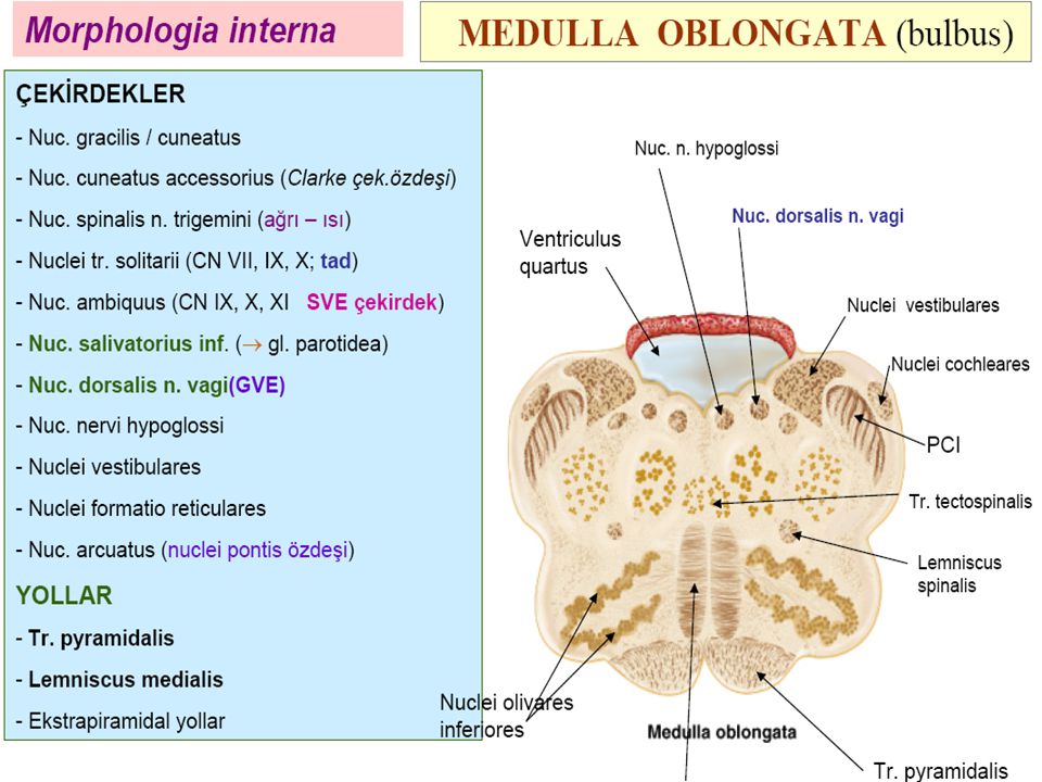
35
Medulla oblongata (bulbus) is the lower half of the brainstem. The medulla contains the cardiac, respiratory, vomiting and vasomotor centers and deals with autonomic functions, such as breathing, heart rate and blood pressure.

36
Functions The medulla oblongata controls autonomic functions, and relays nerve signals between the brain and spinal cord. It is also responsible for controlling several major points and autonomic functions of the body: respiration– chemoreceptors cardiac center – sympathetic, parasympathetic system vasomotor center – baroreceptors reflex centers of vomiting, coughing, sneezing, and swallowing balancing the human body.

37
Nuclei of Medulla oblongata Last five cranial nerve nuclei: Nuc. Grasilis ve nuc. kuneatus: second neurons of conscious proprioceptive, vibration, two point discrimination sensations Nuc. Traktus solitarius: Related to VII,IX,X. cranial nerves. Upper part is called nuc. Gustatorius and involves neurons of taste Nuc. Spinalis nervi trigemini: Pain and temparature sensation of face Nuc. Ambiguus: motor nuclei of IX,X,XI cranial nerves Nuc. Salivatorius inferior: Parasempatik nuclei of XI. CN. The fibres from this nuclei goes to glandula parotidea

39
N. Glossofaryngeus(CN9 3 nuclei, 2 ganglions, Motor: nüc.ambiguus…. İnnervates m.stylofaringeus Nüc salivatorius inferior parasempatik nucleus, innervates glandula parotidea Nuc traktus solitarius 1/3 posterior of taste sensation of tongue, tonsilla palatina and middle ear sense. Lesion: uvula deviates to healthy side. The integrity of the glossopharyngeal nerve may be evaluated by testing the patient's general sensation and that of taste on the posterior third of the tongue. The gag reflex can also be used to evaluate the glossphyaryngeal nerve

40
N.vagus(CNX) 3 nuclei, 2 ganglion Motor nuc: nuc.ambiguus, Parasympatic nuc: nuc posterior nervi vagi, Taste : nuc. ractus solitarius The vagus nerve supplies motor parasympathetic fibers to all the organs except the suprarenal (adrenal) glands, from the neck down to the second segment of the transverse colon. The vagus also controls a few skeletal muscles, namely: parasympatheticadrenalnecktransverse colonskeletal muscles Cricothyroid muscle Levator veli palatini muscle Salpingopharyngeus muscle Palatoglossus muscle Palatopharyngeus muscle Superior, middle and inferior pharyngeal constrictorspharyngeal constrictors Muscles of the larynx (speech).larynxspeech
 glands, from the neck down to the second segment of the transverse colon. The vagus also controls a few skeletal muscles, namely: parasympatheticadrenalnecktransverse colonskeletal muscles Cricothyroid muscle Levator veli palatini muscle Salpingopharyngeus muscle Palatoglossus muscle Palatopharyngeus muscle Superior, middle and inferior pharyngeal constrictorspharyngeal constrictors Muscles of the larynx (speech).larynxspeech.")
41
This means that the vagus nerve is responsible for such varied tasks as heart rate, gastrointestinal peristalsis, sweating, and quite a few muscle movements in the mouth, including speech (via the recurrent laryngeal nerve) and keeping the larynx open for breathing (via action of the posterior cricoarytenoid muscle, the only abductor of the vocal folds). It also has some afferent fibers that innervate the inner (canal) portion of the outer ear, via the Auricular branch (also known as Alderman's nerve) and part of the meninges. This explains why a person may cough when tickled on their ear (such as when trying to remove ear wax with a cotton swab)heart rateperistalsis sweatingspeechrecurrent laryngeal nerveposterior cricoarytenoid muscle outer ear Alderman's nervemeninges
 portion of the outer ear, via the Auricular branch (also known as Alderman s nerve) and part of the meninges. This explains why a person may cough when tickled on their ear (such as when trying to remove ear wax with a cotton swab)heart rateperistalsis sweatingspeechrecurrent laryngeal nerveposterior cricoarytenoid muscle outer ear Alderman s nervemeninges.")
42
N.accessorius(CNXI) Pure motor. Some fibres from bulbus some from servikal m. Spinalis sup. Segment ant horn cells. Leaves cranial cavity from foramen jugulare very close to IX. & X. CNs The nerve functions to control the sternocleidomastoid and trapezius muscles.sternocleidomastoid trapeziusmuscles

43
N. Hypoglossus (CNXII) Motor nerve of tongue. Nucleus in bulbus. Lesion: tongue deviates to the paralysed side, atrophy at that side Unilateral involment: No tongue movements

44
IŞIK REFLEXİ

45
1. Inferior vestibular nucleus, nystagmus and ipsilateral inclination to fall 2. Dorsal nucleus of vagus nerve, tachycardia and dyspnea 3. Inferior cerebellar peduncle, ataxia and ipsilateral asynergia 4. Nucleus of solitary tract, ageusia ipsilateral 5. Ambiguus nucleus, ipsilateral paralysis of palate, larynx, and pharynx 6. Nucleus of cochlear nerve, hypacusia 7. Nucleus of trigeminal spinal tract, ipsilateral analgesia and thermanesthesia of face 8. Central sympathetic pathway, Horner's syndrome. Hypohidrosis, ipsilateral vasodilation in face 9. Anterior spinocerebellar tract, ataxia, ipsilateral hypotonia 10. Lateral spinothalamic tract, analgesia and thermanesthesia contralateral over body WALLENBERG’S SYNDROME (lateral medulla syndrome) It is the clinical manifestation resulting from occlusion of the posterior inferior cerebellar artery (PICA) or one of its branches or of the vertebral artery, in which the lateral part of the medulla oblongata infarcts, resulting in a typical pattern.
 It is the clinical manifestation resulting from occlusion of the posterior inferior cerebellar artery (PICA) or one of its branches or of the vertebral artery, in which the lateral part of the medulla oblongata infarcts, resulting in a typical pattern..")
46
Horner sendromu

47
Medial medüller syndrome(Dejerine’in anterior medüller sendrom: Obstruction of a. spinalis anterior veya a. vertebralis 1. Medial longitudinal fasciculus, nystagmus 2. Medial lemniscus, contralateral decrease of touch, vibration, and position sensations 3. Olive, ipsilateral myorhythmia in velum and pharynx 4. Hypoglossal nerve, ipsilateral flaccid paralysis of hypoglossal muscle with atrophy 5. Pyramidal tract, contralateral, spastic hemiplegia with positive Babinski reflex

49
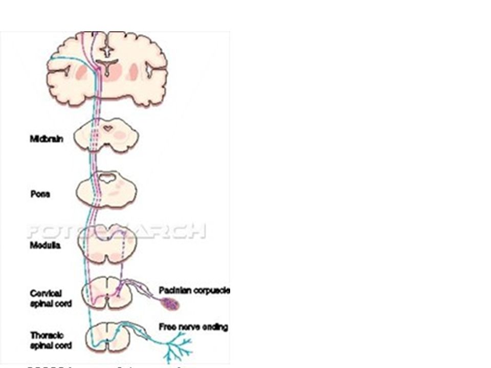
Spinothalamic tract: pain, tempartature, itch, crude touch Posterior column fine touch, vibration, conscious proprioceptive Cortico spinal tract: motor

50
Cranial nerve reflexes

51
Kranyal Sinirler (devam)
")
55
Kranyal Sinirler N.olfaktorius(CN1): Bipolar nöronlardandır. İnsanda çoğalabilme özelliği gösteren tek nöron grubudur. Küçük çaplı ve myelinsiz aksonlara sahip yani en düşük ileti hızı olan nöronlardır. Primer olfaktör korteks Brodmann'ın 34. alanı (temporal lob) olup kokuların ayırt edildiği yerdir. Harabiyetinde koku halüsinasyonları olur.
 olup kokuların ayırt edildiği yerdir. Harabiyetinde koku halüsinasyonları olur..")
56
Kranyal Sinirler (devam) N.optikus(CN2): Retinada bulunan fotoreseptör hücreler görme yolunun birinci nöronları olan bipolar hücrelerle sinaps yapar. Bipolar hücreler görme yolunun ikinci nöronları olan retinal ganglion hücreleri ile sinaps yapar. Retinal ganglion hücrelerinin uzantıları optik siniri oluşturur. Ganglion hücrelerinin uzantıları korpus geniculatum lateralde bulunan üçüncü nöronlarla sinaps yapar. Üçüncü nöron uzantıları primer vizüel kortekse gider.

57
Görme Yolları

58
N.optikus beyin zarları ile sarılıdır. Çevresinde subaraknoid boşluk vardır. Bu nedenle BOS daki basınç artışları diskus nervi optici de ödeme (papil ödem) neden olabilir. Kranyal Sinirler (devam)
 neden olabilir. Kranyal Sinirler (devam).")
59
PAPİL ÖDEM PAPİL ATROFİ NORMAL PAPİL

60
Cranial nerve reflexes

Similar presentations


>")


 contains three components: fiber bundles of the corticospinal tracts, pontine nuclei.>")


