Download presentation
Presentation is loading. Please wait.

1
NEONATAL THROMBOCYTOPAENIA

2
History Baby DP, white male Born 30 March 2011 (now 5 weeks old)
Normal vaginal delivery Birth weight 2830g Length 45cm Head circumference 33cm APGARS 8/10, 9/10, 10/10 Given oxygen via face mask at delivery, but no need for resuscitation

3
Mother 21 years old Apparently 32 weeks pregnant by dates, but unsure of dates, birthweight suggests closer to term Primigravida Rhesus positive, RPR negative, HIV negative Had a ?pustular rash throughout pregnancy, otherwise well during pregnancy No medications taken other than routine vitamins, folate

4
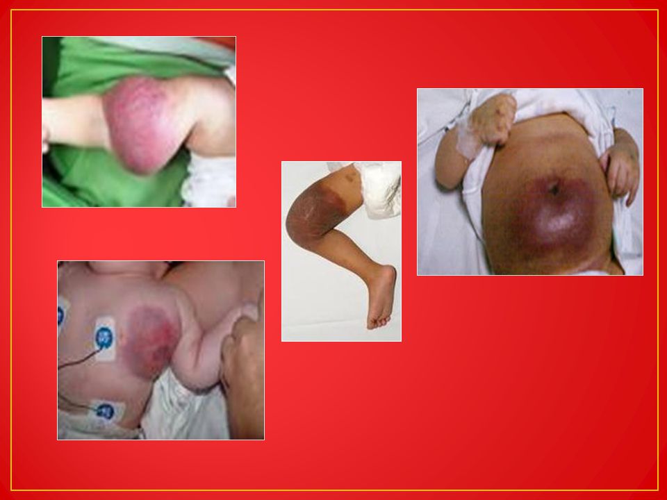
Intern called to see baby on Day 1 of life for a ‘rash’ on the body
On examination Baby looked like a term neonate by Ballard score Pink in room air No distress Not obviously dysmorphic Petechial rash over trunk, arms, legs Bruising on trunk (right upper quadrant), palms and soles Rest of examination noted to be normal Assessment Term male neonate with a suspected clotting abnormality/ bleeding tendency presenting on Day 1 of life Mother not known to be hypertensive or diabetic, no history of perinatal asphyxia
, palms and soles. Rest of examination noted to be normal. Assessment. Term male neonate with a suspected clotting abnormality/ bleeding tendency presenting on Day 1 of life. Mother not known to be hypertensive or diabetic, no history of perinatal asphyxia.")
5
Investigations FBC INR, PTT White cell count 22.35 Haemoglobin 18.2
Platelet count 10 (not clumped) MCV 112.4 Platelet volume normal INR, PTT 1.26, 39.9 (normal)
 MCV Platelet volume normal. INR, PTT. 1.26, 39.9 (normal)")
6
Mother’s results FBC (31/03/2011) WCC 20.8 Hb 12.0 Plts 293 (normal)
 WCC 20.8 Hb 12.0 Plts 293 (normal)")
7
Management and course Admitted to neonatal ward Antibiotics, Vitamin K
Platelet (random donor) infusion ordered immediately Cranial sonar (1 April 2011) showed possible intracerebral haemorrhage CT brain confirmed a haemorrhage in right frontal lobe (suspected to be about a week old already)
 infusion ordered immediately. Cranial sonar (1 April 2011) showed possible intracerebral haemorrhage. CT brain confirmed a haemorrhage in right frontal lobe (suspected to be about a week old already)")
9
Baby developed jaundice on day 3, phototherapy started, resolved quickly
No seizures in ward Blood cultures remained negative CRP, U&E day 3 unremarkable Fed well Gained weight (latest 3.36kg) Much later noted to have abnormal ‘webbed’ toes on left foot – Xrays taken – showed polydactyly of middle and terminal phalanx 5th and 6th toes
 Much later noted to have abnormal ‘webbed’ toes on left foot – Xrays taken – showed polydactyly of middle and terminal phalanx 5th and 6th toes.")
10
Platelet count trend 21/04 24/04 25/04 26/04 28/04 04/05 12.6 10.1 1
37 6 44 22 Platelets given Platelets + IVIG Platelets + steroids

11
Expert advice sought Oncologists (08/04)
Considered alloimmune thrombocytopenia Suggested testing mother for HPA-1a antibodies Advised IVIG (intravenous immunoglobulin) if platelets not improving Plasma serum screen for antibodies to platelet glycoproteins GPIIb/IIIa, GPIa/IIa and GPIb/IX and platelet antigens HPA 1a, -Ib, -3a, -3b, -4a, -5a and -5b: NEGATIVE Plasma serum screen for HLA Class I antibodies:
 if platelets not improving. Plasma serum screen for antibodies to platelet glycoproteins GPIIb/IIIa, GPIa/IIa and GPIb/IX and platelet antigens HPA 1a, -Ib, -3a, -3b, -4a, -5a and -5b: NEGATIVE. Plasma serum screen for HLA Class I antibodies:")
12
Head of SA National Blood Transfusion Service
Oncologist (21/04) May be false negative results Advised to test father’s blood also Give normal platelets again, but would need specific platelets in near future Consider steroids lood Head of SA National Blood Transfusion Service Unusual to have no antibodies on screen performed if alloimmune thrombocytopenia AIT unlikely Not for HPA-1a negative platelets as diagnosis not confirmed Head of Neonatal ICU Try steroids HPA-1a negative platelets/ mother’s platelets
 May be false negative results. Advised to test father’s blood also. Give normal platelets again, but would need specific platelets in near future. Consider steroids. lood. Head of SA National Blood Transfusion Service. Unusual to have no antibodies on screen performed if alloimmune thrombocytopenia. AIT unlikely. Not for HPA-1a negative platelets as diagnosis not confirmed. Head of Neonatal ICU. Try steroids. HPA-1a negative platelets/ mother’s platelets.")
13
Other results Urine CMV Shell vial culture – negative HIV negative Rubella serology – IgG positive, IgM negative – suggestive of maternal antibodies Xrays long bones – no periosteal reactions or features of congenital infections (rubella, syphilis) Xray skull – no calcifications noted suggestive of CMV, Toxoplasmosis
 Xray skull – no calcifications noted suggestive of CMV, Toxoplasmosis.")
19
Platelet production Process similar in neonates and adults
Important developmental differences Tpo concentrations higher in neonate But neonates with thrombocytopenia have lower Tpo concentrations than adults with similar degrees of thrombocytopenia Megakaryocyte progenitors have higher proliferative potential, more sensitive to Tpo, and are present in BM and blood Each megakaryocyte produces fewer platelets (smaller) Can increase number of megakaryocytes
 Can increase number of megakaryocytes.")
20
Neonatal Thrombocytopenia (NT)
Relatively common haematological problem in neonates Defined as count < 150 x 10⁹/L Occurs in 1-5% of all newborns Depends on population – sick prems, ICU – more common (22-35% of all admissions) Most have mild-moderate thrombocytopenia 5-10% - severe (<30 x10⁹/L) urgent investigation and management needed List of causes Until recently many called “idiopathic” Reduced platelet production main underlying mechanism Newer classifications based on timing of onset
 Most have mild-moderate thrombocytopenia. 5-10% - severe (<30 x10⁹/L) urgent investigation and management needed. List of causes. Until recently many called idiopathic Reduced platelet production main underlying mechanism. Newer classifications based on timing of onset.")
21
Classification based on timing of onset (more common causes)
Early (< 72 hours) Late (> 72 hours) Placental insufficiency (IUGR, PIH) Perinatal asphyxia Perinatal infection (GBS, E coli) DIC Alloimmune (severe NT) Autoimmune (ITP, SLE) Late-onset sepsis NEC Account for >80% cases
 Late (> 72 hours) Placental insufficiency (IUGR, PIH) Perinatal asphyxia. Perinatal infection (GBS, E coli) DIC. Alloimmune (severe NT) Autoimmune (ITP, SLE) Late-onset sepsis. NEC. Account for >80% cases.")
22
Less common causes Late Early Congenital infection
Kasabach-Merritt syndrome Metabolic disease Congenital/ inherited Autoimmune Congenital infection (TORCH) Kasabach-Merritt syndrome Metabolic disease Congenital/ inherited (TAR, CAMT) Thrombosis Bone marrow replacement
 Kasabach-Merritt syndrome. Metabolic disease. Congenital/ inherited (TAR, CAMT) Thrombosis. Bone marrow replacement.")
23
Fetal causes Alloimmune Autoimmune Congenital infection Aneuploidy Severe Rhesus disease Congenital/ inherited

24
Classifying causes helps guide towards investigations and management
Often a dilemma – when to transfuse platelets? Variation in platelet transfusion practice between centres No study has yet shown benefit of platelet transfusion in NT Possibly even adverse effects – increased mortality, increased risk of short-bowel syndrome in transfused neonates surviving NEC

25
Chronic fetal hypoxia Occurs in infants of mothers with PIH (pregnancy-induced hypertension), diabetes Manifest in fetus by IUGR Mild-moderate thrombocytopenia Self-limiting, usually resolves within 10 days Mechanism Reduced megakaryopoeisis Associated neutropenia, increased circulating nucleated red cells, +/- polycythaemia Tpo normal/ only minimally elevated – inadequate up-regulation of Tpo production If severe/ not resolving after 2 weeks, look for other causes of thrombocytopenia

26
Thrombocytopenia develops very rapidly over 1-2 days
Sepsis, NEC Thrombocytopenia develops very rapidly over 1-2 days Often very severe (< 30 x 10⁹/L) May recover over weeks Uncommon causes of NT in first few days of life Mechanism – peripheral consumption Elevated levels of Tpo Increased numbers of circulating megakaryocyte progenitors Actually up-regulation of thrombopoiesis
 May recover over weeks. Uncommon causes of NT in first few days of life. Mechanism – peripheral consumption. Elevated levels of Tpo. Increased numbers of circulating megakaryocyte progenitors. Actually up-regulation of thrombopoiesis.")
27
Neonatal Autoimmune Thrombocytopenia
Transplacental passage maternal platelet auto-antibodies Maternal platelets low Immune thrombocytopenic purpura, SLE 1-2:1000 pregnancies Much less clinically problematic than NAIT thrombocytopenia only occurs in 10% of those with auto-antibodies Incidence of ICH only 1% Caesarean section not indicated Maternal disease severity, thrombocytopenia, previous severe NT most useful indicators of likelihood of significant NT in current pregnancy

28
Infant of mother with AI disease
Platelet count checked at birth (cord/peripheral blood – NOT heel-prick) If >150 x 10⁹/L – no further action If platelet count low – repeat after 2-3 days – drop to lowest at this time, then rise spontaneously by day 7 Thrombocytopenia may persist for several weeks in few cases – if severe (< 30)/ prolonged, treat with IVIG – most respond promptly
 If >150 x 10⁹/L – no further action. If platelet count low – repeat after 2-3 days – drop to lowest at this time, then rise spontaneously by day 7. Thrombocytopenia may persist for several weeks in few cases – if severe (< 30)/ prolonged, treat with IVIG – most respond promptly.")
29
Congenital infections
HIV, CMV, enterovirus, Rubella can cause NT HIV Immune-mediated destruction Sequestration Suppression of production, though increased megakaryocytes (ineffective thrombopoiesis) CMV Quite common Thrombocytopenia 50-75% of cases Massive splenomegaly causes sequestration Decreased production
 CMV. Quite common. Thrombocytopenia 50-75% of cases. Massive splenomegaly causes sequestration. Decreased production.")
30
Inherited thrombocytopenia
Numerous, rare Present in fetus/neonate Unexplained, persistent thrombocytopenia due to reduced platelet production Due to abnormal haemopoeitic stem cell development Often associated congenital abnormalities > guide investigations, diagnosis Advances in molecular techniques aid in diagnosis and management

31
Bernard-Soulier Syndrome
May present in neonatal period though bleeding not usually severe Mild-moderate thrombocytopenia Giant platelets AR pattern of inheritance Defect in glycoprotein Ib-IX-V complex Platelet transfusion effective, but reserve for life-threatening haemorrhage Transfused patients may form allo-antibodies to GPIb, GPIX, GPV Offspring of mothers with BSS may present as NAIT with severe fetal ICH due to formation of such allo-antibodies

32
Blood film showing giant platelets

33
Wiskott-Aldrich Syndrome
WAS, X-linked thrombocytopenia spectrum of disorders Mutation in WAS protein gene, short arm X chromosome Over 100 different mutations Microthrombocytopenia, eczema, recurrent bacterial, viral infections, propensity to autoimmune conditions Present in first year, rarely neonatal period unless known family history Bleeding due to abnormal platelet function and reduced platelet survival, number X-linked thrombocytopenia – other clinical features absent, thrombocytopenia milder

34
Fanconi Anaemia Usually presents after infancy, but thrombocytopenia reported in neonates Result of a genetic defect in a cluster of proteins responsible for DNA repair Consider diagnosis Unexplained thrombocytopenia Especially if typical dysmorphic features – malformations of skin, thumb, face, eyes If parental consanguinity Diepioxybutane test diagnostic Treatment rarely necessary in neonatal period

36
Thrombocytopenia absent radii (TAR) syndrome
Bilateral absence of radii Thrombocytopenia – present at birth/ within first 4 months Both thumbs present and normal Platelets usually < 50 x 10⁹/L WCC elevated (sometimes > 100 x 10⁹/L) in > 90% of patients, may mimic congenital leukaemia Associated abnormalities Cow’s milk protein intolerance Lower limb abnormalities Renal, cardiac abnormalities Platelet count seems to improve spontaneously AR inheritance Tpo increased, megakaryocytes decreased > presumed defect in Tpo signalling pathway
 in > 90% of patients, may mimic congenital leukaemia. Associated abnormalities. Cow’s milk protein intolerance. Lower limb abnormalities. Renal, cardiac abnormalities. Platelet count seems to improve spontaneously. AR inheritance. Tpo increased, megakaryocytes decreased > presumed defect in Tpo signalling pathway.")
38
Some other rare inherited conditions
Amegakaryocytic thrombocytopenia with radio-ulnar synostosis Severe thrombocytopenia at birth, absent megakaryocytes in BM Congenital amegakaryocytic thrombocytopenia (CAMT) Nearly always in neonatal period Platelet count < 20 x 10⁹/L Evidence of bleeding Physical anomalies present in 50% 50% later develop aplastic anaemia, leukaemia, myelodysplasia AR inheritance – mutations in Tpo receptor (c-mpl) Stem cell transplant curative
 Nearly always in neonatal period. Platelet count < 20 x 10⁹/L. Evidence of bleeding. Physical anomalies present in 50% 50% later develop aplastic anaemia, leukaemia, myelodysplasia. AR inheritance – mutations in Tpo receptor (c-mpl) Stem cell transplant curative.")
39
Giant platelet syndromes
Many, rare Present in neonatal period/fetus May-Hegglin anomaly Giant platelets, thrombocytopenia, leucocyte Dohle-like inclusion bodies Rare cause of fetal/neonatal intracerebral haemorrhage

40
Kasabach-Merritt Syndrome
Neonatal period 50% vascular tumour diagnosed at birth, 90% diagnosed by 1 year of age Life-threatening consumptive coagulopathy Profound thrombocytopenia PLUS microangiopathic anaemia, disseminated intravascular coagulation Due to enlarging vascular lesion – usually obvious – cutaneous, involving face, neck, trunk or extremities 20% visceral/retroperitoneal involvement (liver) without cutaneous signs; abdominal distension, organ dysfunction, high-output cardiac failure
 without cutaneous signs; abdominal distension, organ dysfunction, high-output cardiac failure.")
41
Previously thought to be haemangioma, actually histological features of kaposiform haemangioendothelioma or tufted angioma Locally aggressive vascular tumours Trapping of platelets on endothelium, exacerbated occasionally by DIC Diagnosis – MRI most frequently used modality Diffusely enhancing Significant morbidity and mortality Haemorrhage Invasion/compression of vital structures 10-30% mortality

42
Maintain haemostasis Platelets for active bleeding/ prior to surgery only, as can increase size of tumour or even exacerbate KMP Aminocaproic acid Antiplatelet agents (Aspirin, diprridamole) may reduce platelet aggregation Fresh frozen plasma if clinically indicated Curative therapy – treat underlying tumour No treatment uniformly effective Surgery not usually possible Tumour embolization with medical/ surgical therapy Corticosteroids, alpha interferon, vincristine – alone/in combination
 may reduce platelet aggregation. Fresh frozen plasma if clinically indicated. Curative therapy – treat underlying tumour. No treatment uniformly effective. Surgery not usually possible. Tumour embolization with medical/ surgical therapy. Corticosteroids, alpha interferon, vincristine – alone/in combination.")
44
Thrombotic disorders Seen in adults, older children, but reported in neonates TTP, HUS, Heparin-induced thrombocytopenia Inherited deficiency of von Willebrand factor cleaving protease Thrombocytopenia, hyperbilirubinaemia, anaemia Diagnosis delayed as condition rare, signs common in sick neonates HUS reported due to B pertussis NT may occur after thrombosis of major vessel (renal vein thrombosis) Consider in neonate with thrombocytopenia and renal failure
 Consider in neonate with thrombocytopenia and renal failure.")
45
Metabolic disorders Thrombocytopenia common presenting feature in certain inborn errors of metabolism Methylmalonic acidaemia, propionic acidaemia, Gaucher disease Can also complicate induced hypothermia used to treat HIE

46
Aneuploidies Trisomy 18, 13, 21, Turner syndrome and triploidy can be associated with thrombocytopenia Down syndrome frequently associated with mild thrombocytopenia Mechanisms Similar to that seen in IUGR infants – chronic fetal hypoxia 10% neonates develop a pre-leukaemic disorder (Transient Abnormal Myelopoiesis); increased myeloblasts, variable degrees of thrombocytopenia Usually resolves spontaneously but 20-30% develop AMKL (acute megakaryoblastic leukaemia)
; increased myeloblasts, variable degrees of thrombocytopenia. Usually resolves spontaneously but 20-30% develop AMKL (acute megakaryoblastic leukaemia)")
47
Neonatal Alloimmune Thrombocytopenia (NAIT)
Also referred to as AIT, FM(fetomaternal)AIT Life-threatening bleeding disorder Represents <5% cases of early NT, most common cause of severe thrombocytopenia in well, term neonates Due to maternal platelet antibodies produced in response to fetal platelet antigens inherited from the father Antibodies cross placenta Destroy fetal platelets Cause severe thrombocytopenia, bleeding, ICH
AIT. Life-threatening bleeding disorder. Represents <5% cases of early NT, most common cause of severe thrombocytopenia in well, term neonates. Due to maternal platelet antibodies produced in response to fetal platelet antigens inherited from the father. Antibodies cross placenta. Destroy fetal platelets. Cause severe thrombocytopenia, bleeding, ICH.")
48
Incompatibility between maternal and fetal platelets for HPA 1a accounts for most cases of NAIT

49
Clinical presentation
Clinical picture varies from mild thrombocytopenia at birth to intracerebral haemorrhage (ICH) in utero/ at delivery/ in first days life Characterised by low platelets and bleeding in an otherwise healthy neonate Severe thrombocytopenia (often < 20x10⁹/L)
 in utero/ at delivery/ in first days life. Characterised by low platelets and bleeding in an otherwise healthy neonate. Severe thrombocytopenia. (often < 20x10⁹/L)")
50
Incidence NAIT affects approximately 1:2000 newborns
Approximately 2% of women are at risk (HPA-1a negative) 10% of these have detectable antibodies 30% of these have affected fetus 20% of these have intracerebral haemorrhage
 10% of these have detectable antibodies. 30% of these have affected fetus. 20% of these have intracerebral haemorrhage.")
51
. Fig 1. Pyramid model of NAT. Each tier shows the number of affected pregnancies and infants expected out of a sample of white women. Each tier represents a proportion of the tier below, shown in parentheses along the right side of the pyramid

52
Pathophysiology 3 major platelet antigen systems on surface of platelets HLA class uncertain role in NAIT ABH blood group antigens Specific platelet antigens (HPA’s) HPA system assigns a name to platelet antigens based on recognition by specific antisera, in chronological order in which they were identified; high- and low-frequency alleles designated a and b respectively 16 unique polymorphisms been described Human platelet antigens are epitopes on platelet glycoproteins Most polymorphisms expressed on GPIIIa Polymorphisms readily detected by PCR techniques
 HPA system assigns a name to platelet antigens based on recognition by specific antisera, in chronological order in which they were identified; high- and low-frequency alleles designated a and b respectively. 16 unique polymorphisms been described. Human platelet antigens are epitopes on platelet glycoproteins. Most polymorphisms expressed on GPIIIa. Polymorphisms readily detected by PCR techniques.")
53
Table 3. Platelet Antigens Implicated in NAT by Frequency
Major platelet antigens (high frequency of NAIT) HPA-1a HPA-5a HPA-5b HPA-15a HPA-15b Platelet antigens accounting for up to 2% of NAT collectively HPA-3a HPA-2a HPA-2b Platelet antigens implicated in NAT but found on very few individuals in the general population HPA-6b HPA-7b HPA-8b HPA-9b HPA-10b HPA-11b HPA-12b HPA-13b HPA-14b HPA-16b Platelet antigens rarely associated with NAT HPA-1b HPA-3b Platelet antigens commonly implicated in NAT in the Asian population HPA-4a
 HPA-1a HPA-5a HPA-5b HPA-15a HPA-15b. Platelet antigens accounting for. up to 2% of NAT collectively. HPA-3a HPA-2a HPA-2b. Platelet antigens implicated in NAT. but found on very few individuals. in the general population HPA-6b HPA-7b HPA-8b HPA-9b HPA-10b HPA-11b HPA-12b HPA-13b HPA-14b HPA-16b. Platelet antigens rarely associated. with NAT. HPA-1b HPA-3b. Platelet antigens commonly. implicated in NAT in the. Asian population HPA-4a.")
54
HPA-1a implicated in +/- 80% of serologically confirmed NAIT – white population
Japanese – HPA-4a alloimmunization far more common HPA-5b, HPA-15a implicated in up to 20% of cases of NAIT All other specificities account for remaining 2% Because of relatively high expression of both alleles in population, genetic incompatibilities are common However not sufficient for NAIT – observed syndrome far less common than would be expected Other factors involved (HLA haplotypes)
")
55
Many antibodies exist to low-frequency antigens (6b-14b, 16b) – antigen expressed in very few individuals Antibodies to these will recognize the GP target on paternal platelets, but not on most random donor target platelets Implication – diagnostic testing – platelets from FATHER optimal platelet targets for detection of NAIT antibodies Fetal platelets/ platelet antigens enter maternal circulation early in pregnancy (unknown mechanism) First pregnancy usually affected
 First pregnancy usually affected.")
56
Complications High risk of bleeding
?not only due to low platelet numbers but also defect in platelet function, antibodies to endothelial cells > loss of vessel integrity Generalised petechiae, mucocutaneous purpura common ICH most feared complication 20% of those with anti HPA-1a antibodies Risk not same for all antigen incompatilities 1/3 of ICH events fatal

57
Up to 80% occur in utero, as early as 16 weeks
Non-fatal often associated with serious neurological sequelae, mental retardation, cerebral palsy, seizures Up to 80% occur in utero, as early as 16 weeks Lead to migrational disorders, porencephalic cysts, hydrocephalus Bleeding elsewhere unusual Thrombocytopenia may last weeks – antibody-mediated destruction of megakaryocytes also

58
Predictors of severity
Most NB predictor history of NAIT in sibling Severity worsens with each subsequent pregnancy and increasing gestational age Depends on antigen incompatibility – HPA-1a severe, but HPA-5a/5b and HPA-15a/15b rarely cause severe disease Maternal antibody titre not consistently reliable as predictor of severity NAIT CAN OCCUR IN ABSENCE OF DETECTABLE ANTIBODIES

59
Lab testing for NAIT Glycoprotein assays – monoclonal antibody immobilisation of platelet antigen (MAIPA) More sensitive than previous tests Enzyme immunoassay Uses GP-specific monoclonal antibodies to identify antigenic target of maternal alloantibodies Goals of investigations Determine if maternal-fetal platelet antigen incompatibilty exists Detect platelet alloantibodies in maternal serum Risk to fetus then determined

60
What do you need to send? Whole blood samples from mother and father
Genotyping and platelet phenotyping Genotyping by PCR techniques Use known ‘anti-sera’ for all relevant alloantigens ELISA kits for common platelet antigens – BUT limited in ability to detect new antigen targets, and high cost Maternal serum For antibody investigations (MAIPA, RIP) Carried out with paternal platelets as ‘target platelets’ where possible, to allow for detection of antibodies against rare / new antigens
 Carried out with paternal platelets as ‘target platelets’ where possible, to allow for detection of antibodies against rare / new antigens.")
61
Management of NAIT Multidisciplinary – haematologists, obstetricians, neonatologists, fetal medicine specialists, transfusion services, specialised platelet testing lab Antenatal Previously affected infant Start treatment by as early as 12 weeks’ gestation – if previous intrauterine ICH Serial fetal sonars – ICH, signs of distress – need for treatment escalation IVIG weekly 2g/kg/week if previous fetus had ICH in 2nd trimester 1-2g/kg/week if previous fetus had ICH in 3rd trimester/perinatally Start at weeks if no history of ICH

62
Headaches, fever most common Steroids Not dexamethasone
Side effects Headaches, fever most common Steroids Not dexamethasone Used in combination with IVIG in high-risk mothers/ when treatment escalation required Mood alterations, diabetes, fluid retention Fetal blood sampling, intrauterine platelet transfusion Complicated by exsanguination, worsening of alloimmunisation, induction of labour Risks outweigh benefits Not advised

63
Mode of delivery Summary Screening
Caesarian section often used, but evidence to support this lacking Use of fetal platelet count impractical Advantage is planning ahead – have platelets ready, personnel Summary Weekly IVIG 1g/kg/week from 24 weeks Benefit of steroids not proven, but considered FBS, intrauterine procedures too risky Screening Shown to be cost effective Lack of well-defined pre-clinical phase, insufficient specificity of diagnostic tests, high risk and cost of treatment

64
Treatment of newborn with NAIT

65
Once suspected, treat before having results of workup
Need to prevent ICH – ‘safe’ platelet count not really defined IVIG (2g/kg over 2-5 days) effective in 75% Platelet transfusions - for severe thrombocytopenia and/or bleeding Random donor platelets if antigen-negative (HPA-1a and 5b negative) or maternal platelets not available Latter platelets ideal Platelet count response higher, lasts longer Maternal platelets must be washed to get rid of antibodies Generally threshold to transfuse platelets – 30 x 10⁹/L (some say 50 x 10⁹/L), higher if ICH or other bleeding No proven role for steroids
 effective in 75% Platelet transfusions - for severe thrombocytopenia and/or bleeding. Random donor platelets if antigen-negative (HPA-1a and 5b negative) or maternal platelets not available. Latter platelets ideal. Platelet count response higher, lasts longer. Maternal platelets must be washed to get rid of antibodies. Generally threshold to transfuse platelets – 30 x 10⁹/L (some say 50 x 10⁹/L), higher if ICH or other bleeding. No proven role for steroids.")
67
??? TERM OTHERWISE WELL SEVERE THROMBOCYTOPENIA DAY 1 OF LIFE (EARLY)
1ST BABY HIGHLY SUGGESTIVE OF NAIT BUT NO RESPONSE TO IVIG NOT IMPROVING AFTER 5 WEEKS MATERNAL ANTIBODY TESTS NEG ?THUMB/ TOE ABNORMALITIES > ?INHERITED THROMBOCYTOPENIA - but plt count should rise after transfusion

68
WHAT’S NEXT? BMAT today Finally to receive HPA-1a neg plts (?will they help) Mother’s platelets ideal
 Mother’s platelets ideal.")
Similar presentations