Download presentation
Presentation is loading. Please wait.

1
The limping child an orthopedic perspective
Devin Peterson, MD, FRCSC, Dip Sport Med Associate Professor, McMaster University McMaster University Medical Centre David Braley Sport Medicine & Rehabilitation Centre

2
Faculty/Presenter Disclosure
Faculty: Devin Peterson Program: 51st Annual Scientific Assembly Relationships with commercial interests: None 2

3
Disclosure of Commercial Support
This program has received no financial support This program has received no in-kind support Potential for conflict(s) of interest: None 3
 of interest: None. 3.")
4
Mitigating Potential Bias
N/A 4

5
objectives To assess and diagnose common causes of childhood limping
To understand the management principles of the limping child including timely referral

6
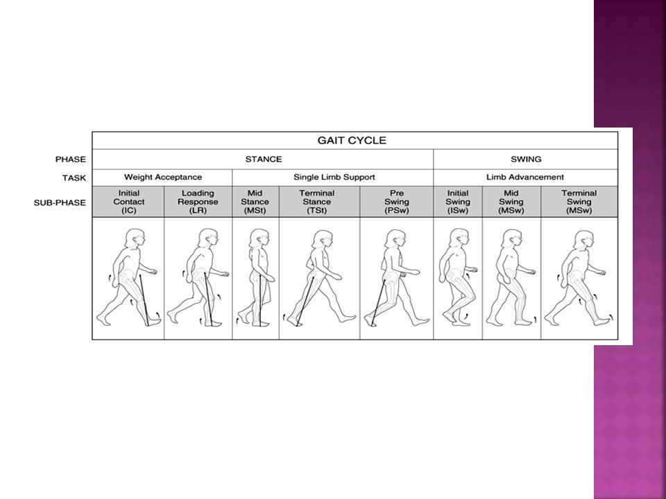
Normal Gait Smooth energy-efficient transfer of the body through space

8
Limp: “to walk with a halting or irregular step”
Funk & Wagnall's

9
Pathological Gaits

10
Antalgic Gait: body’s effort to compensate for pain or instability in the stance-phase limb by minimizing the duration and magnitude of loading

11
Trendelenburg Gait: leaning of the head and trunk toward the lower extremity affected by the pathology Pain Weakness in the hip muscles

12
Common causes of limping seen in early childhood

13
19 month old female referred because of limping

14
Fifth born Normal delivery/presentation Walking at 14 months Always limped No pain Healthy Negative Family history

15
Differential diagnosis
Top three: Hip dysplasia Neuromuscular disease Leg length discrepancy

17
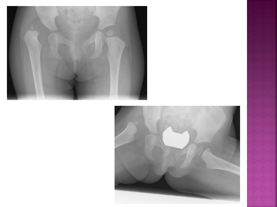
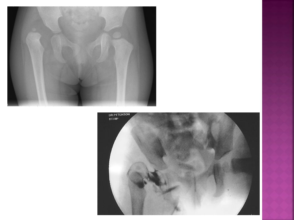
Developmental Dysplasia of the Hip (DDH)
")
18
Dislocated: Dislocatable Subluxed Subluxable Dysplastic
the femoral head is not in contact with the acetabulum Dislocatable the femoral head is within the acetabulum but can be forced out Subluxed the femoral head is within the acetabulum but not in its proper position Subluxable the femoral head can be moved beyond its physiologic limits within the acetabulum Dysplastic although the femoral head is in the proper position the acetabulum or head are abnormally developed

19
History Risk Factors Female Firstborn Breech Large baby Low amniotic fluid Family history

20
Physical Associated Conditions Foot deformity, Torticollis Neuromuscular disorders Syndromes Hip Examination Trendelenburg gait Skin folds Galeazzi sign Abduction Provocative maneuvers Ortolani, Barlow

21
Imaging Ultrasound < 6 months X-ray

23
Treatment URGENT REFERRAL Pavlik Harness Closed Reduction Safe Zone
Open reduction Extra-articular blocks Intra-articular blocks Osteotomies Pelvic Femoral + Shortening

24
Transient synovitis Most common cause of hip pain in childhood
3% childhood risk Idiopathic Frequently associated with concurrent or antecedent illness Right hip = left hip NEVER BILATERAL 2:1 male:female

25
History Age varies (9 months to adolescence) Most between 3 and 8 years old Unilateral hip pain Can present with knee or thigh pain Limp vs non-weight bearing

26
Physical May have a low grade temperature Antalgic or Trendelenburg gait Flexed and externally rotated position Decreased ROM Especially abduction and internal rotation Laboratory tests Non-specific

27
Imaging Radiographs usually normal Ultrasound may show effusion Diagnosis of exclusion

28
Treatment urgent referral Differential includes a septic joint Self limiting disorder May have symptoms for up to 10 days or longer Bed rest until full ROM, no pain, limp free Crutches for older patients NSAIDS Gradual return to activity

29
Legg-Calve-Perthes Disease (perthes)
")
30
Self limiting hip disorder
Caused by ischemia and subsequent necrosis of the femoral head Usually 4 to 8 years of age Male to female ratio: 4-5 to 1 Usually unilateral (88%) Age and lateral head involvement are the key to prognosis 8 years of age seems to be the watershed <50% of lateral pillar involvement better prognosis
 Age and lateral head involvement are the key to prognosis. 8 years of age seems to be the watershed. <50% of lateral pillar involvement better prognosis.")
31
Differential Diagnosis
Unilateral Perthes: septic arthritis sickle cell disease spondyloepiphyseal dysplasia tarda Gaucher’s disease Bilateral Perthes: Hypothyroidism Multiple epiphyseal dysplasia

32
History May be painless at first present with a limp only symptoms occur with subchondral collapse/fracture Can present with knee or thigh pain Positive family history 1.6% – 20% 1% - 3% of patients with transient synovitis will develop Perthes

33
Physical Imaging gait: Trendelenburg
decreased abduction/ internal hip rotation thigh, calf, and buttock atrophy LLD Imaging X-ray, bone scan, MRI

34
Treatment Timely referral Principles of treatment are maintenance of ROM & containment (good coverage of the head by the acetabulum) of the femoral head through the evolution of healing May be obtained by non-operative means relative rest pain control physiotherapy traction abduction splinting at night
 of the femoral head through the evolution of healing. May be obtained by non-operative means. relative rest. pain control. physiotherapy. traction. abduction splinting at night.")
35
Operative Treatments Containable Hip adductor release
Femoral varus/pelvic osteotomies Non-Containable Hip Hip/Late-presenting patient with deformity Hinge abduction Chiari/lateral shelf Cheilectomy Femoral abduction/extension osteotomy OCD, non-operative, revascularization, removal, ORIF

36
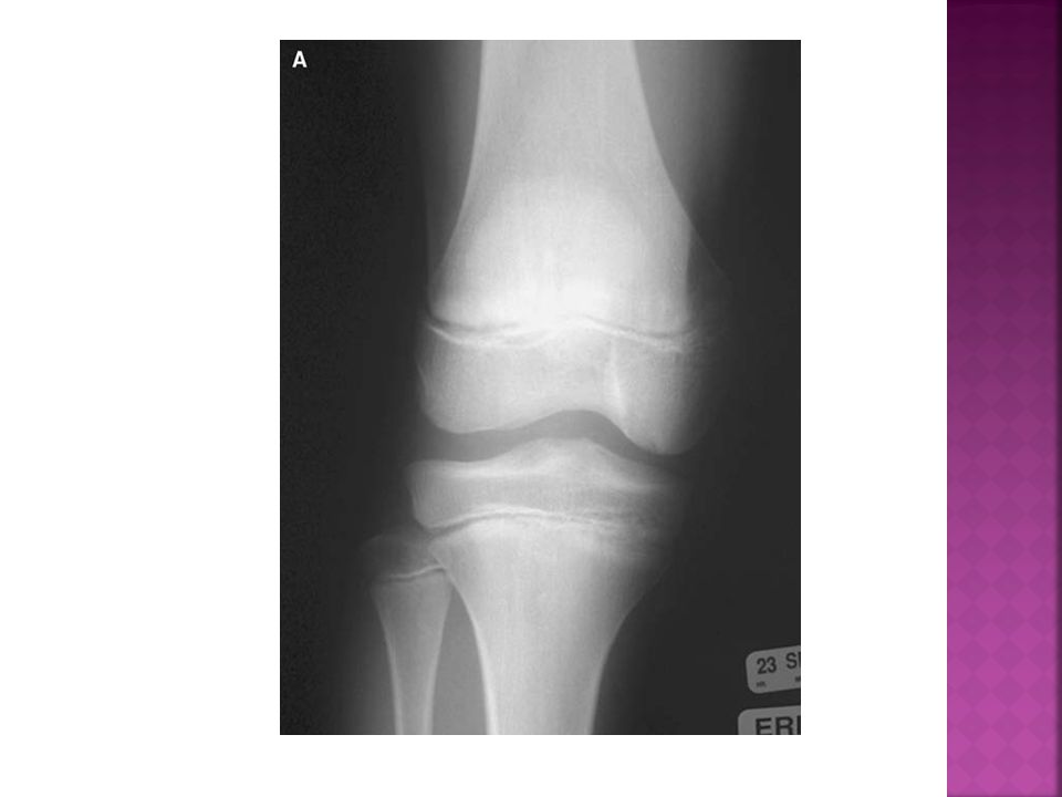
Discoid Meniscus First description: Young 1889
3-5% in general population have a larger than normal meniscus Almost all in lateral, but reported in medial 20% bilateral and 10% associated with OCD of lateral femoral condyle

37
Anatomy Three Segments Anterior horn Body Posterior horn
Attached to tibial plateau, primarily through Coronary Ligament Figure 1 Attached to the capsule except at popliteal hiatus

38
Diagnosis Clinical Presentation History: Asymptomatic
“Snapping knee syndrome” Meniscal tear symptoms Physical: Snapping knee with gait Meniscal signs

39
X-ray Widened lateral joint space, squaring of lateral femoral condyle, cupping of lateral tibial plateau MRI Verify diagnosis and assess damage

40
Treatment Options Asymptomatic: observe Symptomatic:
TIMELY REFERRAL UNLESS LOCKED KNEE THEN URGENT Non-operative: restricted activity, bracing, physiotherapy Operative: Partial meniscal “saucerization” Repair of tear

41
Common causes of limping seen in LATE childhood

42
Apophyseal Conditions

43
Apophysitis of the Hip and Pelvis
Sinding-Larsen-Johansson: inferior pole of patella Osgood-Schlatter Disease: tibial tuberosity disturbance Sever Disease: calcaneal apophysitis Iselin Disease: apophysitis of the fifth metatarsal

44
Osgood Schlatter Disease
Tibial tuberosity disturbance Partial avulsion (microscopic fractures) of the ossification center and overlying hyaline cartilage Epidemiology 10 – 15 years old Boys > girls > 10% of teenagers
 of the ossification center and overlying hyaline cartilage. Epidemiology. 10 – 15 years old. Boys > girls. > 10% of teenagers.")
45
History Pain localized to tubercle Worse with direct blows to the are and activity Physical Antalgic gait may be present Prominent tubercle + local swelling Tenderness localized to tubercle

46
Lovell and Winter’s Pediatric Orthopaedics 5th edition

47
Treatment Spontaneous resolution at maturity 20% may have pain with kneeling surgery for loose ossicles Reassurance Symptomatic treatment/activity modification NSAIDS, stretching, knee pads/braces, foot orthosis, casts TIMELY REFERRAL

48
Osteochondritis dissecans (OCD)
")
49
Introduction Acquired potentially reversible lesion of subchondral bone resulting in delamination and sequestration with or without articular cartilage involvement and instability Juvenile and Adult forms Adult form is typically progressive and unremitting May occur in almost any joint in upper or lower extremity Very common in the knee

50
Epidemiology 15-29 per 100,000 May be bilateral in 25% of cases
Male: female ratio 5:3 >70% are in the classical area Posterolateral aspect of the medial femoral condyle

51
Etiology Idiopathic Theories include: Genetics Inflammation Ischemia
Ossification Repetitive trauma (stress reaction causing a stress fracture in the underlying subchondral bone)
")
52
Clinical Presentation
Physical Antalgic gait Effusion Crepitus Painful ROM Quads atrophy Maximum tenderness usually anteromedial knee Wilson sign Pain with internal tibial rotation History Juvenile Poorly localized pain Exacerbated by exercise May present with symptoms of instability (swelling, stiffness, catching, locking) Limp
 Limp.")
53
Diagnostic Studies X-rays AP Lateral Notch MRI + gadolinium
Lesion size Status of the cartilage and subchondral bone Bone edema and high signal zone beneath fragment Loose bodies Technetium bone scan

56
Management Nonoperative Open physis = good prognosis?
Activity modification Immobilization? Rehabilitation Local and systemic pain management Review every 3-6 months or sooner if symptoms worsening Repeat MRI every 6 months? TIMELY REFERRAL if no improvement or worsening, URGENT IF LOCKED KNEE

57
Operative Indications Lesions not responding to nonoperative management Unstable lesions? Detached lesions

58
The Painful flatFOOT: tarsal coalition

59
HISTORY Tarsal coalition is an abnormal connection between some of the tarsal bones May be painful Can be associated with increased ankle sprains

60
physical Gait Antalgic Flexibility Toe standing Sitting/supine
Subtalar ROM

61
Flexible Flatfoot Arch returns with sitting or tiptoe standing Normal subtalar and midtarsal motion

62
Tarsal Coalition Arch may not return with sitting or tiptoe standing May be painful to move or palpate subtalar joint or other tarsal bones Subtalar motion often decreased

63
imAGING Normal in flexible flatfoot
Oblique views and Harris view may help view a coalition May need an MRI or CT to make diagnosis

64
TREATMENT Tarsal Coalition TIMELY REFERRAL Rest/activity modification
Antiinflammatories Physiotherapy? Orthotics Casts Surgery: resection or fusion

65
Slipped Capital Femoral Epiphysis (SCFE)
")
66
The slip normally occurs during adolescent growth phase
Mechanical or systemic factors may be present Commonly obese Endocrinopathies (eg. 1o & 2o hypothyroidism, panhypopituitarism, GH, hypogonadal conditions, & renal osteodystrophy

67
Male > female Left > right Bilateral involvement may occur Second slip presents within 18 months in 88%

68
History Chronic and/or acute Limp
May present with knee or thigh pain instead of hip/groin pain

69
Physical Gait: Trendelenburg Shortened/external rotation Decreased abduction/internal hip rotation Passive flexion leads to thigh abduction and external rotation Imaging X-ray, CT, MRI

70
X-rays Physeal plate widening & irregularity
Decrease in epiphyseal height Blanch sign of Steel Crescent-shaped area of increased density in the proximal femoral neck Femoral metaphysis appears laterally displaced Klein’s line Southwick angles

71
Imaging Frog-leg lateral avoid in acute situation Cross-table lateral

72
Treatment - Acute Prophylactic Pinning EMERGENT REFERRAL
Immediate bed rest Insertion of one or more screws in situ fixation Designed to fuse the epiphysis on the metaphysis to prevent further slipping Prophylactic Pinning Known metabolic/endocrine disorders? Inability to follow-up

73
Stress fractures

74
Stress fractures Stress Fractures in Skeletally Immature Patients
Walker et. al.: JPO 1996 34 stress fractures Tibia (47%), fibula, femur, radius, humerus, MT
, fibula, femur, radius, humerus, MT.")
75
History Pain often associated with an increase in activity Be wary of female triad Physical Antalgic gait may be present Tenderness localized

76
Radiographs Rapid bony response may be present Bone Scan Helpful in questionable situations Treatment (depends on causative factors) Urgent referral Modification of activities Immobilization

77
Common causes of limping seen at various ages

78
Bone and soft tissue Tumors

79
Bone and Soft Tissue Tumors
History Pain Night pain History of trauma may delay diagnosis Osteoid Osteoma pain relieved by NSAIDs Constitutional Symptoms Fever, night sweats, anorexia, weight loss eg. Ewing sarcoma Soft tissue mass may not be symptomatic

80
Physical Exam Gait disturbance Muscle atrophy Neurovascular exam Range of motion Mass Size, tenderness, pulsation, mobility, bruits, tenderness, erythema, consistency Lymph nodes

81
Investigations Bloodwork CBC, ESR, CRP, serum alkaline phosphatase, serum and urine calcium & phosphorus, LDH Imaging X-ray Bone Scan CT/MRI

82
Management Referral Urgency dependent on tumor type

83
Septic joints

84
History Pain Refusal to bear weight Limping Recent illness Decreased immunity eg. chickenpox Trauma

85
Physical Exam Temperature Antalgic gait Disuse of a part Erythema/swelling Tenderness Decreased ROM

86
Laboratory tests CBC WBC CRP ESR Blood cultures Aspirates (Gram stain, Culture) Imaging X-rays Ultrasound Bone Scan CT MRI

87
Treatment Emergent referral Stop tissue destruction ASAP Decrease bacterial load and irrigation of the joint Identify the Organism Select appropriate antibiotic

Similar presentations


>")


 MB BS BSc MSc (SEM) MRCS (Eng) Diploma in MM (UIAA)>")
>")


