Download presentation
Presentation is loading. Please wait.

1
Bone Tissue

2
Bone Markings Bulges, depressions, and holes serve as
Sites of attachment for muscles, ligaments, and tendons Joint surfaces Conduits for blood vessels and nerves

3
Bone Markings: Projections
Sites of muscle and ligament attachment Tuberosity—rounded projection Crest—narrow, prominent ridge Trochanter—large, blunt, irregular surface Line—narrow ridge of bone Tubercle—small rounded projection Epicondyle—raised area above a condyle Spine—sharp, slender projection Process—any bony prominence

4
Table 6.1

5
Bone Markings: Projections
Projections that help to form joints Head Bony expansion carried on a narrow neck Facet Smooth, nearly flat articular surface Condyle Rounded articular projection Ramus Armlike bar

6
Table 6.1

7
Bone Markings: Depressions and Openings
Meatus Canal-like passageway Sinus Cavity within a bone Fossa Shallow, basinlike depression Groove Furrow Fissure Narrow, slitlike opening Foramen Round or oval opening through a bone

8
Table 6.1

9
Cartilage – Three types
Hyaline – Most abundant Articular cartilages – cover the ends of bones at moveable joints Costal cartilages – connect the ribs and sternum Laryngeal cartilages – for the skeleton of the larynx (voice box) Tracheal and bronchial cartilages – reinforce the respiratory passages Nasal cartilages – support the external nose.
 Tracheal and bronchial cartilages – reinforce the respiratory passages. Nasal cartilages – support the external nose.")
10
Cartilage Elastic – Contains more stretchy elastic fibers.
Found in the external ear and form the epiglottis, the flap that closes on the larynx when you swallow. Fibrocartilage – Rows of chondrocytes and thick collagen fibers Very high tensile strength and compressible. Found between the vertebral disks, the pad-like cartilages of the knee, and the pubic symphysis.

11
Cartilage Cartilage grows in two ways:
Appositional growth – growth from the outside. Interstitial growth – growth from within.

12
Functions of Bones Support – Bones give shape and support to the entire body and provide places for organs to attach. Allows standing, etc. Protection – Cranial bones protect the brain, vertebrae protect the spinal cord, rib cage protects the thoracic organs. Movement – Skeletal muscles, which are attached to bone by tendons, use bones to move the body and its parts.

13
Functions continued Mineral storage - Calcium and phosphorus are stored in bones and are constantly being deposited and withdrawn. Blood cell formation – AKA hematopoiesis occurs in the marrow of certain bones.

14
Four Classes of Bone (bases on shape)
Long Bones – Longer than they are wide. Consist of a shaft and 2 ends. All bones of the limbs are long bones except the carpals, tarsals, and patella. Short Bones – Roughly cube-like. Tarsals and carpals. Flat Bones – Thin, flat, and usually somewhat curved. Sternum, ribs, scapula, and cranial bones.

15
Four Classes of Bone Irregular Bones – Bones that don’t fit any of the previous shapes. Vertebrae and pelvic bones.

16
Structure of Bones Two layers. The external layer that appears smooth and solid is compact bone. Internal to this is the spongy bone. Spongy bone is comprised of small, needle-like pieces called trabeculae that form a honeycomb. The spaces between trabeculae are filled with red or yellow bone marrow. Long bones are primarily compact bone, but may have a fair amount of spongy bone. Flat bones have two parallel layers of compact bone with a layer of spongy bone between.

17
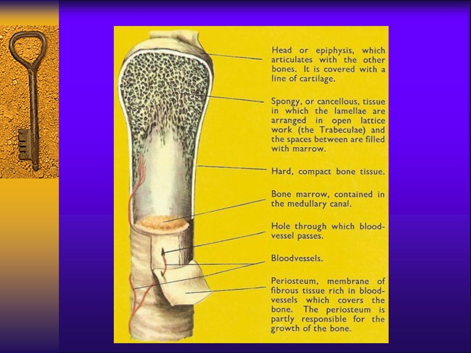
General Structure of Long Bones
Diaphysis – The shaft or long axis of the bone. It has a thick collar of compact bone that surrounds the central medullary cavity that contains fat (yellow bone marrow). Epiphysis – The ends of the bone consisting of the distal epiphysis and proximal epiphysis. The exterior is compact bone while the interior is spongy bone. The joint surfaces of each are covered with a thin layer of articluar (hyaline) cartilage which absorbs stress and cushions during movement.
. Epiphysis – The ends of the bone consisting of the distal epiphysis and proximal epiphysis. The exterior is compact bone while the interior is spongy bone. The joint surfaces of each are covered with a thin layer of articluar (hyaline) cartilage which absorbs stress and cushions during movement.")
21
Structure of a Flat Bone
Figure 6.4

22
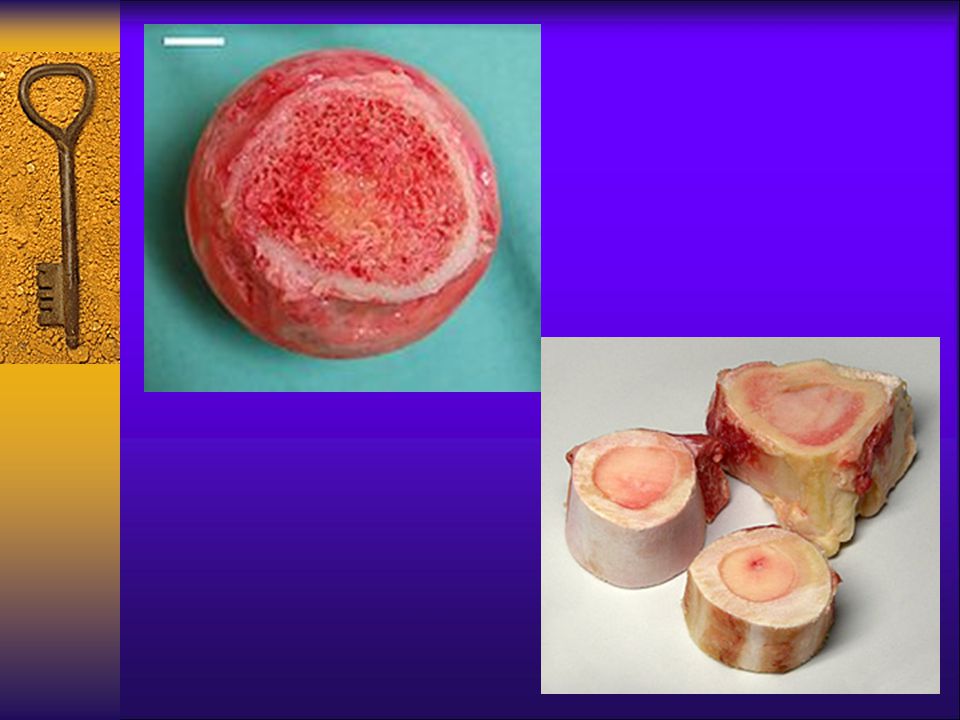
Location of Red Bone Marrow
Red marrow is known as hematopoietic tissue because it gives rise to blood cells. Typically found within the cavities of spongy bone. In adults, the medullary cavity extends into the epiphyses, and a little red marrow is found in most long bones. Blood cell production is limited to the head of the femur and humorous.

23
Location of Red Bone Marrow
More important are the flat bones (sternum) and irregular bones (pelvic bones). Yellow marrow can be converted to red marrow if a person becomes anemic and needs enhanced Red Blood Cell production.
 and irregular bones (pelvic bones). Yellow marrow can be converted to red marrow if a person becomes anemic and needs enhanced Red Blood Cell production.")
27
Pages 3&4 of notes will be on a WS
Bone Tissue Pages 3&4 of notes will be on a WS

28
Composition of bone tissue
Bone tissue is composed of 2 types of tissues Organic Inorganic

29
Organic portion: 35% of mass
The organic portion consists of the bone cells and the organic matrix The Bone cells are the: Osteocytes Osteoblasts Osteoclasts FYI: There are also osteoprogenitor cells that are the precursers to blasts & cytes. They are derived from mesenchyme & found on all bone surfaces.

30
Blasts, clasts & Cytes

31
Organic portion The Organic Matrix aka. Osteoid
is produced by the osteoblasts Analogy: The organic matrix is the portion that is deposited first as the “grillwork” or framework of the bone during the process of OSTEOGENESIS It consists of ground substance and collagen fibers produced by CT cells Its function is to provide the bone with tensile strength and resilience – in other words, to make the bone a little flexible Review: ground substance, collagen fibers & EC matrix functions!!

32
Inorganic matrix: 65% of mass
The inorganic matrix consists of inorganic salt compounds mainly: Calcium & phosphorus salt compounds Its function is to give Strength to the bone Analogy: It is formed during OSTEOGENESIS by the process of Mineralization The inorganic matrix minerals are deposited into the organic matrix “grillwork” The enzyme alkaline phosphatase mediates this process

33
Two types of bone tissue
Know slides, locations, functions

34
Compact bone tissue (cbt)
Arranged in OSTEONS aka Haversian system Contains a series of openings that permit exchange of materials between osteocytes (& other bone cells) and the blood. Location: look at diagrams
 and the blood. Location: look at diagrams.")
35
Osteon diagram – cross section

36
osteocytes Lacunae, osteocytes, & canaliculi

37
Osteon diagram – sagittal section

38
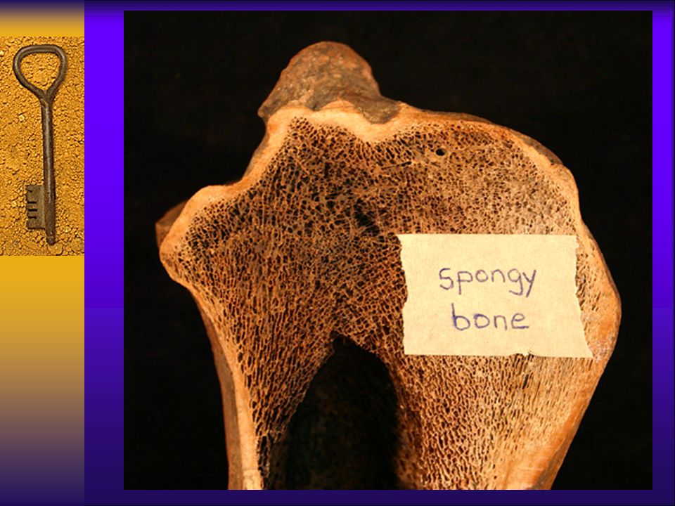
Cancellous (spongy) bone tissue
Main structures are the trabeculae which are needlelike structures of minerals that are arranged along stress lines to provide strength Materials are exchanged by diffusion since there are NO canals for passage Location: ends of long bones & middle of flat, short, and irregular bones

41
Bone marrow Aka myeloid tissue Yellow bone marrow Red bone marrow
Fat storage Found in medullary canal of long bones Red bone marrow Found in spongy bone (ends of long bones, flat bones, irregular bones) Hematopoiesis (formation of all blood cells)
 Hematopoiesis (formation of all blood cells)")
44
Types of growth Longitudinal growth – bone growth in length at epiphyseal plates (till plates ossify) Appositional growth – bone growth in diameter (throughout life) Known as remodeling These 2 types work together to make the bones long enough & strong enough
 Known as remodeling. These 2 types work together to make the bones long enough & strong enough.")
45
Regulation of bone growth
Bone is a dynamic and active tissue. They are constantly being remodeled according to the activities that we do. Main factor = Ca levels in blood Ca imp for bone strength but also for nervous & muscular system to work correctly!!!

46
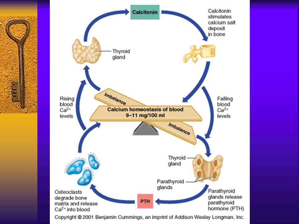
Regulation by hormonal feedback
Purpose: to maintain optimal ionic calcium levels in blood This is your body’s TOP priority!!! A main factor that affects what our bone tissue does is our blood calcium level Optimum blood Ca 2+ level = 9-11 mg/100 ml of blood Calcium ions are VERY important for muscle & nervous function – our body cares more about this level than it does our bone strength!!

48
Regulation by hormonal feedback
PTH (parathyroid gland) – activated when Ca levels in blood are too low (hypocalcemia) - promotes calcium reabsorption Calcium will go from bone to blood Calcitonin (thyroid gland) activated when Ca levels in blood are too high (hypercalcemia) - promotes calcium deposition Calcium will go from blood to bone
 – activated when Ca levels in blood are too low. (hypocalcemia) - promotes calcium reabsorption. Calcium will go from bone to blood. Calcitonin (thyroid gland) activated when Ca levels in blood are too high. (hypercalcemia) - promotes calcium deposition. Calcium will go from blood to bone.")
49
Regulation by mechanical stress
Purpose: keep bones strong This is the secondary purpose Wolff’s law states that bones will grow according to the stresses placed upon them So activities that compress bones and pull on muscles which pull on bones can make bones stronger

50
How they work together to regulate
PTH & calcitonin (hormones) determine WHEN the remodeling will occur Primary purpose = Ca 2+ regulation in blood The Mechanical stresses determine WHERE the remodeling will occur Secondary purpose = where will the calcium ions be deposited or reabsorbed from
 determine WHEN the remodeling will occur. Primary purpose = Ca 2+ regulation in blood. The Mechanical stresses determine WHERE the remodeling will occur. Secondary purpose = where will the calcium ions be deposited or reabsorbed from.")
51
Fractures & Disorders This section of info will be part of a lab practical quiz along with the tissue slides & functions from earlier in the notes

52
Instructions Construct a chart or other type of graphic organizer for the 9 types of fractures You will also need to be able to identify the X-rays or pictures of each. See Lab practical slide study on webpage for quiz review.

53
Common types of fractures
1. Comminuted Bone breaks into many fragments Common in aged whose bones are more brittle (contain less organic matrix)
")
54
Common types of fractures
2. Compression Bone is crushed Common on osteoporotic bones (or bones that are porous for other reasons)
")
55
Common types of fractures
3. Depressed Broken bone portion is pressed inward Typical of skull fracture

56
Common types of fractures
4. Impacted Broken bone ends are forced into each other Commonly occurs when one attempts to break a fall with outstretched arms Or when one jumps off something too high

57
Impacted

58
Common types of fractures
5. Spiral Ragged break occurs when excessive twisting forces are applied to a bone Common sports fracture

59
Spiral fractures

60
Spiral fracture

61
Common types of fractures
6. Greenstick Bone breaks incompletely, much in the way a green twig breaks Common in children whose bones are more flexible (more organic matrix present)
")
62
7. Simple fracture Bone does not protrude through skin

63
8. Compound Fractures Bone protrudes through skin

64
9. Epiphyseal fracture Bone shears off at growth plate
Can cause problems with early ossification at location of break

65
Example:Clavicle fracture

66
Example: Fracture

67
Example: dislocation

68
Disorders

69
Osteoporosis Define: Who is affected? reabsorption outpaces deposition
chemical composition remains the same BUT less total bone mass SO bones become more porous & lighter Which leads to fractures & deformities from body weight Who is affected? Mainly: Aged, post-menopausal women

70
osteoporosis Contributing factors Treatments Decreased estrogen
Decreased calcium & protein Vitamin D metabolic disorders Hormone conditions Insufficient exercise Immobility Treatments Replace what is missing Medication for metabolic disorders

71
osteoporosis

72
Osteomalacia (rickets)
Define: Chemical composition is abnormal (% are not right) Increase in organic matrix – decrease in inorganic matrix Indequate mineralization so bones are too soft Lack of calcium deposition Who is affected? More severe effects in children since they are still growingbut primarily a nutritional disorder
 Increase in organic matrix – decrease in inorganic matrix. Indequate mineralization so bones are too soft. Lack of calcium deposition. Who is affected More severe effects in children since they are still growingbut primarily a nutritional disorder.")
73
osteomalacia Contributing factors: Treatments:
Vitamin D &/or calcium deficiency Poor nutrition Treatments: Supplementation Sunlight: think about why Vitamin D deficiency would lead to this disorder!

74
osteomalacia

75
Other disorders – add to notes
Osteomyolitis: inflammation of bone and muscle Gigantism: excess of growth hormone before epiphyseal plates have ossified Acromegaly: excess of growth hormone after epiphyseal plates have ossified Dwarfism: deficit of growth hormone as well as other factors Achondroplasia: most common form of dwarfism– autosomal dominant but can be from mutation

76
gigantism Robert Wadlow
Lots of internal health problems including joint problems

77
acromegaly Epiphyseal plates are sealed so bones can only grow in diameter/thickness

78
achondroplasia Disproportionate dwarfism Normal torso & head
Short arms & legs

79
Hypopituitary dwarfism
More proportionate dwarfism

Similar presentations


 Nervous tissue Cartilage.>")

 Short bones – Cube-shaped bones (i.e. wrist and ankle) – Bones.>")


 - Tendons (muscle to bone)>")

 2.Protection: skull, vertebrae,>")

 Joints ► Cartilages Ligaments ► Divided.>")


