Download presentation
Presentation is loading. Please wait.

1
COMUTED TOMOGRAHY Dr. Amr A. Abd-Elghany 1

2
COMPUTED TOMOGRAPHY It is a non-invasive medical imaging modality that combines the use of X-rays and computer processing to generate tomographic (‘slices’) of the area scanned. Four classifications of the types of scans: -Abdominal -Bone -Head -Vascular 2
 of the area scanned. Four classifications of the types of scans: -Abdominal -Bone -Head -Vascular. 2.")
4
Tomo = image // to long axis of the body CT = image is transverse to the body

5
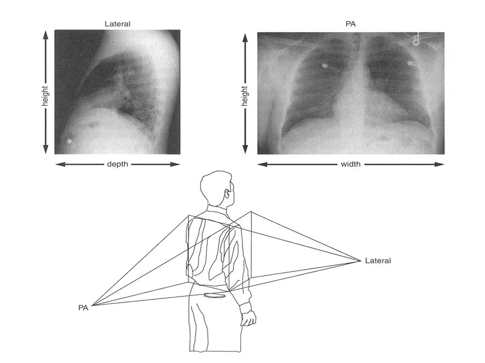
X-ray image of the thorax CT scan of the thorax
5

6
Why computed tomography?
Mathematical principles of CT were first developed in 1917 by Radon. Proved that an image of an unknown object could be produced if one had an infinite number of projections through the object. Plain film imaging reduces the 3D patient anatomy to a 2D projection image. Used to determine: the extent of Trauma, Location and type of the tumor, status of blood vessels, pre surgical planning.

7
Why computed tomography? Cont.
Density at a given point on an image represents the x-ray attenuation properties within the patient along a line between the x-ray focal spot and the point on the detector corresponding to the point on the image. With a conventional radiograph information with respect to the dimension parallel to the x-ray beam is lost. Large area x-ray beams used in conventional radiography produces considerable scattered radiation that causes blurring images.

9
CT the beginning Was formally introduced in 1972 by a British engineer Sir Godfrey Hounsfield The first scan was done in 1971, of the brain Physicist Allan Cormack also invented a similar machine in the United States Both Cormack and Hounsfield were awarded the Nobel Prize in 1979 First clinical scanners were installed in 1974 Was a revolutionary invention in terms of medical imaging

11
PRINCIPLE OF COMPUTED TOMOGRAPHIC IMAGING
In early CT imaging devices (“scanners”) a narrow x-ray beam is scanned across a patient in synchrony with a radiation detector on the opposite side of the patient. If the beam is mono-energetic or nearly so, the transmission of x rays I through the patient is I = I0e−μx 11
 a narrow x-ray beam is scanned across a patient in synchrony with a radiation detector on the opposite side of the patient. If the beam is mono-energetic or nearly so, the transmission of x rays I through the patient is. I = I0e−μx. 11.")
12
x-ray tube

13
Basic CT scanner component
Gantry. X-ray tube. Detector. Control console. Computed Tomography Machine

14
X

15
Y

16
Z

17
ISOCENTER

18
Gantry CT X-ray tube. High voltage generator. Detector array.
Data acquisition system. Slip ring: eliminated the need of cables and enables the continuous rotation of the gantry components (electromechanical technology).
.")
19
Detector Elements Capture energy that has not been attenuated by the patient

20
CT Generations 1st Generation: Rotate/ translate, pencil beam 1972
1 to 3 x-ray detectors used. Parallel ray geometry. X-ray tube only able to rotate180 degrees at 1-degree intervals. About 4.5 min/slice. Whole chest can be scanned at 1 hr. Pencil beam allowed very efficient scatter reduction, best of all scanner generations.

21
1st generation cont.

22
Second Generation 1973 Fan-shaped x-ray beam 30 or more detectors
20 seconds per slice or 10 minutes for a 40 slice exam. 180 degree rotation. Long data reconstruction time.\.

23
Third Generation 1974 Fan-shaped x-ray beam.
960 detectors opposite the x-ray tube. Complete 360 degree rotation Rotate/Rotate movement. One rotation = one slice. Second data acquisition could be made as the tube and detectors move in the opposite direction. Time reduced to 1 sec per slice.

24
3rd generation configuration

25
Fourth Generation 1975 Developed in 1980’s.
Fixed ring of as many as 4800 detectors, completely surrounding the patient, Rotate only movement Rotating x-ray tube provides short bursts of radiation Detectors collect the remnant radiation to reconstruct into an image 1 minute for multiple slices

26
4th generation configuration

27
Fifth Generation Stationary/stationary 1984
Developed specifically for cardiac tomographic imaging. No conventional x-ray tube; large arc of tungsten encircles patient and lies directly opposite to the detector ring. Electron beam steered around the patient to strike the annular tungsten target. Capable of 50msec scan times; can produce fast frame rat CT movies of the beating heart

29
Modern Scanners No longer categorize into Generations
Contemporary (modern) CT scanners are either third or fourth generation designs. Scanners are categorized by tube and detector movement Slip Ring Technology: connects generator with tube (no cables)
 CT scanners are either third or fourth generation designs. Scanners are categorized by tube and detector movement. Slip Ring Technology: connects generator with tube (no cables)")
30
Spiral CT 1986 (Helical CT-volumetric CT) 6th generation
Helical (continuous rotation) CT scanners acquire data while the table is moving By avoiding the time required to translate the patient table, the total scan time required to image the patient can be much shorter Allows the use of less contrast agent. In some instances the entire scan be done within a single breath-hold of the patient
 CT scanners acquire data while the table is moving. By avoiding the time required to translate the patient table, the total scan time required to image the patient can be much shorter. Allows the use of less contrast agent. In some instances the entire scan be done within a single breath-hold of the patient.")
31
Multiple detector array (Multi-slice CT)1999 7th generation
When using multiple detector arrays, the collimator spacing is wider and more of the x-rays that are produced by the tube are used in producing image data Opening up the collimator in a single array scanner increases the slice thickness, reducing spatial resolution in the slice thickness dimension With multiple detector array scanners, slice thickness is determined by detector size, not by the collimator

33
Procedure Indications Patient preparation. Patient positioning.
If physician could not diagnose by plain x-ray. CT can detect masses, fluids, mediastina and chest wall lesions, pulmonary embolism (blocking of main artery of the lung or one of its branches). Patient preparation. Contrast media administration, fasting 4-6 hours. Patient positioning. Chest, abdomen, pelvis=AP. Brain, spine=lateral. Scanogram
. Patient preparation. Contrast media administration, fasting 4-6 hours. Patient positioning. Chest, abdomen, pelvis=AP. Brain, spine=lateral. Scanogram.")
34
Scanogram cont. CT will ask you slice thickness 1cm or more or less.
CT will ask you if you will inject contrast or not (lung =no, mediastinum=yes). Press the button. The table will be moved 1cm. Each slice will have 2 images (mediastinal window, lung window) 1 = root of aorta 2 = pulmonary outflow 3 = left atrium 4 = left pulmonary vein 5 = superior vena cava 6 = descending aorta
. Press the button. The table will be moved 1cm. Each slice will have 2 images. (mediastinal window, lung window) 1 = root of aorta 2 = pulmonary outflow 3 = left atrium 4 = left pulmonary vein 5 = superior vena cava 6 = descending aorta.")
35
Benefit of CT scan Unlike other imaging methods, fast.
Painless, non-invasive & Accurate CT Scanning offers detailed views. Diagnosis made with the assistance of CT can eliminate the need for invasive exploratory surgery & surgical biopsy. Information to show cross section of body tissue & organ Use wide range of clinical problems.

36
Risks of CT scan CT scan is not generally indicated for pregnant women. CT scan does involves high radiation exposure Risk of serious allergic reaction to iodine containing contrast media.

37
C.T Scan of Blood Vessel

38
CT angiogram shows pulmonary vessels
Normal Abdomen CT Oral & IV contrast level of spleen & liver CT angiogram shows pulmonary vessels

Similar presentations


 theory was developed 1972: The CT scan was invented by Godfrey.>")

>")


 of radiation directed toward the pt. and the remnant radiation emitted from the pt.>")


 Dynamic scanning implies 15 or more scans in rapid sequence within one.>")
