Download presentation
Presentation is loading. Please wait.

1
Diagnostic tools – imaging and lung function (humans)
David A Lynch, MB

2
Disclosures Consultant: Research support:
Perceptive Imaging Centocor, Inc Boehringer Ingelheim Siemens Inc Genentech NHLBI Gilead Intermune Veracyte Pfizer

3
I ain’t going near no CT scanner
Conflict of interest I ain’t going near no CT scanner Jack

4
CT in human fibrotic lung disease
Technique Terminology Patterns of fibrosis Quantification

5
High Resolution CT technique
Thin section ( mm) Small field of view Edge-enhancing reconstruction Adequate inspiration Absent motion Prone and expiratory images very helpful Multiplanar images Standard window width (1500) and level (-700) Mayo JR. CT evaluation of diffuse infiltrative lung disease: dose considerations and optimal technique. Journal of thoracic imaging. 2009;24(4):252-9.
 Small field of view. Edge-enhancing reconstruction. Adequate inspiration. Absent motion. Prone and expiratory images very helpful. Multiplanar images. Standard window width (1500) and level (-700) Mayo JR. CT evaluation of diffuse infiltrative lung disease: dose considerations and optimal technique. Journal of thoracic imaging. 2009;24(4):")
8
Features of lung fibrosis
Reticular lines Honeycombing Ground glass attenuation or consolidation (uncommon) Traction bronchiectasis/architectural distortion Lobar volume loss Hansell DM, et al. Fleischner Society: glossary of terms for thoracic imaging. Radiology. 2008;246(3):
 Traction bronchiectasis/architectural distortion. Lobar volume loss. Hansell DM, et al. Fleischner Society: glossary of terms for thoracic imaging. Radiology. 2008;246(3):")
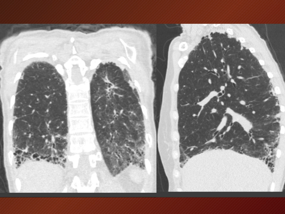
9
Reticular abnormality with traction bronchiectasis

10
Reticular abnormality with honeycombing

11
Ground glass abnormality

12
Consolidation with traction bronchiectasis

13
Architectural distortion

14
Lobar volume loss

15
CT distribution Craniocaudal Upper lung predominant
Lower lung predominant Mid lung predominant Axial Peripheral/subpleural Peribronchovascular

16
Patterns of lung fibrosis in humans
UIP NSIP Hypersensitivity pneumonitis Sarcoidosis Organizing pneumonia Idiopathic pleuroparenchymal fibroelastosis

18
UIP pattern Subpleural, basal predominance Reticular abnormality
Honeycombing with or without traction bronchiectasis Absence of features inconsistent with UIP pattern ATS/ERS/JRS/ALAT Statement: Idiopathic Pulmonary Fibrosis: Evidence-based Guidelines for Diagnosis and Management. AJRCCM, 2011 Mar 15;183(6):
:")
19
Possible UIP Subpleural, basal predominance Reticular abnormality
Absence of features inconsistent with UIP pattern ATS/ERS/JRS/ALAT Statement: Idiopathic Pulmonary Fibrosis: Evidence-based Guidelines for Diagnosis and Management. AJRCCM, 2011 Mar 15;183(6):
:")
20
IPFNET: Concordance between CT and pathologic diagnoses
Pathology diagnosis CT diagnosis Definite UIP Probable UIP Possible UIP Not UIP Total UIP 82 17 1 2 102 51 9 4 64 Inconsistent with UIP 55 16 75 188 42 241 Yagihashi et al. Soc Thorac Radiol 2014

21
NSIP Basal predominance Peribronchovascular/ subpleural sparing
Confluent pattern Volume loss Ground glass Reticular Traction bronchiectasis Consolidation +/- Honeycombing rare Travis et al. Am J Respir Crit Care Med. 2008;177:1338

22
Fibrotic sarcoidosis

23
Fibrotic hypersensitivity pneumonitis
Upper, mid or lower lung predominance Infiltrative Centrilobular nodules Irregular lines Ground glass Reticular opacity Honeycombing Obstructive Air trapping Cysts Emphysema

24
Organizing pneumonia

25
Idiopathic pleuropulmonary fibroelastosis (IPPFE)
Rare entity Upper lobe predominant Dense pleural/subpleural fibrosis Traction bronchiectasis and volume loss Frankel SK. Chest 2004;126:

26
Bleomycin induced fibrosis in rabbits
Hirose et al. Am Rev Respir Dis Mar;147(3):730-8. Lynch et al. Acad Radiol Feb;4(2):102-7
: Lynch et al. Acad Radiol Feb;4(2):")
27
Bleomycin induced fibrosis in rabbits
Lynch et al. Acad Radiol Feb;4(2):102-7
:")
28
Quantification of lung fibrosis
Semiquantitative Densitometry/CT histogram Texture-based methods

29
Relationship between semiquantitative assessment and physiologic impairment
Wells et al. Am J Respir Crit Care Med Vol 167. pp 962–969, 2003

30
Relationship between semiquantitative assessment and mortality: Multivariate
Baseline Variable Hazard ratio 95% Confidence Interval p Value HRCT features Overall extent of fibrosis 2.71 1.61, 4.55 < % predicted DLCO 0.94 0.90, 0.98 0.004 Treatment assignment to IFN-γ1b 0.53 0.28, 0.99 0.04 Lynch DA, et al. Am J Respir Crit Care Med2005;172:

31
Histogram-based parameters
Courtesy: Stephen Humphries, National Jewish Health

32
Relationship between quantitative histogram assessment and physiologic impairment
Best et al., Radiology; 2003;208:

33
Multivariate analysis of predictors of mortality in IPF (n=167, 35 deaths)
Effect Odds Ratio Estimates 95% Confidence Limits Wald Chi-Square Pr > ChiSq Kurtosis at Baseline 0.579 0.32 to 1.049 3.249 0.0715 Mean Visual Fibrosis at Baseline CT 1.104 1.018 to 1.198 5.7171 0.0168 Best et al., Radiology; 2008 Mar;246(3): 33
:")
34
Textural analysis Normal lung Reticular Abnormality
Courtesy: Stephen Humphries, National Jewish Health

35
Critical features of CT assessment of fibrosis
High resolution technique, with minimal motion Standardized descriptive lexicon Systematic categorization of fibrotic patterns Quantification Visual Histogram based Textural Validation against physiology, pathology, outcome

36
THANKS!

Similar presentations


 Infections (pneumonia, airways disease)>")

 – The need for early recognition and referral PRC-2128.>")

 and its importance to recognize Abstract ID -1188.>")


 is a specific form of chronic, progressive, fibrosing interstitial pneumonia of unknown cause that occurs.>")
,>")