Download presentation
Presentation is loading. Please wait.

1
Chapter 10 Muscular Tissue
Alternating contraction and relaxation of cells Chemical energy changed into mechanical energy

2
3 Types of Muscle Tissue Skeletal muscle
attaches to bone, skin or fascia striated with light & dark bands visible with scope voluntary control of contraction & relaxation

3
3 Types of Muscle Tissue Cardiac muscle striated in appearance
involuntary control autorhythmic because of built in pacemaker

4
3 Types of Muscle Tissue Smooth muscle
attached to hair follicles in skin in walls of hollow organs -- blood vessels & GI nonstriated in appearance involuntary

5
Functions of Muscle Tissue
Producing body movements Stabilizing body positions Regulating organ volumes bands of smooth muscle called sphincters Movement of substances within the body blood, lymph, urine, air, food and fluids, sperm Producing heat involuntary contractions of skeletal muscle (shivering)
")
6
Connective Tissue Components

8
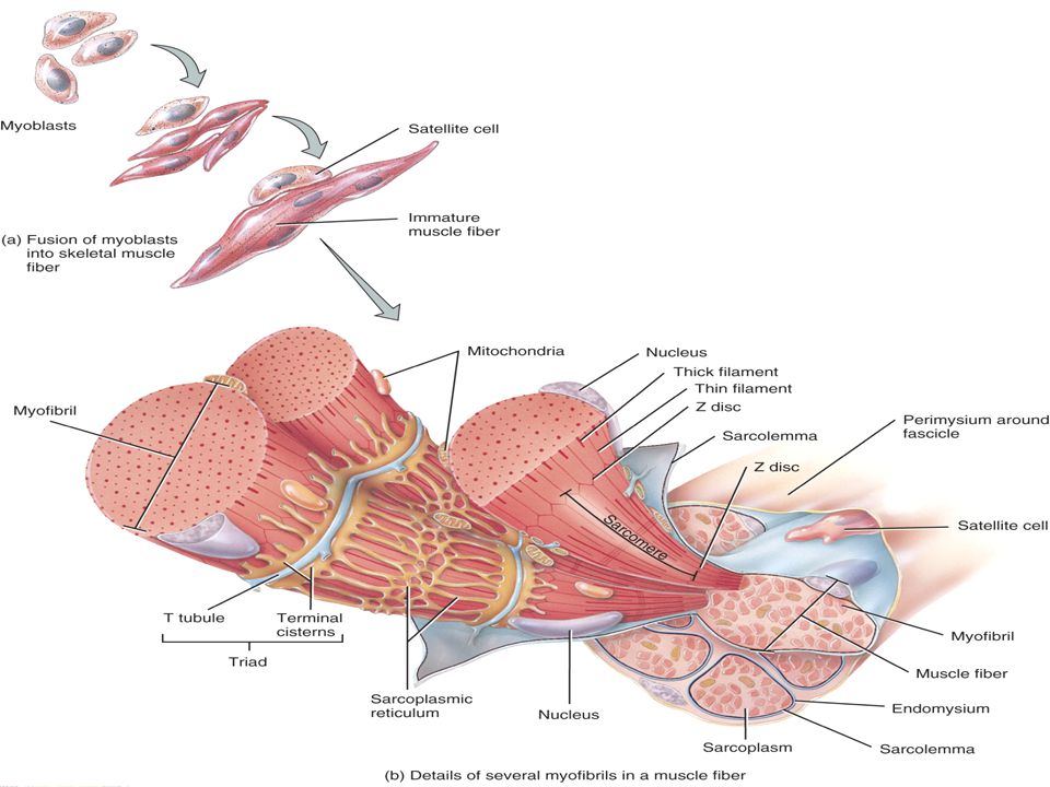
Muscle Fiber or Myofibers
Muscle cells are long, cylindrical & multinucleated Sarcolemma = muscle cell membrane Sarcoplasm filled with tiny threads called myofibrils & myoglobin (red-colored, oxygen-binding protein)
")
9
Transverse Tubules T (transverse) tubules are invaginations of the sarcolemma into the center of the cell filled with extracellular fluid carry muscle action potentials down into cell Mitochondria lie in rows throughout the cell near the muscle proteins that use ATP during contraction

10
Myofibrils & Myofilaments
Muscle fibers are filled with threads called myofibrils separated by SR (sarcoplasmic reticulum) Myofilaments (thick & thin filaments) are the contractile proteins of muscle
 Myofilaments (thick & thin filaments) are the contractile proteins of muscle.")
11
Sarcoplasmic Reticulum (SR)
System of tubular sacs similar to smooth ER in nonmuscle cells Stores Ca+2 in a relaxed muscle Release of Ca+2 triggers muscle contraction

12
Filaments and the Sarcomere
Thick and thin filaments overlap each other in a pattern that creates striations (light I bands and dark A bands) They are arranged in compartments called sarcomeres, separated by Z discs. In the overlap region, six thin filaments surround each thick filament
 They are arranged in compartments called sarcomeres, separated by Z discs. In the overlap region, six thin filaments surround each thick filament.")
13
Rigor Mortis Rigor mortis is a state of muscular rigidity that begins 3-4 hours after death and lasts about 24 hours After death, Ca+2 ions leak out of the SR and allow myosin heads to bind to actin Since ATP synthesis has ceased, crossbridges cannot detach from actin until proteolytic enzymes begin to digest the decomposing cells.

14
Neuromuscular Junction (NMJ) or Synapse
NMJ = myoneural junction end of axon nears the surface of a muscle fiber at its motor end plate region (remain separated by synaptic cleft or gap)
")
15
Motor units

16
Structures of NMJ Region
Synaptic end bulbs are swellings of axon terminals End bulbs contain synaptic vesicles filled with acetylcholine (ACh) Motor end plate membrane contains 30 million ACh receptors.
 Motor end plate membrane contains 30 million ACh receptors.")
17
Events Occurring After a Nerve Signal
Arrival of nerve impulse at nerve terminal causes release of ACh from synaptic vesicles ACh binds to receptors on muscle motor end plate opening the gated ion channels so that Na+ can rush into the muscle cell Inside of muscle cell becomes more positive, triggering a muscle action potential that travels over the cell and down the T tubules The release of Ca+2 from the SR is triggered and the muscle cell will shorten & generate force Acetylcholinesterase breaks down the ACh attached to the receptors on the motor end plate so the muscle action potential will cease and the muscle cell will relax.

18
Isotonic and Isometric Contraction
Isotonic contractions = a load is moved concentric contraction = a muscle shortens to produce force and movement eccentric contractions = a muscle lengthens while maintaining force and movement Isometric contraction = no movement occurs tension is generated without muscle shortening maintaining posture & supports objects in a fixed position

19
Anatomy of Cardiac Muscle
Striated , short, quadrangular-shaped, branching fibers Single centrally located nucleus Cells connected by intercalated discs with gap junctions Same arrangement of thick & thin filaments as skeletal

20
Histology of cardiac muscle

21
Appearance of Cardiac Muscle
Striated muscle containing thick & thin filaments T tubules located at Z discs & less SR

22
Microscopic Anatomy of Smooth Muscle
Small, involuntary muscle cell -- tapering at ends Single, oval, centrally located nucleus Lack T tubules & have little SR for Ca+2 storage

23
Microscopic Anatomy of Smooth Muscle
Thick & thin myofilaments not orderly arranged so lacks sarcomeres Sliding of thick & thin filaments generates tension Transferred to intermediate filaments & dense bodies attached to sarcolemma Muscle fiber contracts and twists into a helix as it shortens -- relaxes by untwisting

Similar presentations


 –sheet or band of fibrous C.T. under.>")

