Download presentation
Presentation is loading. Please wait.

1
Leishmania

2
Leishmania Life cycle and transmission ecology of Leishmania
Three clinical syndromes caused by these parasites Understanding host-pathogen interaction at the molecular level - LPG and its role for survival in the insect vector

4
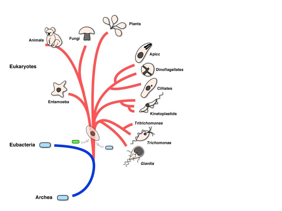
The phylum Kinetoplastida
Group of protozoans at the basis of the eukaryotic tree Move using a single flagellum Harbor name-giving mitochondrion with large genome which is always associated with the basal body of the single flagellum All kinetoplastids are parasites Trypanosoma & Leishmania are the medically important and best studied genera in this group (but a huge variety of animals and even plants harbor kinetoplastid parasites)
")
5
Leishmania belong to the order kinetoplastida
Different life cycle stages in this group show characteristic morphological differences and are distinguished by the position of the basal body within the cell However, the differences are not limited to morphology Different stages express different set of genes from their genomes which adapt their surface, internal structure and metabolism to survival in insect or mammalian hosts trypomastigote epimastigote promastigote amastigote

6
Leishmania parasites exist as pro- and amastigotes
The parasite lives in the digestive tract of sandflies as extracellular promastigote In the mammalian host parasites multiply as intracellular amastiogotes

7
procyclics and metacyclics
Infected macrophages are taken up with the blood meal and amastigotes are released by digestion, transform into procyclic promastigotes and attach to the midgut epithelium Attached promastigotes divide rapidly (procyclics are not infective to mammals) Metacyclic (infective) promastigotes cease replication, detach and pass forward into the pharynx from where they are regurgitated into the bite site (detached) (attached)
 Metacyclic (infective) promastigotes cease replication, detach and pass forward into the pharynx from where they are regurgitated into the bite site. (detached) (attached)")
8
Leishmania infects and thrives in macrophages
Within its mammalian host Leishmania infects macrophages and replicates as intracellular amastigoe Macrophages are “professional” phagocytic cells and an important part of the innate immune system (they also play a role in initiating the adaptive response by antigen presentation) The parasite invades its host cell passively by triggering phagocytosis
 The parasite invades its host cell passively by triggering phagocytosis.")
9
Leishmania infects and thrives in macrophages
Leishmania amazonensis metacyclic promastigote by a mouse macrophage. The parasite is phagocytosed with the cell body entering first and through the formation of a long tubular pseudopod. Images were captured every 0.5 seconds over the course of 367 seconds. Uptake of Leishmania amazonensis metacyclic promastigote by a mouse macrophage. The parasite is phagocytosed with the cell body entering first and through the formation of a long tubular pseudopod. Images were captured every 0.5 seconds over the course of 367 seconds. Courret et al

10
Leishmania infects and thrives in macrophages
Leishmania amazonensis metacyclic promastigote by a mouse macrophage. The parasite is phagocytosed with the cell body entering first and through the formation of a long tubular pseudopod. Images were captured every 0.5 seconds over the course of 367 seconds. Phagocytosis of a Leishmania amazonensis metacyclic promastigote by a mouse macrophage. The parasite binds to the macrophage plasma membrane by the tip of the flagellum. It then turns around and is finally ingested via the cell body. Images were captured every 0.5 seconds over the course of 125 seconds. Courret et al

11
Leishmania infects and thrives in macrophages
Leishmania stimulates this process by binding elements of the complement system to its surface Binding of complement can destroy pathogens but also tags them for phagocytosis (opsonisation: pathogen bound 3Cb is a potent ‘eat me’ signal for macrophages & neutrophils) However, the parasite prevents the formation of the fully functional membrane attack complex A molecule on the surface of the parasite seems to be responsible both for complement activation and prevention of the final attack
 However, the parasite prevents the formation of the fully functional membrane attack complex. A molecule on the surface of the parasite seems to be responsible both for complement activation and prevention of the final attack.")
12
Leishmania infects and thrives in macrophages
Once a pathogen is taken up by phagocytosis the phagosome usually fuses with primary lysosomes to form secondary lysosomes Within secondary lysosomes pathogens are killed and digested by the action of lytic enzymes and an acidic environment produced by membrane proton pumps Additional mechanisms of killing include the generation of oxygen radicals (‘biological bleach’)
")
13
Leishmania infects and thrives in macrophages
Macrophages are important “microbe killers”, however several pathogens have found ways to escape killing Trypansoma cruzi -- induces phagocytosis but then escapes into the cytoplasm Toxoplasma -- active invasion, parasitophorous vacuole is never part of the endocytic pathway Mycobacterium tuberculosis -- induce phagocytosis and block lysosomal maturation Leishmania ...

14
Leishmania infects and thrives in macrophages
… Leishmania just doesn’t seem to care Amastigotes thrive in what looks like a fully matured lysosome with acidic pH and abundant lysosomal hydrolases Amastigotes rapidly divide and will infect new macrophages after rupture of host cell The dense surface coat covering Leishmania seems to protect the parasite from the action of the lytic enzymes However, with help from T cells macrophages can be stimulated to kill the parasite

15
A TH1 response is required for parasite control and healing
Stimmulation with different cytokines leads to the development of two types of T-cells specialized for different immune responses Th1 and Th2 strongly downregulate each other This polarization has important consequences for the downstream response and can spell life or death Non healing Leishmania infections are characterized by a strong TH2 response (remember this was the response useful to get rid of worms by antibody and hypersensitivity) Healing infections are characterized by TH1 The parasites seems to manipulate this balance in his favor, we don’t understand yet how that is done
 Healing infections are characterized by TH1. The parasites seems to manipulate this balance in his favor, we don’t understand yet how that is done.")
16
Leishmania distribution (all species)
")
17
American soldiers contracted Leishmaniasis in Iraq & Afghanistan
A REGION INFLAMED: Hundreds of U.S. Troops Infected by Parasite Borne by Sand Flies, Army Says By DONALD G. MCNEIL JR. Published: December 6, 2003 Hundreds of American troops in Iraq have been infected with a parasite spread by biting sand flies, and the long-term consequences are still unknown, Army doctors said Friday … Skin Disease Strikes Iraqi Children Thursday, February 14, 2008 By MARIA CHENG, AP Medical Writer LONDON — At least 275 children in southern Iraq have been infected with a disfiguring skin disease, an outbreak some health officials are blaming on the war's devastating effect on the public health system.

18
Leishmania is transmitted by sand flies (Phlebotomidae)
Sand flies are minute diptera (they are more closely related to mosquitoes than “real” flies and only females bite) Only mosquito nets with fine meshwork hold them off In the wild sand flies often breed in rodent burrows Old world Phlebotomus, new world Lutzomya Can also transmit Bartonella bacilliformis and Papatsi virus
 Only mosquito nets with fine meshwork hold them off. In the wild sand flies often breed in rodent burrows. Old world Phlebotomus, new world Lutzomya. Can also transmit Bartonella bacilliformis and Papatsi virus.")
19
Ecology of old world Leismaniasis
Close contact of humans and their domestic animals can provide optimal conditions for sand flies and Leishmania transmission (stables provide good breeding ground for larvae) In urban environments infection is mostly human to human In rural areas Leishmaniasis can be a zoonosis Infection in dogs is quite frequent in the Mediterranean
 In urban environments infection is mostly human to human. In rural areas Leishmaniasis can be a zoonosis. Infection in dogs is quite frequent in the Mediterranean.")
20
Ecology of new world leishmaniasis
In the new world most people get infected while working or hunting in the forest Here wild animals including rodents, monkeys and sloths provide a reservoir for the parasite A transmission pattern within a population of wild animals that result in occasional infection of humans is called sylvatic

21
A variety of species and species complexes causes disease in humans
Visceral Leishmaniasis Leishmania donovani Leishmania infantum Leishmania chagasi Cutaneous Leishmaniasis Leishmania tropica Leishmania major Leishmania aethiopica Mucocutaneous Leishmaniasis Leishmania brazieliensis Leishmania mexicana Leishmania amazoniensis

22
Three syndromes associated with Leishmania infection in humans

23
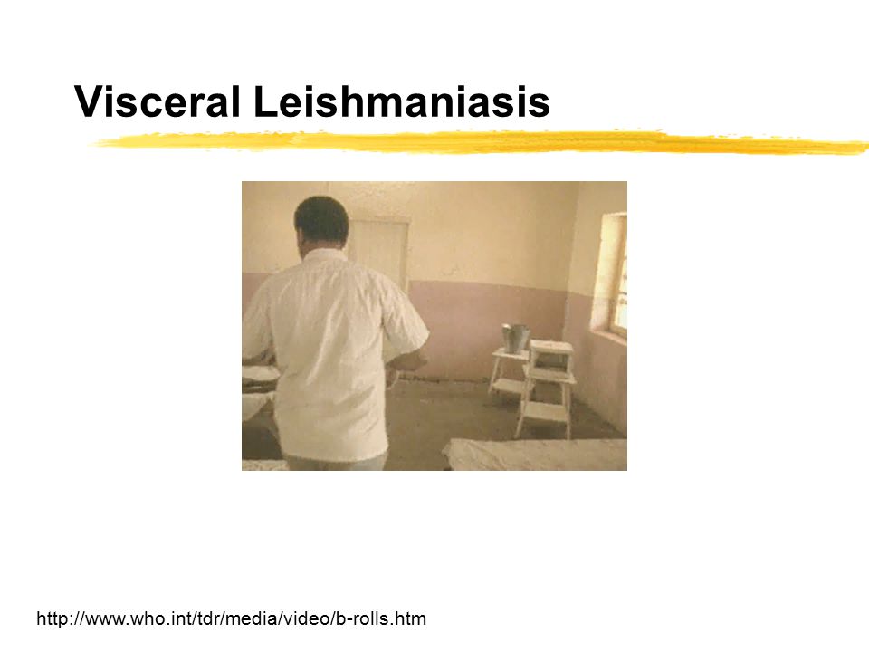
Visceral Leishmaniasis or Kala Azar
Systemic infection of reticulo-entdothelial cells (mostly macrophages) throughout multiple internal organs and the blood
 throughout multiple internal organs and the blood.")
24
Visceral Leishmaniasis
25
Kala Azar - Visceral Leishmaniasis
Caused by the L. donovani complex General infection of macrophages in the entire RES Weeks to months incubation period The lead symptom is abdominal swelling due to hepato- and splenomegaly High fever. Fever often oscillates with a peak every second day Progressive drastic weight loss (kachexia) Darkening of the skin Mortality of untreated disease 75-95%
 Darkening of the skin. Mortality of untreated disease 75-95%")
26
Post Kala-Azar Dermal Leishmaniasis
Sequel of visceral leishmaniasis which may manifest years after successful treatment and resolution of Kala Azar. Dermal lesions may contain parasites in great numbers

27
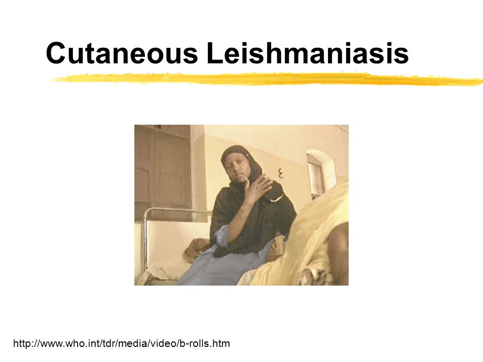
Cutaneous Leishmaniasis
Infection remains restricted to the initial site of infection (the bite site)
")
28
Cutaneous Leishmaniasis
29
Cutaneous Leishmaniasis is usually self-limiting
Old world oriental sore is caused by parasites of the L. tropica complex. (similar disease in the new world is caused by L. mexicana) A chronic but self-limiting dry ulceration at the site of the bite Ulceration starts months after infection Parasites are not found outside the lesion A granuloma is formed which finally leads to healing leaving a depressed scar Nearly absolute resistance to reinfection Inoculation to vaccinate has long been practiced in the middle east
 A chronic but self-limiting dry ulceration at the site of the bite. Ulceration starts months after infection. Parasites are not found outside the lesion. A granuloma is formed which finally leads to healing leaving a depressed scar. Nearly absolute resistance to reinfection. Inoculation to vaccinate has long been practiced in the middle east.")
30
Espundia or mucocutaenous leishmaniasis
Caused by L. braziliensis ~20% of infected patients develop ulcers of the oral and nasal mucosa

31
Espundia or mucocutaenous leishmaniasis
Caused by L. braziliensis ~20% of infected patients develop ulcers of the oral and nasal mucosa Progression of the ulceration is slow but steady, ultimately destroying all soft parts of the nose, the lips, and the soft palate Death can occur through secondary bacterial infection

32
Diagnosis & Treatment Gold standard of diagnosisis demonstration of parasites in the blood or lymph (kala azar) or in scrapings of the ulcer (cutaneous versions) Treatment for all forms of Leishmaniasis is not satisfactory at the moment Visceral Leishmaniasis (pentavalent antimonials) cutaneous (amphotheracin B, a liposome formulation is better tolerated but much more expensive) The available drugs have significant side effects and resistance has emerged (pentavalent antimonials) HIV coinfection further complicates treatment Better and more affordable drugs are urgently needed
 or in scrapings of the ulcer (cutaneous versions) Treatment for all forms of Leishmaniasis is not satisfactory at the moment. Visceral Leishmaniasis (pentavalent antimonials) cutaneous (amphotheracin B, a liposome formulation is better tolerated but much more expensive) The available drugs have significant side effects and resistance has emerged (pentavalent antimonials) HIV coinfection further complicates treatment. Better and more affordable drugs are urgently needed.")
33
A not-for-profit drug company?
New hope for better treatment includes paramomycin an old off-patent antibiotic which has shown promising activity in studies conducted by the Institute of One World Health Paramomycin interferes with the growth of a number of bacteria and protozoa (the mode of action is not fully understood) Intravenous injection has shown significant cure in kala azar patients in India (this was the first large scale clinical trial for this disease) One World Health develops this drug for kala azar together with an indian drug maker and the indian health authorities (
 Intravenous injection has shown significant cure in kala azar patients in India (this was the first large scale clinical trial for this disease) One World Health develops this drug for kala azar together with an indian drug maker and the indian health authorities. (")
34
procyclics and metacyclics

35
Parasite interacts with the sand fly midgut
Promastigotes attach and detach to the midgut epithelium Attachment helps them to remain in the insect gut when the blood meal is passed However, later they need to detach to move to the pharynx and proboscis for infection How is this accomplished at the molecular level?

36
Lipophosphoglycans play important roles in Leishmania pathogenesis
Lipophosphglycan or LPG is the dominant molecule on the surface of Leishmania LPG is not a protein but a glycolipid The molecule is anchored in the membrane by a lipid to which a long chain of highly hydrophilic sugar-phosphate repeats are attached The structure of LPG changes over the life cycle to adapt to various functions LPG is a pathogenesis factor in the mammalian host and important for the life cycle within the sandfly

37
Structural modification of LPG during the sand fly cycle
LPG is structurally modified during the developmental process of metacylogenesis (parasites decide to stop replicating and begin to pre-adjust to the mammalian host) LPG in metacylics has 2-3 times the number of repeat units Sidechains with terminal galactose (squares) are downregulated and the remaining galactose residues are capped with another terminal sugar (arabinopyranose) Are these chemical changes responsible for the pathogens attachment phenotype?
 LPG in metacylics has 2-3 times the number of repeat units. Sidechains with terminal galactose (squares) are downregulated and the remaining galactose residues are capped with another terminal sugar (arabinopyranose) Are these chemical changes responsible for the pathogens attachment phenotype")
38
Only LPG from procyclics attaches to the midgut
Phosphoglycans were isolated from procyclics (those that attach) and metacyclics (those that detach and infect) and were labeled with a dye (resulting in fluorescence seen as ‘white’ in the lower panels) Opened sandfly midguts were incubated with PG from procyclics (A/B) and metacyclics (C/D) and detected with an antibody Only procyclic phosphoglycan binds to the midgut
 and metacyclics (those that detach and infect) and were labeled with a dye (resulting in fluorescence seen as ‘white’ in the lower panels) Opened sandfly midguts were incubated with PG from procyclics (A/B) and metacyclics (C/D) and detected with an antibody. Only procyclic phosphoglycan binds to the midgut.")
39
LPG binds to the microvilli of the sandfly midgut epithelium
OK, LPG appears to be the molecule on the parasite responsible for the attachment of parasites to the sandfly midgut, but what kind of host molecule does it bind to?

40
LPG binds to a species specific galectin in the sandfly midgut
A gene for an abundantly expressed galactose binding protein or lectin (galectin) was identified in a sandfly sequencing project A specific antibody against the galectin reacts with the midgut of the sandfly species from which it was isolated (but not from other species) High resolution microscopy shows that the protein(red in lower panel) is found on the luminal side of the midgut epithelium Figure 4. In Vitro Binding of Leishmania Species to His-Tagged Recombinant PpGalec(A) Mean fluorescence of L. major (V1) incubated with FITC anti-His alone (black) or with recombinant PpGalec (rPpGalec) followed by FITC anti-His (gray).(B) Mean fluorescence of various Leishmania strains bound to His-tagged rPpGalec shown as fold-over controls incubated with FITC anti-His alone. Strains used were the following: V1, L. major V1; LV39, L. major "LV39"; NIH/SD, L. major "Seidman"; 1S, L. donovani "1S"; KK27, L. tropica "KK27"; Spock, L. major "Spock"; V1 met, L. major metacyclics. On average, 50,000 cells were acquired for each sample. Mean of four experiments ア SEM.(C) Mean fluorescence of the parasites noted above stained with monoclonal anti-LPG WIC79.3, which specifically recognizes the galactose-containing side chains of L. major LPG. Figure 5. Distribution and Surface Expression of PpGalec on Phlebotomus papatasi Midgut(A) Midguts incubated with preimmune serum or with anti-PpGalec. One of three representative experiments.(B) Midguts of P. papatasi and Lu. longipalpis incubated with anti-PpGalec. A Dapi staining of Lu. longipalpis midgut is shown. One of three representative experiments.(C) Midguts were incubated with either preimmune or anti-PpGalec sera and detected with Cy-3-conjugated anti-mouse (red). Alexa 488-conjugated phalloidin (green) and Dapi were used to stain actin and nuclei, respectively. Multiple confocal sections were obtained and merged into four groups, representing the lumen, middle, basal, and muscle planes of the midgut epithelium. Scale bars, 5 μm. Arrow indicates the position of the horizontal section shown as a side view in the bottom panel.(D) Side view of merged sections from the luminal surface (L) to the basal layer (B) of the midgut epithelium. Scale bar, 5 μm. Cell 119:329-41
 was identified in a sandfly sequencing project. A specific antibody against the galectin reacts with the midgut of the sandfly species from which it was isolated (but not from other species) High resolution microscopy shows that the protein(red in lower panel) is found on the luminal side of the midgut epithelium. Figure 4. In Vitro Binding of Leishmania Species to His-Tagged Recombinant PpGalec(A) Mean fluorescence of L. major (V1) incubated with FITC anti-His alone (black) or with recombinant PpGalec (rPpGalec) followed by FITC anti-His (gray).(B) Mean fluorescence of various Leishmania strains bound to His-tagged rPpGalec shown as fold-over controls incubated with FITC anti-His alone. Strains used were the following: V1, L. major V1; LV39, L. major LV39 ; NIH/SD, L. major Seidman ; 1S, L. donovani 1S ; KK27, L. tropica KK27 ; Spock, L. major Spock ; V1 met, L. major metacyclics. On average, 50,000 cells were acquired for each sample. Mean of four experiments ア SEM.(C) Mean fluorescence of the parasites noted above stained with monoclonal anti-LPG WIC79.3, which specifically recognizes the galactose-containing side chains of L. major LPG. Figure 5. Distribution and Surface Expression of PpGalec on Phlebotomus papatasi Midgut(A) Midguts incubated with preimmune serum or with anti-PpGalec. One of three representative experiments.(B) Midguts of P. papatasi and Lu. longipalpis incubated with anti-PpGalec. A Dapi staining of Lu. longipalpis midgut is shown. One of three representative experiments.(C) Midguts were incubated with either preimmune or anti-PpGalec sera and detected with Cy-3-conjugated anti-mouse (red). Alexa 488-conjugated phalloidin (green) and Dapi were used to stain actin and nuclei, respectively. Multiple confocal sections were obtained and merged into four groups, representing the lumen, middle, basal, and muscle planes of the midgut epithelium. Scale bars, 5 μm. Arrow indicates the position of the horizontal section shown as a side view in the bottom panel.(D) Side view of merged sections from the luminal surface (L) to the basal layer (B) of the midgut epithelium. Scale bar, 5 μm. Cell 119:")
41
LPG binds to a species specific galectin in the sandfly midgut
Galectin (labeled with a fluorescent dye) binds specifically to procyclic Leishmania major parasites However little binding is detected when incubated with metacyclic parasites (the stage that detaches and moves to the the proboscis to infect the mammalian host, V1met) There is little binding to a mutant parasite (Spock) which lacks LPG (B lower) anti-galectin also blocks binding of procyclic parasites to the midgut epithelium New studies show that differences in galectins and LPG in different sandfly and parasite species might govern host specificity Figure 4. In Vitro Binding of Leishmania Species to His-Tagged Recombinant PpGalec(A) Mean fluorescence of L. major (V1) incubated with FITC anti-His alone (black) or with recombinant PpGalec (rPpGalec) followed by FITC anti-His (gray).(B) Mean fluorescence of various Leishmania strains bound to His-tagged rPpGalec shown as fold-over controls incubated with FITC anti-His alone. Strains used were the following: V1, L. major V1; LV39, L. major "LV39"; NIH/SD, L. major "Seidman"; 1S, L. donovani "1S"; KK27, L. tropica "KK27"; Spock, L. major "Spock"; V1 met, L. major metacyclics. On average, 50,000 cells were acquired for each sample. Mean of four experiments ア SEM.(C) Mean fluorescence of the parasites noted above stained with monoclonal anti-LPG WIC79.3, which specifically recognizes the galactose-containing side chains of L. major LPG. Figure 5. Distribution and Surface Expression of PpGalec on Phlebotomus papatasi Midgut(A) Midguts incubated with preimmune serum or with anti-PpGalec. One of three representative experiments.(B) Midguts of P. papatasi and Lu. longipalpis incubated with anti-PpGalec. A Dapi staining of Lu. longipalpis midgut is shown. One of three representative experiments.(C) Midguts were incubated with either preimmune or anti-PpGalec sera and detected with Cy-3-conjugated anti-mouse (red). Alexa 488-conjugated phalloidin (green) and Dapi were used to stain actin and nuclei, respectively. Multiple confocal sections were obtained and merged into four groups, representing the lumen, middle, basal, and muscle planes of the midgut epithelium. Scale bars, 5 μm. Arrow indicates the position of the horizontal section shown as a side view in the bottom panel.(D) Side view of merged sections from the luminal surface (L) to the basal layer (B) of the midgut epithelium. Scale bar, 5 μm. Cell 119:329-41
 binds specifically to procyclic Leishmania major parasites. However little binding is detected when incubated with metacyclic parasites (the stage that detaches and moves to the the proboscis to infect the mammalian host, V1met) There is little binding to a mutant parasite (Spock) which lacks LPG. (B lower) anti-galectin also blocks binding of procyclic parasites to the midgut epithelium. New studies show that differences in galectins and LPG in different sandfly and parasite species might govern host specificity. Figure 4. In Vitro Binding of Leishmania Species to His-Tagged Recombinant PpGalec(A) Mean fluorescence of L. major (V1) incubated with FITC anti-His alone (black) or with recombinant PpGalec (rPpGalec) followed by FITC anti-His (gray).(B) Mean fluorescence of various Leishmania strains bound to His-tagged rPpGalec shown as fold-over controls incubated with FITC anti-His alone. Strains used were the following: V1, L. major V1; LV39, L. major LV39 ; NIH/SD, L. major Seidman ; 1S, L. donovani 1S ; KK27, L. tropica KK27 ; Spock, L. major Spock ; V1 met, L. major metacyclics. On average, 50,000 cells were acquired for each sample. Mean of four experiments ア SEM.(C) Mean fluorescence of the parasites noted above stained with monoclonal anti-LPG WIC79.3, which specifically recognizes the galactose-containing side chains of L. major LPG. Figure 5. Distribution and Surface Expression of PpGalec on Phlebotomus papatasi Midgut(A) Midguts incubated with preimmune serum or with anti-PpGalec. One of three representative experiments.(B) Midguts of P. papatasi and Lu. longipalpis incubated with anti-PpGalec. A Dapi staining of Lu. longipalpis midgut is shown. One of three representative experiments.(C) Midguts were incubated with either preimmune or anti-PpGalec sera and detected with Cy-3-conjugated anti-mouse (red). Alexa 488-conjugated phalloidin (green) and Dapi were used to stain actin and nuclei, respectively. Multiple confocal sections were obtained and merged into four groups, representing the lumen, middle, basal, and muscle planes of the midgut epithelium. Scale bars, 5 μm. Arrow indicates the position of the horizontal section shown as a side view in the bottom panel.(D) Side view of merged sections from the luminal surface (L) to the basal layer (B) of the midgut epithelium. Scale bar, 5 μm. Cell 119:")
42
Modification of LPG modulate interaction of the parasite and midgut galectin

43
Summary Leishmania species cause three clinical syndromes depending on the spread of the infection in the body Leishmania ‘provoke’ phagocytosis by macrophages and develop intracellular in an fully acidified lysosome LPG-galectin interaction and modification of LPG regulate attachment and detachment of parasites in the sandfly host

Similar presentations


>")
>")


 (VL) Leishmania tropica (CL) Leishmania major (CL) Leishmania aethiopica (CL) Leishmania mexicana (Complex)>")
 ◦ Pathogen: ◦ Reservoir: ◦ Portal of.>")