Download presentation
Presentation is loading. Please wait.

1
Blunt and penetrating neck injury

2
reference B.J.Bailey ,et al. Head & Neck surgery Otolaryngology.4th edition.2006 Charles W. Cummings, et al, Cummings Otolaryngology, Head & Neck Surgery, 5th ed D.V. Feliciano ,et al. Trauma, 6th Edition.2008

3
Zones of the Neck . Zone I: thoracic inlet to cricoid cartilage Zone II: cricoid cartilage to the angle of mandible Zone III: angle of the mandible to skull base

4
CLASSIFICATION

5
Zone I Superior Mediastinum Thoracic Duct Spinal Cord Brachial Plexus
From the clavicles to the cricoid Trachea Lungs Proximal carotid and vertebral arteries Jugular veins Thoracic Vessels Esophagus Superior Mediastinum Thoracic Duct Spinal Cord Brachial Plexus

6
Zone II From cricoid to angle of mandible Trachea Larynx
Carotid and vertebral aa. Jugular Vein Esophagus Spinal Cord

7
Zone III Angle of mandible to base of skull
Distal carotid and vertebral arteries Pharynx Spinal cord

8
PENETRATING NECK TRAUMA
Presently, penetrating neck injury comprises 5% to 10% of all trauma cases. All penetrating neck wounds are potentially dangerous and require emergency treatment.

9
Physical properties of penetrating objects
handgun Rifle Shotguns Knife and stab injuries

10
Physical properties of penetrating objects
Kinetic energy= ½ mv2 m = mass V = velocity Degree of wound Firearm Low velocity ( < 1,000 ft/sec) handgun ft/sec high velocity ( > 1,000 ft/sec) shotgun 1,200 ft/sec , rifle 2,200 ft/sec
 handgun ft/sec. high velocity ( > 1,000 ft/sec) shotgun 1,200 ft/sec , rifle 2,200 ft/sec.")
11
Physical properties of penetrating objects
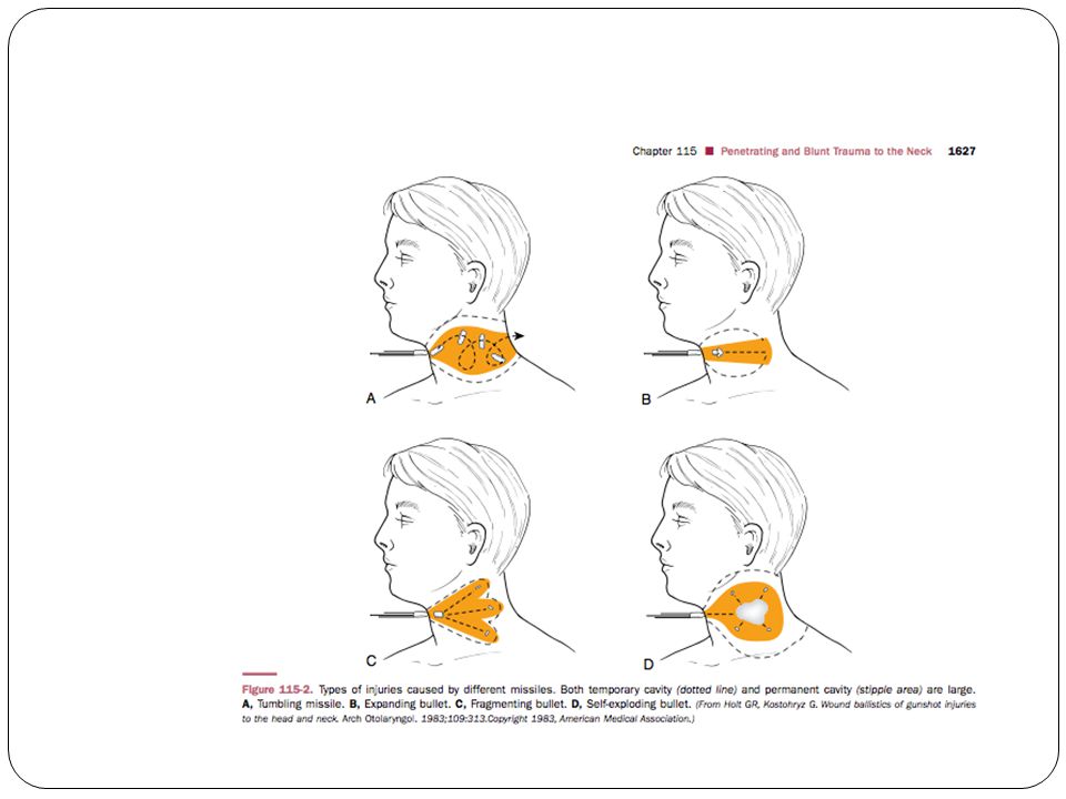
Gunshot wound tissue injury from 2 mechanism Direct tissue injury Temporary caviation Low velocity tissue damage High velocity tissue loss

13
KNIFE and STAB Knife, ice-pick, cut-glass, or razor-blade
more predictable pathways single-entry wound may be from multiple stab wounds cervical stab wounds have a higher incidence of subclavian vessel laceration because stabbings to the neck often occur in a downward direction with the knife slipping over the clavicle and into the subclavian vessels. spinal injuries, neck stab wounds have a lower incidence than cervical bullet wounds.

14
Genaral trauma principle
A : airway with C-spine control B : breathing and ventilation C : circulation D : disability and neurologic status E : exposure and evaluation other injury

15
A : Airway Most casecarefully intubated transorally
If C –spine injury is suspected intubate with neck stabilized Unstable airway with sig. bleed or edema in oral cavity or pharynx cricothyroidotomy or urgent tracheostomy

16
A : Airway Multiple blind intubation attempts will risk enlarging a lacerated piriform sinus wound and extending it iatrogenically into the mediastinum. Tracheal tear may be exacerbated by extending the neck

17
A : Airway Obvious tracheal injury carefully intubated through entry wound using armored/reinforced ETT

18
B: Breathing Administer high-flow oxygen Monitor : pulse oximetry
Difficulty ventilation may upper airway or thoracic injury Unequal breath sounds & asymmetric chest movement inadequate ventilation Pneumothorax Hemothorax Tension pneumothorax

19
C : Circulation Control active bleeding with direct pressure
Do not clamp bleeding vessels Subsequent injury to vascular or nervous structure Avoid placing IV access at a location where the IV fluid would flow toward the site of injury Avoid inserting NG tube at the initial resuscitation : gag & retching cause dislodge a clot & cause hemorrhage

20
D : Disability Neurodeficit indicate
directed nerve or spinal cord injury cerebral ischemia cause by carotid artery injury Need rapid sedation and paralysis for intubation Immobilize the cervical spine in a neutral position

21
Vital structures of the neck
four groups: the air passages (trachea, larynx, pharynx, lung); vascular (carotid, jugular, subclavian, innominate, aortic arch vessels); gastrointestinal (pharynx, esophagus) neurologic (spinal cord, brachial plexus, peripheral nerves, cranial nerves [CNs])
; vascular (carotid, jugular, subclavian, innominate, aortic arch vessels); gastrointestinal (pharynx, esophagus) neurologic (spinal cord, brachial plexus, peripheral nerves, cranial nerves [CNs])")
22
SYMPTOM Airway Vascular System Reparatory distress Stridor Hemoptysis
Hoarseness Tracheal deviation Subcutaneous emphysema Sucking wound Vascular System Hematoma Persistent bleeding Neurologic deficit Absent pulse Hypovolemic shock Bruit Thrill Change of sensorium From Stiernberg C, Jahrsdoerfer RA, Gillenwater A, et al. Gunshot wounds to the head and neck. Arch Otolaryngol Head Neck Surg. 1992;118:592

23
SYMPTOM Nervous System
Hemiplegia Quadriplegia Coma Cranial nerve deficit Change of sensorium Hoarseness Esophagus/Hypopharynx Subcutaneous emphysema Dysphagia Odynophagia Hematemesis Hemoptysis Tachycardia Fever From Stiernberg C, Jahrsdoerfer RA, Gillenwater A, et al. Gunshot wounds to the head and neck. Arch Otolaryngol Head Neck Surg. 1992;118:592

24
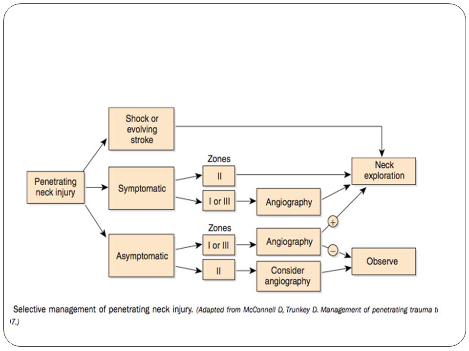
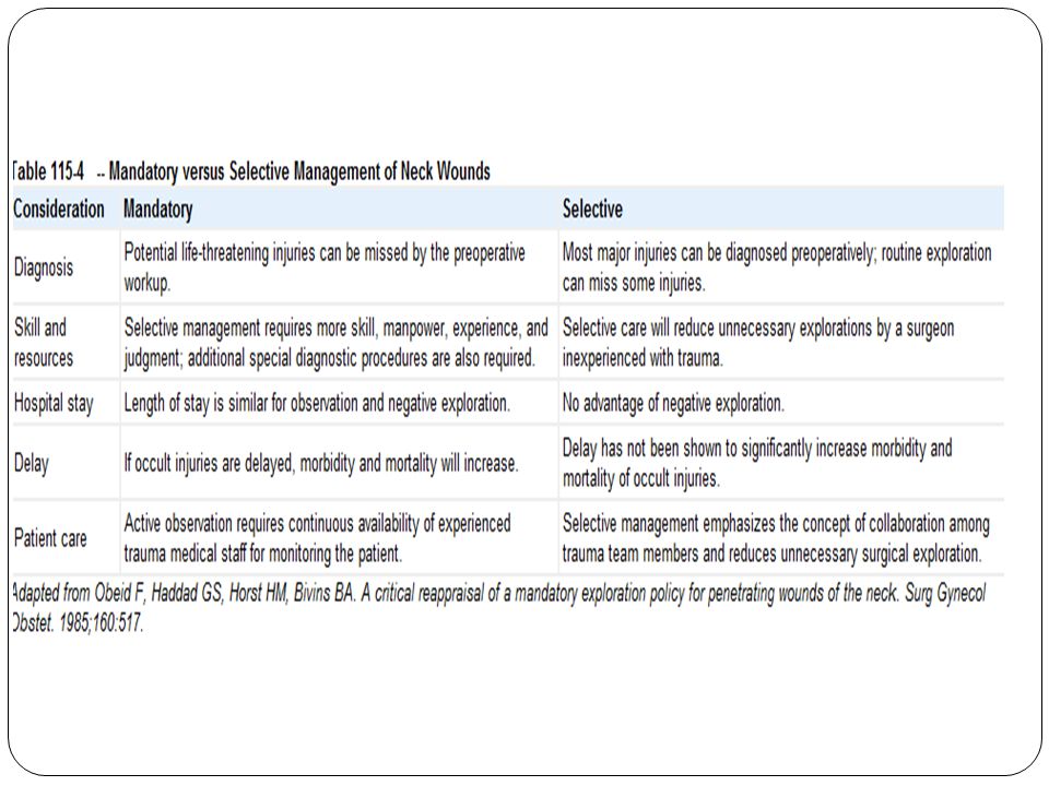
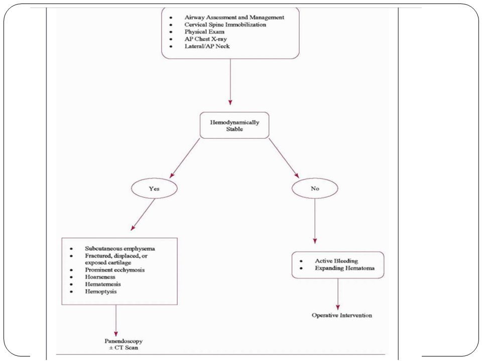
Mandatory versus Elective Exploration
Immediately life threatening: massive bleeding, expanding hematoma, hemodynamic instability, hemomediastinum, hemothorax, and hypovolemic shock.Immediate surgical exploration Hemodynamically stable ,non–life-threatening features can undergo thorough imaging investigations to determine the extent of injury.

25
Injury

26
Zone 1 injury Below cricoid, dangerous area
Protect zone bony thorax and clavicle Motality rate 12 % Potential for injury to great vessel and mediastinum Mandatory exploration : not recommend Angiography and esophageal evaluation: usually suggest > 1/3 no symptom at presentation

27
Zone 1 injury Esophageal evaluation endoscopy , contrast esophagogram
Contrast medium Barium- based Gastrografin ( meglumine diatrizoate) Combination tests should not miss an njury CT scan Determine the path of projectile
 Combination tests should not miss an njury. CT scan. Determine the path of projectile.")
28
Zone 2 injury Largest zone,most common site of trauma 60-75%
Between angle of mandible & inf border of cricoid cartilage Isolate venous injury & pharyngoesophageal injury most common structure missed clinically All pt. are admitted for observation and 24 hr re-evaluation 50% of death hemorrhage from vascular structure

29
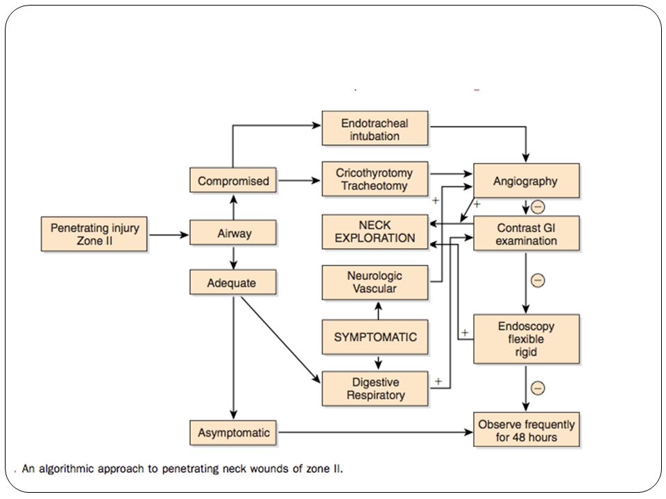
Zone 2 injury Symptomatic neck exploration Asymptomatic
Directed evaluation and serial exam Arteriography, Laryngotraheoscopy flexible esophagoscopy barium swallow Requires adequate physician ,24 hr facility prepared for emergency testing and Surgery

31
Zone 3 injury Superior to angle mandible to skull base
Potential for injury to major blood vessel and CN near skull base Arterial injury may be asymptomatic at presentation Surgical exposure and control bleeding may be difficult amenable to definitive treatment by an interventional radiologist Vertebral artery injury appear to be relatively rare Should be imaged if bullet path is near the vertebral column Four vessel angiography

33
Angiography : Zone1 & 3 Routine preoperative arteriography in stable case Surgical approach is more difficult than zone 2 If wound involve both side of neck ( stable but symptomatic) four vessel angiography
 four vessel angiography.")
34
Angiography : Zone1 & 3 1Arteriogram demonstrating common carotid artery injury with small hematoma 2extravasation of the internal carotid artery near the base of the skull (arrow). 3. A follow-up arteriogram of the internal carotid artery 1 week later shows enlargement of the pseudoaneurysm.
. 3. A follow-up arteriogram of the internal carotid artery 1 week later shows enlargement of the pseudoaneurysm.")
35
Angiography : Zone2 Easy accessible,low risk for exploration
Certain indication for an angiogram in zone 2 Stable pt. who has persistent hemorrhage Neurodeficit compatible with adjacent vascular structure damage eg. Horner’s syndrome , hoarseness Need exploration Positive arteriography Negative arteriography but positive clinical sign Asymptomatic in zone 2 Controversy, No sig difference btw. Clinical exam & angiography CTA fast ,minimal invasive in hemostatic stable

36
Management of vascular injury zone 1
Vascular perforation requires thoracic Sx Mediastinotomy extension or formal lateral thoracotomy

37
Management of vascular injury zone 3
Injury at the skull base can be temporalize by pressure Mandibulectomy in midline Temporaly arteral bypass of carotid artery

38
Management of vascular injury
All vein in the neck can be safely ligated to control hemorrhage injury both internal jugular vein try repair All external carotid artery suture ligation Good collateral circulation

39
Management of vascular injury
Common carotid artery/internal carotid artery in zone 2 Approach along SCM if no pulsating followed retrograde from facial artery/sup thyroid artery

40
Technique of vascular repair
End to end or autogenous graft reccomended when stenosis is evident by arteriography Ligation of common or internal carotid a.reserved for irreparable injury and in pt, who are in a profound coma state Delayed complication from unrepaired vascular injury Aneurysm formation Dissecting aneurysm AV fistulas

41
Technique of vascular repair
Intervention radiologists used angiography technique to treat vascular injury Embolization Zone 3 high incidence of multiple vascular injury event Complication of intervention angiography Blood vessel injury Inadvertent balloon detachment Ischemic events Pseudoaneurysm formation Treatment failure

42
Pharynx and esophageal injury
Clinical sign and symptom neck exploration subcutaneous emphysema Hematemesis Hypopharyngeal blood >50%of Pt. asymptomatic at presentation Combination of esophagoscopy and contrast esophagography Most sensitive for detected injury Delayed explore & repair beyond 24 hrs after injury poorer outcome

43
Digestive tract evaluation
Possible esophageal perforation gastrografin swallow Barium : extravasation & distort soft tissue plane and toxic

44
Digestive tract evaluation
Flexible esophagoscopy Missed perforation : cricopharyngeus, hypopharynx Negative endoscopy but air leak in soft tissue mandatory neck explore Infiltrate methylene blue : localize injury size Combination of flexible and rigid endoscopy Exam entire cervial and upper esophagus No perforation missed

45
Digestive tract evaluation
Suspicious pharyngeal perforation NPO for several days S&S : fever , tachycardia,widening of mediastinum Repeat endoscopy or neck exploration Esophageal injury in the early phase Two layer closure with wound irrigation Debridement Adequate drainage Extensive esophageal injury lateral cervical esophagostomy

46
Digestive tract evaluation
C-spine fx omitted rigid esophagoscopy Clinical exam F/U exam frequently Monitor V/S Observe period hrs

47
Penetrating of hypopharynx
Superior to the level of arytenoid cartilage IV ABO NPO ทางปาก 5-7 days Primary closure not always necessary Inferior to the level of arytenoid cartilage Dependent portion Exploration with primary watertight closure Use absorbable suture with drainage of adjacent neck space NPO 5-7 days Treat liked esophageal injury

48
Treatment Conservative Medical therapy
Adequate ventilation & oxygenation Fluid resuscitation Monitor neurolodic status Pain control ABO Tetanus prophylaxis

49
Treatment Surgical approach Zone 1 Zone 2 Zone 3 Median sternotomy
Thoracotomy Zone 2 Collar incision Apron incision Zone 3 Consult neuroSx

51
Blunt neck trauma motor vehicle accidents and sports
result in laryngeal, vascular, and digestive injury easily underdiagnosed because their onset can be delayed occult cervical spine injury

52
Blunt neck trauma careful observation : delayed onset
slow progression of airway edema airway obstruction may not occur until several hours after the injur CT may be helpful to determine degrees of injury to the larynx and vessels

53
Blunt neck trauma Blunt injury to the cervical vessels can lead to
thrombosis, intimal tears, dissection, and pseudoaneurysm Treatment options for blunt artery injuries are based on the mechanism, type of injury, and location

54
Blunt neck trauma Treatments for blunt artery injuries include
surgery, anticoagulation, and observation. Surgical intervention for blunt vascular injuries includes ligation, resection, thrombectomy, and stent placement

Similar presentations