Download presentation
Presentation is loading. Please wait.

1
DISORDERS OF MALABSORPTION DR SIMIN PARTOVI

2
© 2007 Thomson - Wadsworth

3
Table 330-1 -- MALABSORPTION DISORDERS AND CHRONIC DIARRHEA ASSOCIATED WITH GENERALIZED MUCOSAL DEFECT

5
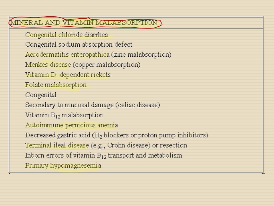
Table 330-2 -- CLASSIFICATION OF MALABSORPTION DISORDERS AND CHRONIC DIARRHEA BASED ON THE PREDOMINANT NUTRIENT MALABSORBED

9
Table 330-3 -- DIARRHEAL DISEASES APPEARING IN THE NEONATAL PERIOD CLINICAL FEATURESCONDITION Secretory watery diarrheaMicrovillus inclusion disease Secretory watery diarrheaTufting enteropathy Acidic diarrheaCongenital glucose-galactose malabsorption Acidic diarrheaCongenital lactase deficiency Hydramnion, secretory watery diarrhea Metabolic alkalosis Congenital chloride diarrhea Hydramnion, secretory watery diarrheaCongenital defective jejunal Na + /H + exchange SteatorrheaCongenital bile acid malabsorption Failure to thrive, edemaCongenital enterokinase deficiency Failure to thrive, edemaCongenital trypsinogen deficiency Failure to thrive, oily stoolCongenital lipase and/or co-lipase deficiency Hyperchloremic acidosis, failure to thriveEnteric anendocrinosis (NEUROG 3 mutation)
")
10
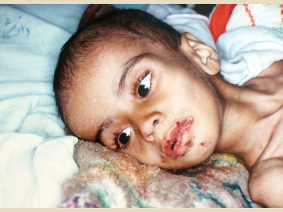
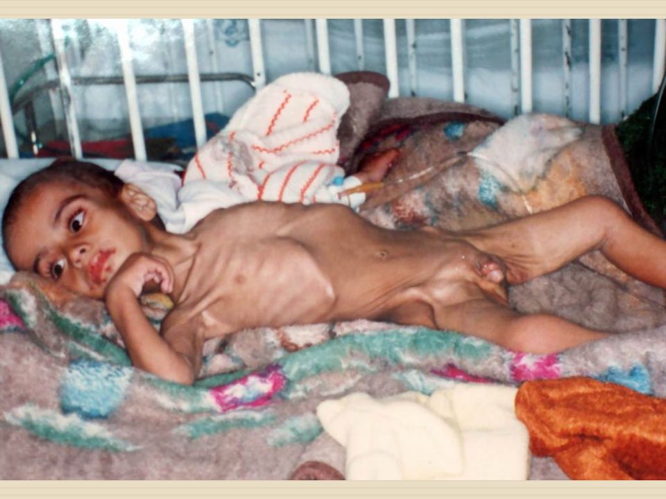
Malabsorption Clinical manifestations : -Diarrhea -Abdominal distention -Failure to thrive -edema -digital clubbing -abnormal hair -muscle wasting -stomatitis and glossitis -signs of rickets -skin bruises

11
Evaluation of children with malabsorption CBC and blood film anemia, lymphopenia (lymphangiectasia), neutropenia (shwachman syndrome), acanthocytosis (abetalipoproteinemia) Stool: Leukocytes and occult blood Parasites PH and reducing substances
, neutropenia (shwachman syndrome), acanthocytosis (abetalipoproteinemia) Stool: Leukocytes and occult blood Parasites PH and reducing substances")
12
Evaluation of children with malabsorption Celiac serology Albumin level Ca, Mg, zinc Iron level, folic acid level, Vit B12 Vit D, E, A Prothrombin time Upper endoscopy

13
Investigations for Carbohydrate malabsorption Clinitest: Detect reducing substances in the stool stool PH less than 5.6 Carbohydrate reach the bowel where they are degraded to Hydrogen gas+ CO2+ organic acids

14
Investigations for Carbohydrate malabsorption 3-Breath hydrogen test Ingestion of carbohydrate load (sucrose or lactose)1-2g/kg, sugar will not be ingested in the small bowel and passes to the colon and then metabolized by normal flora into hydrogen gas which will be detected in the breath
1-2g/kg, sugar will not be ingested in the small bowel and passes to the colon and then metabolized by normal flora into hydrogen gas which will be detected in the breath")
15
Investigations for Carbohydrate malabsorption 4-Small bowel mucosal biopsies Low mucosal disaccharidase levels in primary disaccharidase deficiency (lactase, sucrase, maltase)
")
16
Investigations for fat malabsorption Sudan test - Best screening method -Mixing the stool with sudan red stain, fat droplets will separate and be identified, more than 6-8 droplets / low power field is abnormal 72-hr quantitative fecal fat test - The gold standard to confirm steatorrhea - Dietary record is used to calculate fat intake for 3 days, stool is collected, excretion of more than 7% is abnormal

17
Investigations for Gastrointestinal Protein loss Dietary and endogenous proteins are almost absorbed Majority of stool nitrogen is derived from gut bacterial proteins Albumin Level: - GI loss of protein manifests as hypoalbuminemia -low albumin occur due to other factors α 1-antitrypsin: -Useful screening test for protein losing enteropathy -Unlike albumin, is resistant to digestion in the GIT -High levels in the stool indicate protein losing enteropathy

18
Investigations for Pancreatic Exocrine function Most common is Cystic fibrosis - Sweat chloride test - Genetic testing - Fecal elastase: -Sensitive test to assess exocrine pancreatic function -endoprotease that is human and pancreas specific, not altered by pancreatic enzyme replacement - Serum Trypsinogen - Duodenal aspirate Analysis of bicarbonate, trypsinogen and lipase after secretin stimulation

19
Celiac Disease Diagnosis Serological markers Antigliadin antibodies IgG & IgA Antiendomysial antibodies IgA Anti tissue transglutaminase IgA and IgG

20
Celiac Disease Small bowel biopsy -Short, flat villi -increased number of lymphocytes in the epithelial layer -crypt hyperplasia

21
Stool Odor Indole and sketole are the substances that produce normal odor formed by intestinal bacteria putrefaction and fermentation Clinical implication. 1.A foul odor is caused by degradation of undigested protein. 2.A foul odor is produced by excessive carbohydrate ingestion. 3.A sickly sweet odor is produced by volatile fatty acids and undigested lactose

22
Stool pH Normal value : Neutral to acid or alkaline Clinical implication 1. Increased pH ( alkaline) b. protein break downa. Villous adenoma d.Colitisc.Antibiotic use 2. Decreased pH ( acid) a. Carbohydrate malabsorption b. Fat malabsorption c. Disaccharidase deficiency
 b. protein break downa. Villous adenoma d.Colitisc.Antibiotic use 2. Decreased pH ( acid) a. Carbohydrate malabsorption b. Fat malabsorption c. Disaccharidase deficiency.")
23
Stool color Normal value : Brown Clinical implication: 1. Yellow to yellow-green : severe diarrhea 2. Green : severe diarrhea bile Black: resulting from bleeding into the upper gastrointestinal tract (>100 ml blood) 3. Tan or Clay colored : blockage of the common bile duct. 4. Pale greasy acholic (no bile secretion) stool found in pancreatic insufficiency.
 3. Tan or Clay colored : blockage of the common bile duct. 4. Pale greasy acholic (no bile secretion) stool found in pancreatic insufficiency..")
24
Stool color(con) 4. Maroon-to-red-to-pink : possible result of bleeding from the lower gastrointestinal tract (eg. Tumors, hemorrhoids, fissures,inflammatory process) 5. Blood streak on the outer surface of usually indicates hemorrhoids or anal abnormalities. 6. Blood in stool can arise from abnormalities higher in the colon. In some case the transit time is rapid blood from stomach or duodenum can appear as bright or dark red or maroon in stool
 5. Blood streak on the outer surface of usually indicates hemorrhoids or anal abnormalities. 6. Blood in stool can arise from abnormalities higher in the colon. In some case the transit time is rapid blood from stomach or duodenum can appear as bright or dark red or maroon in stool.")
25
Blood in Stool Normal value : Negative Clinical Implication : 1. Dark red to tarry black indicates a loss of 0.50 to 0.75 ml of blood from the upper GI tract. 2. Positive for occult blood may be caused by a. Carcinoma of colonb. Ulcerative colitis c. Adenoma d. Diaphramatic hernia e. Gastric carcinoma f. Diverticulitis g. Ulcers

26
Mucous in Stool Normal value : Negative for mucous Cli nical Implication: 1. Translucent gelatinous mucous clinging to the surface of formed stool occurs in a. Spastic constipationb. Mucous colitis c. Emotionally disturbed patients stool d. Excessive straining 2. Bloody mucous suggests a. Neoplasm

27
Fat in Stool Normal value : fat in stool will account for up to 20 % of total solids. Lipids are measured as fatty acids (0-6.0 g/24hr) Clinical Implication : 1. Increased fat or fatty acids is associated with the malabsorption syndromes b.Nontropical spruea.Crohn’s disease d. Whipple’s diseasec. Cystic fibrosis e. Enteritis and pancreatic diseases f. Surgical removal of a section of the intestine
 Clinical Implication : 1. Increased fat or fatty acids is associated with the malabsorption syndromes b.Nontropical spruea.Crohn’s disease d. Whipple’s diseasec. Cystic fibrosis e. Enteritis and pancreatic diseases f. Surgical removal of a section of the intestine.")
28
Urobilinogen in Stool Normal value : 125-400 Ehrlich units / 24 hr 75-350 Ehrlich units/100 g Clinical Implication: 1.Increased values are associated with Hemolytic anemias 2.Decreased values are associated with a. Complete biliary obstruction b. Severe liver disease, infectious hepatitis c. Oral antibiotic therapy that alters intestinal bacteria flora d. Infants are negative up to 6 months of age

29
Bile in Stool : Adults –negative : Children may be positive Clinical Implication Clinical Implication: 1. Bile may be present in diarrheal stools. 2. Increased bile levels occur in Hemolytic anemia

30
Trypsin in Stool : Positive in small amounts in 95 % of normal persons. Clinical Implication : Decreased amounts :occur in Pancreatic deficiency Malabsorption syndromes Screen for cystic fibrosis

31
Leukocytes in Stool Normal value : Negative Clinical Implication 1. Large amounts of leukocytes b. Chronic ulcerative colitis a. Chronic bacilliary dysentery c. Localized abscess d. Fistulas of sigmoid rectum or anus 2. Mononuclear leukocytes appear in Typhoid

32
Leukocytes in Stool (con) 3. Polymorphonuclear leukocytes appear in b. Shigellosisa. Salmonellosis d.Yersiniac. Invasive Escherichia coli diarrhea e. Ulcerative colitis 4. Absence of leukocytes is associated with b. Choleraa. Non specific diarrhea d. Viral diarrheac. Amebic colitis e. Noninvasive E.coli diarrhea f. Toxigenic bacteria Staphylococci spp., Clostidium. Parasites-Giardia g,

Similar presentations


 are not my.>")


