Download presentation
Presentation is loading. Please wait.

1
Mitochondria Prof. Daniel C. Hoessli, Geneva, Switzerland,
Dr. Waqar Hameed, PCMD March 2013 1

2
2

3
Figure 14-4 Essential Cell Biology (© Garland Science 2010)
3

4
Figure 14-3 Essential Cell Biology (© Garland Science 2010)
")
5
Mitochondria: where do they come from ?
Mitochondria probably originate from archeo- bacteria that were engulfed by a phagocytic cell. The two have lived in symbiosis ever since. Then what has the bacterium brought in that is beneficial to the host cell ? The capacity to transport electrons and make a proton gradient across the membrane 5

6
How do mitochondria and host cells share resources ?
Mitochondria have their own DNA and express their genes to produce proteins active in the electron transport chain. However, most of the proteins they need are encoded in the nucleus of the cell. They need to import most of their proteins to function. 6

7
Mitochondrial division and segregation
These processes involve both outer and inner mitochondrial membranes. (A) During fusion and fission, both matrix and intermembrane space compartments are maintained. Different membrane fusion machines are thought to operate at the outer and inner membranes. Conceptually, the fission process resembles that of bacterial cell division (discussed in Chapter 18). The pathway shown has been postulated from static views such as that shown in (B). (B) An electron micrograph of a dividing mitochondrion in a liver cell. (B, courtesy of Daniel S. Friend.) Molecular Biology of the Cell. 4th edition. Alberts B, Johnson A, Lewis J, et al 7
 During fusion and fission, both matrix and intermembrane space compartments are maintained. Different membrane fusion machines are thought to operate at the outer and inner membranes. Conceptually, the fission process resembles that of bacterial cell division (discussed in Chapter 18). The pathway shown has been postulated from static views such as that shown in (B). (B) An electron micrograph of a dividing mitochondrion in a liver cell. (B, courtesy of Daniel S. Friend.) Molecular Biology of the Cell. 4th edition. Alberts B, Johnson A, Lewis J, et al. 7.")
8
Mitochondrial genome: evolutional influence

9
Overview of mitochondrial gene expression

10
Mitochondrial genome

11
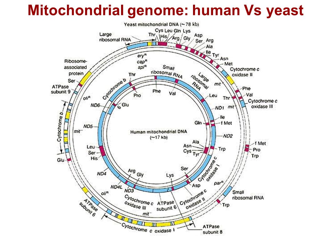
Mitochondrial genome: human Vs yeast
12
Mitochondrial DNA replication fork: critical proteins required for DNA replication
Fig. 1. Schematic diagram of a mitochondrial DNA replication fork showing the critical proteins required for DNA replication. The nascent DNA synthesized by pol γ (green) is shown as a solid red line, while the RNA primer (jagged red line) created by the mitochondrial RNA polymerase (orange) is being degraded by RNase H1 (yellow). The mitochondrial DNA helicase (purple) unwinds the downstream DNA forming a single-stranded loop which is coated with mtSSB (light blue). Topoisomerases (brown) work to relieve torsional tension in the DNA created by unwinding The interface of transcription and DNA replication in the mitochondria ☆ Rajesh Kasiviswanathan1, Tammy R.L. Collins1, William C. Copeland, Laboratory of Molecular Genetics, National Institute of Environmental Health Sciences, National Institutes of Health, Research Triangle Park, NC 27709, USA Copeland W. C. et al., 2012 12
 is shown as a solid red line, while the RNA primer (jagged red line) created by the mitochondrial RNA polymerase (orange) is being degraded by RNase H1 (yellow). The mitochondrial DNA helicase (purple) unwinds the downstream DNA forming a single-stranded loop which is coated with mtSSB (light blue). Topoisomerases (brown) work to relieve torsional tension in the DNA created by unwinding. The interface of transcription and DNA replication in the mitochondria ☆ Rajesh Kasiviswanathan1, Tammy R.L. Collins1, William C. Copeland, Laboratory of Molecular Genetics, National Institute of Environmental Health Sciences, National Institutes of Health, Research Triangle Park, NC 27709, USA. Copeland W. C. et al.,")
13
Mitochondrial DNA replication
Copeland W. C. et al., 2012

14
Mitochondrial DNA transcription initiation machinery and promoters
Mitochondrial transcription is bidirectional and starts in the D-loop region where the promoters HSP1, HSP2 and LSP are located. Transcription initiation requires the coopera... Gene Regulatory Mechanisms Volume 1819, Issues 9? Mitochondrial transcription: Lessons from mouse models 14

15
Mitochondria have a specific genetic code
Alterations in the Standard Genetic Code in Mitochondria Codon Standard Code: Nuclear-Encoded Proteins Mitochondria Mammals Drosophila Neurospora Yeasts Plants UGA Stop Trp AGA, AGG Arg Ser AUA Ile Met AUU UUU, CUG, CUA, CUG Leu Thr

16
Biogenesis of mitochondrial proteins
Pfanner et al., 2009

17
Mitochondrial targeting and sorting signals

18
Pathways of protein import into mitochondria
Preproteins with an amino-terminal presequence (red) as well as preproteins with internal targeting signals (blue) are recognized by receptors (R) and translocated by the general import pore (GIP) of the translocase of the outer membrane (TOM). Preproteins with a presequence are translocated across the inner membrane by the TIM23 complex. This requires the membrane potential (Δψ) and the ATP- dependent action of mitochondrial heat-shock protein 70 (mtHsp70). The presequences are cleaved off by the mitochondrial processing peptidase (MPP) in the matrix. Preproteins with internal signals are guided by tiny Tim proteins across the intermembrane space to the TIM22 complex of the inner membrane and inserted into the membrane in a Δψ-dependent step 18
 as well as preproteins with internal targeting signals (blue) are recognized by receptors (R) and translocated by the general import pore (GIP) of the translocase of the outer membrane (TOM). Preproteins with a presequence are translocated across the inner membrane by the TIM23 complex. This requires the membrane potential (Δψ) and the ATP- dependent action of mitochondrial heat-shock protein 70 (mtHsp70). The presequences are cleaved off by the mitochondrial processing peptidase (MPP) in the matrix. Preproteins with internal signals are guided by tiny Tim proteins across the intermembrane space to the TIM22 complex of the inner membrane and inserted into the membrane in a Δψ-dependent step. 18.")
19
Two models for unfolding and translocation of preproteins across the mitochondrial membranes
Stage 1, early translocation intermediate. The folded domain on the outside of the mitochondria is preceded by an unfolded segment that spans both mitochondrial membranes. The precursor reaches this state by the -dependent translocation of the amino-terminal targeting sequence and cleavage of the unfolded segment by the mitochondrial processing peptidase in the matrix. MtHsp70s are recruited by Tim44 to the exit of the TIM channel. Stage 2. Mitochondrial heat shock protein 70 (mtHsp70) binds to the incoming polypeptide chain as it emerges from the import channel. A conformational change that is triggered by ATP hydrolysis leads to a tight association of mtHsp70 with the preprotein. a | Power-stroke model. Stage 3. MtHsp70 then undergoes a second important conformational change that is perpendicular to the inner mitochondrial membrane. This generates a pulling force that unfolds the folded domain on the surface of mitochondria. Stage 4. The second mtHsp70 hydrolyses ATP and binds tightly to the segment of preprotein that emerges from the channel. Stage 5. This mtHsp70 pulls on the preprotein. b | Brownian-ratchet model. Stage 3. The folded domain unfolds and refolds locally on a timescale of milliseconds ('thermal breathing'). Random thermal fluctuations (Brownian oscillations) allow unfolded segments to move forwards and backwards in the import channel. Stage 4. A second mtHsp70 molecule, which is associated with the Tim44 dimer, traps the newly translocated segment of the incoming polypeptide chain and thereby prevents backsliding and refolding of the preprotein on the surface of mitochondria. Stage 5. Tim44 recruits mtHsp70 from the matrix to the import site. The preprotein slides back and forth in the import channel. The newly recruited mtHsp70 binds to the segment of the translocating polypeptide chain that emerges from the channel. Neupert W. and Brunner M. (2002) 19
 binds to the incoming polypeptide chain as it emerges from the import channel. A conformational change that is triggered by ATP hydrolysis leads to a tight association of mtHsp70 with the preprotein. a | Power-stroke model. Stage 3. MtHsp70 then undergoes a second important conformational change that is perpendicular to the inner mitochondrial membrane. This generates a pulling force that unfolds the folded domain on the surface of mitochondria. Stage 4. The second mtHsp70 hydrolyses ATP and binds tightly to the segment of preprotein that emerges from the channel. Stage 5. This mtHsp70 pulls on the preprotein. b | Brownian-ratchet model. Stage 3. The folded domain unfolds and refolds locally on a timescale of milliseconds ( thermal breathing ). Random thermal fluctuations (Brownian oscillations) allow unfolded segments to move forwards and backwards in the import channel. Stage 4. A second mtHsp70 molecule, which is associated with the Tim44 dimer, traps the newly translocated segment of the incoming polypeptide chain and thereby prevents backsliding and refolding of the preprotein on the surface of mitochondria. Stage 5. Tim44 recruits mtHsp70 from the matrix to the import site. The preprotein slides back and forth in the import channel. The newly recruited mtHsp70 binds to the segment of the translocating polypeptide chain that emerges from the channel. Neupert W. and Brunner M. (2002) 19.")
20
The translocase of the inner mitochondrial membrane for presequence-carrying preproteins
The presequence translocase (TIM23 complex) consists of the transmembrane proteins Tim23 and Tim17 and the peripherally attached import motor. Tim23–Tim17 form a channel (or channels) for preproteins with amino-terminal presequences. The membrane potential () exerts an electrophoretic effect on the positively charged presequences and might activate Tim23. The import motor consists of the ATP-driven mitochondrial heat-shock protein 70 (mtHsp70), its membrane anchor protein Tim44, and the homodimeric co-chaperone mitochondrial GrpE (Mge1) that promotes the reaction cycle of mtHsp70 by supporting nucleotide exchange. Tim23 and Tim44 are probably also present as homodimers20, 100(not shown). The heterodimeric mitochondrial processing peptidase (MPP) removes the presequences 20
 consists of the transmembrane proteins Tim23 and Tim17 and the peripherally attached import motor. Tim23–Tim17 form a channel (or channels) for preproteins with amino-terminal presequences. The membrane potential () exerts an electrophoretic effect on the positively charged presequences and might activate Tim23. The import motor consists of the ATP-driven mitochondrial heat-shock protein 70 (mtHsp70), its membrane anchor protein Tim44, and the homodimeric co-chaperone mitochondrial GrpE (Mge1) that promotes the reaction cycle of mtHsp70 by supporting nucleotide exchange. Tim23 and Tim44 are probably also present as homodimers20, 100(not shown). The heterodimeric mitochondrial processing peptidase (MPP) removes the presequences. 20.")
21
Import of a hydrophobic carrier protein into the inner mitochondrial membrane
Stage I: the preprotein is transported through the cytosol in a complex with molecular chaperones. Stage II: several molecules of the receptor Tom70 bind to the preprotein. Stage IIIa: translocation through the general import pore (GIP) of the outer membrane in a loop formation and interaction with Tim9–Tim10 of the intermembrane space. Stage IIIb: the preprotein binds to the surface of the inner membrane through association of Tim9–Tim10 with Tim12. Stage IV: insertion of the preprotein into the TIM22 complex of the inner membrane requires a membrane potential (). Stage V: the carrier protein assembles into a functional homodimer in the inner membrane 21
 of the outer membrane in a loop formation and interaction with Tim9–Tim10 of the intermembrane space. Stage IIIb: the preprotein binds to the surface of the inner membrane through association of Tim9–Tim10 with Tim12. Stage IV: insertion of the preprotein into the TIM22 complex of the inner membrane requires a membrane potential (). Stage V: the carrier protein assembles into a functional homodimer in the inner membrane. 21.")
22
The mitochondrion Is the site of the citric acid cycle, where the substrates NADH and FADH2 donate electrons to the inner mitochondrial membrane electron transport chain. The result of electron transport is to build up a gradient of H+ protons outside the inner mitochondrial membrane (IMM) The accumulated H+ protons traverse the IMM back to the matrix and in so doing, activate the membrane ATP synthase and generate 80% of the cellular ATP 22
 The accumulated H+ protons traverse the IMM back to the matrix and in so doing, activate the membrane ATP synthase and generate 80% of the cellular ATP. 22.")
23
Oxidative phosphorylations
Oxidative phosphoryations are a chemi osmotic process whereby high energy electrons of NADH are converted into high energy phosphate bonds of ATP Oxidative phosphorylations do not need O2 to produce ATP. O2 is actually combined with the electrons at the end of the transport chain to produce water. 23

24
Figure 14-8 Essential Cell Biology (© Garland Science 2010)
24

25
25

26
The majority of ATP molecules are produced in the inner mitochondrial membranes
Mitochondrial membranes contain highly specialized proteins that: Transport electrons from one protein to another, Thus providing energy to pump H+ protons from one side of the membrane to the other, and Allow the ATP synthase transmembrane protein to produce ATP 26

27
The key bioenergetic molecules
The electron and proton transporters: NADH FADH2 and the final products: ATP 27

28
Figure 14-5 Essential Cell Biology (© Garland Science 2010)
28

29
29

30
Figure 14-7 Essential Cell Biology (© Garland Science 2010)
30

31
31

32
Figure 14-11 Essential Cell Biology (© Garland Science 2010)
32

33
Figure 14-20 Essential Cell Biology (© Garland Science 2010)
33

34
Figure 14-14 Essential Cell Biology (© Garland Science 2010)
34

35
Figure 14-9 Essential Cell Biology (© Garland Science 2010)
35

36
Ubiquinone 36

37
Cytochrome c 37

38
Horse Cytochrome c Stevens. Metallomics, 3:319 (2011) 38
 38")
39
Figure 14-6 Essential Cell Biology (© Garland Science 2010)
39

40
The cytochome oxidase: the 3rd complex in the electron transport chain
Figure Essential Cell Biology (© Garland Science 2010) 40
 40.")
41
The ATP synthase machine
Figure Essential Cell Biology (© Garland Science 2010) 41
 41.")
42
42

43
The last (and most important steps) of cellular energy production occur in mitochondria
43

44
The mitochondrion Maintains cellular homeostasis of energy levels (ATP), Ca and of reactive oxygen species (ROS). The electron transport chain produces most of the ROS present in the cell Cytochrome c is capable of both sensing the ATP-ADP levels and decreasing the level of ROS 44

45
The other side of mitochondria: apoptosis regulation
Mitochondria not only sustain life by producing ATP, they also control the health of the cell and may induce it to die in the controlled manner of apoptosis, without causing inflammatory damages. Apoptosis is therefore called: programmed cell death 45

46
Apoptosis Through the release of Cytochrome c, the mitochondrion controls (induces or prevents) most decisions to undergo programmed cell death by the cell: this implies both mostly the intrinsic and to a lesser extent the extrinsic pathways of apoptosis 46
 most decisions to undergo programmed cell death by the cell: this implies both mostly the intrinsic and to a lesser extent the extrinsic pathways of apoptosis. 46.")
47
Modalities of cell death
necrosis apoptosis phagocytosis 47

48
Nature reviews Mol.Cell Biol. 9; 2008
48

49
The Cytochrome c Is evolutionarily a conserved, nuclear-encoded and highly charged (pI 9.6) mitochondrial protein of 104 aa. Contains a heme group covalently bound to cysteines 14 and 17 via thioether bonds. The heme iron is in a hexacoordinate configuration with histidine 18 and methionine 80 and lies in a very hydrophobic environment Is a multifunctional enzyme involved in: electron transfer, apoptosome formation, cardiolipin peroxidation and which contains 4 phosphorylation sites (Y97, Y48, T28 and S/T 47) 49
 49.")
50
The Cytochrome c Contains 4 phosphorylatable residues (Y97, Y48, T28 and S/T47) that can be modified following yet to be defined extracellular signals. Contains an ATP binding site (Glu69, Asn 70, Lys88 and Lys72, Lys86, Lys87) and may slow down ATP production when the site is occupied. A phospholipid-binding site interacts with cardiolipin, the membrane lipid that attaches Cytc to the membrane, and specific Lys (39, 25 and 7) are involved in contacting Apaf-1. 50
 that can be modified following yet to be defined extracellular signals. Contains an ATP binding site (Glu69, Asn 70, Lys88 and Lys72, Lys86, Lys87) and may slow down ATP production when the site is occupied. A phospholipid-binding site interacts with cardiolipin, the membrane lipid that attaches Cytc to the membrane, and specific Lys (39, 25 and 7) are involved in contacting Apaf")
51
Nature reviews Mol.Cell Biol. 9; 2008
51

52
The many functions of cytochrome c
Hütteman et al. Mitochondrion 11, 369 (2011 52

53
Critical amino acid residues in Cytochrome c
Hütteman et al. Mitochondrion 11, 369 (2011 53

54
Hüttemann et al. 2011. Mitochondrion vol 11
54

55
Unifying hypothesis for mitochondrial function
Phosphorylation of Cytc (Y48 and Y97) ensures controlled ATP production, without excessive ROS generation. Dephosphorylation of Cytc will cause: Increased oxidative phosphorylations, with increased membrane mitochondrial potential (Δψm) and increased ROS production Release of Cytc from the IMM (inner mitochondrial membrane) following oxidation of cardiolipin. A dephosphorylated Cytc is necessary for apoptosome formation and activate caspases. 55
 ensures controlled ATP production, without excessive ROS generation. Dephosphorylation of Cytc will cause: Increased oxidative phosphorylations, with increased membrane mitochondrial potential (Δψm) and increased ROS production. Release of Cytc from the IMM (inner mitochondrial membrane) following oxidation of cardiolipin. A dephosphorylated Cytc is necessary for apoptosome formation and activate caspases. 55.")
56
Which are the cytoplasmic proteins that impact on mitochondria and control apoptosis ?
The Bcl-2 family of proteins, which may be pro- or anti-apoptotic The Ras small GTPases, which may enhance or decrease the action of Bcl-2 proteins 56

57
Bcl-2 family of proteins
The major role of Bcl-2 family of proteins is to control the mitochondrial outer membrane permeability (MOMP). MOMP controls the release into the cytoplasm of proteins contained in the mitochondrial intermembrane space, including cytochrome c. Cytochrome c, when in the cytoplasm, interacts with Apaf-1 and leads to the assembly of the apoptosome to carry out apoptosis 57
. MOMP controls the release into the cytoplasm of proteins contained in the mitochondrial intermembrane space, including cytochrome c. Cytochrome c, when in the cytoplasm, interacts with Apaf-1 and leads to the assembly of the apoptosome to carry out apoptosis. 57.")
58
Developmental Cell 21, 2011 58

59
Groups of Bcl-2 proteins
The anti-apoptotic proteins (Bcl-2, Bcl-XL; Bcl-w; Mcl-1 and A1/Bfl-1) display the BH1, BH2, BH3 and BH4 domains, and a transmembrane domain ™. The pro-apoptotic proteins (Bax; Bak and Bok/Mtd) also display BH1 to BH4 domains and TM domain. The BH3-only proteins (Bid, Bim/Bod, Bad, Bmf, Bik/Nbk, Blk, Noxa, Puma/Bbc3 and Hrk/DP5) lack TM domains and thus directly interact with anti- or pro- apoptotic Bcl-2 family proteins 59
 display the BH1, BH2, BH3 and BH4 domains, and a transmembrane domain ™. The pro-apoptotic proteins (Bax; Bak and Bok/Mtd) also display BH1 to BH4 domains and TM domain. The BH3-only proteins (Bid, Bim/Bod, Bad, Bmf, Bik/Nbk, Blk, Noxa, Puma/Bbc3 and Hrk/DP5) lack TM domains and thus directly interact with anti- or pro- apoptotic Bcl-2 family proteins. 59.")
60
When the Akt kinase phosphorylates Bad, Bcl-2 is released and
Bad (BH3-only protein) sequesters Bcl-2 (an anti-apoptotic protein) in cells responding to apoptotic stimuli. When the Akt kinase phosphorylates Bad, Bcl-2 is released and its anti-apoptotic potential restored. 60
 sequesters Bcl-2 (an anti-apoptotic protein) in cells responding to apoptotic stimuli. When the Akt kinase phosphorylates Bad, Bcl-2 is released and. its anti-apoptotic potential restored. 60.")
61
Interaction of pro-apoptotic Bax with anti-apoptotic Bcl-xL: Bax-Bcl-xL complexes shuttle
from the cytoplasm to the OMM, thus preventing accumulation of Bax in the OMM, oligo- merization of Bax and permeabilization of the OMM (MOMP), and consequently release of cytochrome c Dev. Cell 2011, 21:92-101 61
, and consequently release. of cytochrome c. Dev. Cell 2011, 21:")
62
The BH3-only proteins The BH3-only proteins interact with anti-apoptotic proteins (i.e. Bad with Bcl-2 or Bcl-xL) and allow Bax-Bak oligomerization in the mitochondial membrane, followed by cytochrome C release and apoptosis. The BH3-only proteins have little in common among themselves, aside the BH3 domain. They act as sensors for cellular stress, cell damage, infection, growth factor deprivation, and any other signal that causes apoptosis. Activation of BH3-only proteins occurs by a variety of means such as transcriptional up-regulation, limited proteolysis or dephosphorylation. 62
 and allow Bax-Bak oligomerization in the mitochondial membrane, followed by cytochrome C release and apoptosis. The BH3-only proteins have little in common among themselves, aside the BH3 domain. They act as sensors for cellular stress, cell damage, infection, growth factor deprivation, and any other signal that causes apoptosis. Activation of BH3-only proteins occurs by a variety of means such as transcriptional up-regulation, limited proteolysis or dephosphorylation. 62.")
63
Mol. Cell. 36; 2009 63

64
Other cytoplasmic small GTPases can influence Bcl-2 proteins
Ras proteins constitute a very large family of small GTPases that can influence the function of Bcl-2 proteins in either promoting or inhibiting their action. 64

65
Intracellular switches
65

66
66

67
Mitochondrial dynamics
Highly connected mitochondria support a higher level of ATP production. For instance, starved cells tend to increase the size of their mitochondrial networks and cells undergoing apoptosis fragment their mitochondrial networks. Fusion and fission of mitochondria is carried out by mitofusins (fusion) and Drp1 (fission), proteins, which are dynamin-related GTPases. 67
 and Drp1 (fission), proteins, which are dynamin-related GTPases. 67.")
68
68

69
Mol. Cell. 36; 2009 69

70
70

71
Do mitochondria function differently in cancer cells ?
The metabolic phenotype of cancer cells is the consequence of a remodeling of mitochondria, resulting in: 1) suppressed oxidative phosphorylations, 2) enhanced glycolysis 3) suppressed apoptosis 71
 suppressed oxidative phosphorylations, 2) enhanced glycolysis. 3) suppressed apoptosis. 71.")
72
72

73
Mitochondria in cancer cells
In the initial stages of tumor growth, cancer cells live in relative hypoxic conditions, which favors glycolytic degradation of glucose for energy production. This indeed promotes an acidic cytoplasmic pH (lactate accumulation) which suppresses apoptosis, but also facilitates extracellular matrix breakdown, cell motility and invasion. 73
 which suppresses apoptosis, but also facilitates extracellular matrix breakdown, cell motility and invasion. 73.")
74
Mitochondria in cancer cells
It was considered earlier that the „glycolytic“ phenotype of cancer cells was the consequence (due to damaged mitochondria), rather than the cause of cancer. It is now believed that the bioenergetic cancer defect of mitochondria is not permanent, but reversible, and could be targeted in cancer treatment. 74
, rather than the cause of cancer. It is now believed that the bioenergetic cancer defect of mitochondria is not permanent, but reversible, and could be targeted in cancer treatment. 74.")
75
How to modify the metabolic phenotype of cancer cell mitochondria ?
The enzyme pyruvate dehydrogenase (PDH): uses the pyruvate made by glucose degradation outside the mitochondrion to make acetyl CoA, which will be utilized in the Krebs cycle in the mitochondrion. This key enzyme, which can be activated with the simple molecule dichloracetate to treat children with congenital lactic acidosis, has been tested in cancer cells to target mitochondrial metabolism 75
: uses the pyruvate made by glucose degradation outside the mitochondrion to make acetyl CoA, which will be utilized in the Krebs cycle in the mitochondrion. This key enzyme, which can be activated with the simple molecule dichloracetate to treat children with congenital lactic acidosis, has been tested in cancer cells to target mitochondrial metabolism. 75.")
76
The enzyme pyruvate dehydrogenase (PDH) 76
 76")
77
The enzyme pyruvate dehydrogenase (PDH 77

78
. 78

79
Remodeling of cancer cell mitochondria with DCA
DCA activates PDH and: Shifts mitochondrial metabolism from aerobic glycolysis to glucose oxidation, Decreases the mitochondrial membrane potential and opens the MTP (mitochondrial transition pores), releasing Cytochrome c and apoptosis-inducing factors (AIF). Increases mitochondrial production of H2O2, Decreases intracellular K concentration by upregulating plasma membrane K+ channels Releases inhibition on caspases by lowering K+ concentrations. 79
, releasing Cytochrome c and apoptosis-inducing factors (AIF). Increases mitochondrial production of H2O2, Decreases intracellular K concentration by upregulating plasma membrane K+ channels. Releases inhibition on caspases by lowering K+ concentrations. 79.")
80
Consequences of DCA treatment (I)
By activating PDH, DCA causes increased: acetylCoA influx in the Krebs cycle, delivery of NADH to the ETC, H2O2 production, causing damage to complex I, with reduced H+ efflux, lower Δψm, pore opening in the mitochondrial membrane and release of Cytochrome c and AIF. Cytochrome c and H2O2 open the K+ (Kv1.5) plasma membrane channels, decrease cytoplasmic levels of K+ and ACTIVATE CASPASES AND FORM APOPTOSOMES 80
 plasma membrane channels, decrease cytoplasmic levels of K+ and. ACTIVATE CASPASES AND FORM APOPTOSOMES. 80.")
81
Consequences of DCA treatment (II)
Cytochrome c and H2O2 both open the Kv1.5 channels, hyperpolarizing the cells and inhibiting the voltage-dependent Ca++ entry. Decreased Ca++ desactivates the NFAT transcription factor, removes it from the nucleus, and leads to uncontrolled Kv1.5 expression and further K+ efflux and cytoplasmic decrease, thus FURTHER ENHANCING CASPASE ACTIVATION 81

82
82

83
83

84
Nutrient sensors are influenced by NAD+/NADH and AMP/ATP ratios, as well as AcetylCoA levels
Cell 148, 2012 84

85
Life and death of a mitochondrion and its intracellular interactions
85

86
The cellular environment and contacts of the mitochondrion
86

87
How mitochondria may elicit responses from the whole organism
Cell 148, 2012 87

88
Summary Malfunctioning mitochondria are apoptosis inducers,
essentially through the release of cytochrome c. The hypoactivity of mitochondria in cancer cells may be corrected by forcing acetylCoA into the Krebs cycle and providing excess fuel to the electron transport chain (pyruvate dehydrogenase activation by DCA). Apoptosis is thus induced, mainly by Cytochrome c release and caspase activation by intracellular [K] decrease. The key mitochondrial protein in linking the bioenergetic function of mitochondria with apoptosis in Cytochrome c 88
. Apoptosis is thus induced, mainly by Cytochrome c release and caspase activation by intracellular [K] decrease. The key mitochondrial protein in linking the bioenergetic function of mitochondria with apoptosis in Cytochrome c. 88.")
89
89

90
90

Similar presentations
 pathways Redox reactions release energy when electrons.>")


 2 B) 4 C) 6 D) 8 E) 10.>")


 Dr. Ahmed Sherif Attia https://sites.google.com/site/ahmedsattia/>")


