Download presentation
Presentation is loading. Please wait.

1
www.immunology.unideb.hu Login: student Password: download Esther Bokhobza estherbokhobza@gmail.com Basic Immunology BSc in Physiotherapy Weeks 1-15 Wednesdays 10-12 F.402

2
Immunitas = freedom from (Latin) What is the function of the immune system? How does it recognize, eliminate and remember pathogenic microorganisms?

3
How does it differentiate between harmful and harmless? How does it differentiate between self and non-self? What about its specificity? What about flexibility? (Influenza) Speed? Is there room for failure? (Immunodeficiency) Keep in mind! Harmful self- tumors Harmless non-self- normal flora Keep in mind! Harmful self- tumors Harmless non-self- normal flora Immunitas = freedom from (Latin)
 Speed. Is there room for failure. (Immunodeficiency) Keep in mind. Harmful self- tumors Harmless non-self- normal flora Keep in mind. Harmful self- tumors Harmless non-self- normal flora Immunitas = freedom from (Latin).")
4
We live in a potentially hostile world filled with infectious agents of diverse shape, size and composition which would very happily use us as rich sanctuaries… …had we not developed a series of defense mechanisms.

5
WHY IS THE IMMUNE SYSTEM SO IMPORTANT? Viruses Multicellular parazites (helminths) Monocellular parazites Virus 3 hours Bacteria PATHOGENS Cells of human body: 90% microbes, 10% human Gut bacteria: 10 14 Human cell population: 10 13
 Monocellular parazites Virus 3 hours Bacteria PATHOGENS Cells of human body: 90% microbes, 10% human Gut bacteria: Human cell population:")
6
DEFENCE MECHANISMS

7
Microbiological barriers The normal flora A microbial community that inhabits the skin and mucosal membranes of mouth, gut and vagina. Symbiotic, non-pathogenic microbes, living in „peaceful” commensalisms. In this ecosystem, the fittest survive and there is a competition over nutrients with pathogenic organisms. (Bacteria, Fungi, Protozoa, Viruses and Helminthes).
..")
8
Physical and chemical barriers pH of 3-4 Pepsin Stomach Skin Tight junctions Keratin layer Antibacterial peptides; Defensins pH of 5.5 Fatty acids Burns and susceptibility to infections! pH of 3.8-4.5 Lactobacillus Lactic acid Vagina Eye Tear film (Oils, lactoferin, mucin and lyzosyme) Respiratory tract Impaired cilia movement (CF)! Cilliary movement Coughing, sneezing
 Respiratory tract Impaired cilia movement (CF). Cilliary movement Coughing, sneezing.")
9
Primary lymphoid organs : - Bone marrow - Thymus Secondary lymphoid organs: - Spleen - Lymphatic vessels - Lymph nodes - Adenoids and tonsils - MALT (Mucosal Associated Lymphoid Tissue) GALT (Gut Associated Lymphoid Tissue) BALT (Bronchus Associated Lymphoid Tissue) SALT (Skin Associated Lymphoid Tissue) NALT (Nasal Associated Lymphoid Tissue) LYMPHOID ORGANS !!
 GALT (Gut Associated Lymphoid Tissue) BALT (Bronchus Associated Lymphoid Tissue) SALT (Skin Associated Lymphoid Tissue) NALT (Nasal Associated Lymphoid Tissue) LYMPHOID ORGANS !!")
10
Bone marrow Hematopoietic cells leave the central cavity and drain to a central vein The bone marrow of a 65kg person weights 2.6kg and produces around 500 billion cells per day Red bone marrow The site of hematopoiesis

11
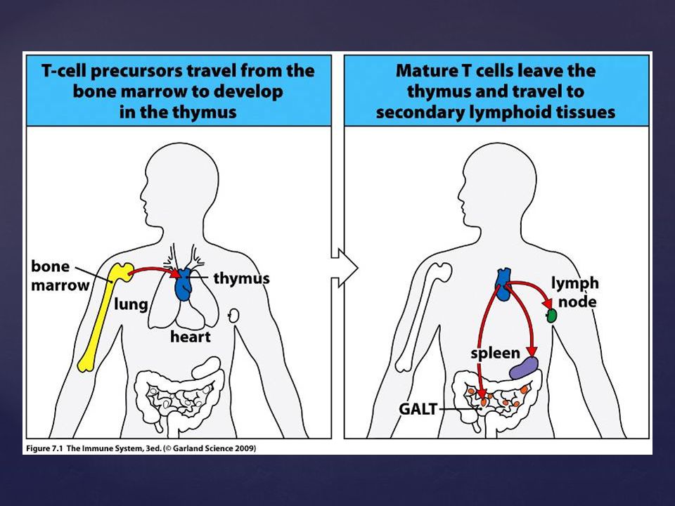
Thymocytes Epithelial cells Dendritic cells Macrophages Thymus Thymocytes from the bone marrow arrive at the thymus and mature into T cells

13
Spleen Weights 150g, in the upper left abdomen. The spleen filters the blood and serves as a secondary lymphoid organ

14
Spleen Lymphocyte aggregations similar to the lymph node only that cells and pathogens enter from the blood Red pulp- filters the blood; from antigens, microorganisms and worn-out RBCs

15
Lymphatic vessels

16
Lymphatic nodes

17
Lymph node

19
Secondary lymphatic tissues

20
Lymphatic tissues that are more diffused are generally known as MALT (Mucosa associated lymphatic tissue). Similar microanatomy as the lymph nodes and spleen Most of the pathogens get into human body through mucosa hin, huge surface, dinamic structure A thin, huge surface, dinamic structure Intense and active immune surveillance mechanisms ensure the protection Intense and active immune surveillance mechanisms ensure the protection Mucus contains glycoproteins, proteoglycans, special enzymes Mucus contains glycoproteins, proteoglycans, special enzymes Anti microbial peptides provide biological defence mecanism against intection Anti microbial peptides provide biological defence mecanism against intection Most of the lymphocyte reside arround the mucosal surface Most of the lymphocyte reside arround the mucosal surface Secondary lymphatic tissues MALT

21
Kripta GALT

22
The small intestine contains lymphoid nodules; the Peyer’s patches and isolated lymphoid follicles. Pathogens are delivered across the mucosa to APCs by specialized mucosal epithelial cells are called the M cells (microfold cells). The Lamina propria contains lymphatic tissue underlying the gastrointestinal tract connective tissue GALT
. The Lamina propria contains lymphatic tissue underlying the gastrointestinal tract connective tissue GALT.")
24
Intra-epithelial lymphocytes

25
GALT Antigens arising from Peyer’s patches and Lamina Propria travel to T cell areas in the GALT or Mesenteric lymph nodes. The large intestine contains isolated lymphoid follicles and the appendix

26
Guarding the gastrointestinal entrance Waldeyer’s ring: Pharyngeal, Tubal, Palatine and Lingual Tonsils Tonsilitis NALT

27
Supplementary material

28
Edward Jenner 1796 The induction of immunity/protection from smallpox (cowpox) FIRST VACCINATION
 FIRST VACCINATION")
30
Louis Pasteur 1880 a a a Immunization with attenuated pathogens against rabies

31
Ilya Mechnikov 1883 Probiotics Paul Ehrlich 1900 Phagocytosis Pathogen recognition by special receptor, clonal proliferation to multiply cells that are able to recognize the pathogen

32
Koch Laboratory, Germany 1890 Diphteria and Tetanus toxin Emil Behring Shimbasaru Kitasato 1.Many diseases occur only once (natural protection) 2.Some diseases can be prevented by vaccination 3.The blood contains anti-bacterial activity (anti-toxins, serum therapy) Protective humoral factors Antibodies in serum bound to relevant pathogens
 2.Some diseases can be prevented by vaccination 3.The blood contains anti-bacterial activity (anti-toxins, serum therapy) Protective humoral factors Antibodies in serum bound to relevant pathogens")
Similar presentations


 Immune system.>")


 The adaptive immune system:>")


 primary defense against disease- causing organisms.>")

 of immunocytes Diffuse cells Follicle organ Patch.>")