Download presentation
Presentation is loading. Please wait.

1
PATHOLOGY OF SMALL & LARGE INTESTINE DIVERTICULAR DISEASE
Diverticulum= blind pouch leading off the alimentary tract, lined by mucosa, communicating with gut lumen Acquired >> congenital (e.g. Meckel’s) May arise anywhere in GIT; colon is the most common site Two main factors: 1) Weakness in the wall: defects at site of bv entry; congenital weakness (e.g. Marfan’s syndrome) 2) Increased peristalsis and intra-luminal pressure: hard stools due to low-fiber diet results in exaggerated peristaltic contractions
 May arise anywhere in GIT; colon is the most common site. Two main factors: 1) Weakness in the wall: defects at site of bv entry; congenital weakness (e.g. Marfan’s syndrome) 2) Increased peristalsis and intra-luminal pressure: hard stools due to low-fiber diet results in exaggerated peristaltic contractions.")
3
PATHOLOGY OF COLONIC DIVERTICULOSIS
Colonic diverticulosis is common in the West (50% of adults >60 yrs); associated with low-fiber diet Multiple small cm Sigmoid colon is involved in 95% of patients Gross examination: Prominent taeniae coli & circular muscle bundles due to muscular hypertrophy; outpouching may be visible from serosal surface Histology: sacs with thin wall made of mucosa & submucosa surrounded by fat or intact peritoneum
; associated with low-fiber diet. Multiple small cm. Sigmoid colon is involved in 95% of patients. Gross examination: Prominent taeniae coli & circular muscle bundles due to muscular hypertrophy; outpouching may be visible from serosal surface. Histology: sacs with thin wall made of mucosa & submucosa surrounded by fat or intact peritoneum.")
4
CLINICAL FEATURES OF COLONIC DIVERTICULOSIS
Usually asymptomatic Intermittent cramping or continuous LLQ discomfort, sensation of incomplete emptying of rectum Complications: Diverticulitis: accentuates symptoms, tenderness, fever Peridiverticulitis, perforation, abscess or fistula Fistula formation Bleeding Rx: High-fiber diet; surgery

5
PATHOLOGY OF SMALL & LARGE INTESTINE BOWEL OBSTRUCTION
May occur at any level, but is more frequent in small intestine May be acute or chronic Clinical features: abdominal distension (+/- pain) , followed by vomiting, then constipation May present at birth, during infancy, childhood or adulthood Obstruction may be in the lumen, wall or extramural Tumors and infarction account for 10-15% of small bowel obstruction Hernias, intestinal adhesions, intussusception, and volvulus account for 80% of cases
 , followed by vomiting, then constipation. May present at birth, during infancy, childhood or adulthood. Obstruction may be in the lumen, wall or extramural. Tumors and infarction account for 10-15% of small bowel obstruction. Hernias, intestinal adhesions, intussusception, and volvulus account for 80% of cases.")
7
CLASSIFICATION OF BOWEL OBSTRUCTION
Mechanical: Congenital: strictures, atresias, bands, imperforate anus, meconium in cystic fibrosis Infants: hernias, intussusception Adults: hernias, adhesions, tumors, inflammatory strictures, obstructive gallstones, fecaliths, foreign bodies, volvulus Functional: Paralytic ileus: electrolyte imbalance, neural injury, or reflex atony secondary to peritonitis bowel infarction, myopathies & neuropathathies (e.g. Hirschsprung’s)
")
8
BOWEL OBSTRUCTION HERNIAS
Weakness or defect in wall of peritoneal cavity with protrusion of a serosa-lined sac of peritoneum Inguinal & femoral canals, umbilicus & surgical scars Segments of viscera may protrude & become entrapped, mostly in inguinal hernias If small bowel is involved, partial or complete obstruction of its lumen may follow Incarceration: permenant trapping of hernial sac contents due to venous stasis & edema Strangulation: Venous & arterial supply to entrapped viscus is compromised leading to infarction or gangrene

9
BOWEL OBSTRUCTION INTESTINAL ADHESIONS
Due to localized or generalized peritonitis secondary to: Surgical procedures Infection Endometriosis Rarely due to congenital fibrous bands May result in intestinal obstruction, incarceration and strangulation Fibrous bridges which develop between bowel segments creating closed loops through which viscera may slide & become entrapped (internal hernias)
")
10
BOWEL OBSTRUCTION INTUSSESCEPTION
Uncommon disorder where a segment of small intestine, constricted by a a wave of peristalsis, suddenly becomes telescoped into the immediately distal segment of bowel Intussusceptum: trapped bowel segment Intussuscipiens: bowel segment which envelops it Mostly in infants & children of unknown pathogenesis In adults, an intraluminal tumor may act as point of traction, pulling along a segment of bowel Intestinal obstruction Intestinal infarction due to trapping of mesenteric blood vessels

11
INTUSSESCEPTION

12
BOWEL OBSTRUCTION VOLVULUS
Complete twisting of a loop of bowel about its mesenteric base of attachment Mostly in small intestine; large intestine especially sigmoid & cecum, and other structures (e.g. ovary) may be involved Constriction of venous outflow &/or arterial supply Intestinal obstruction and infarction Uncommon disorder, occuring in all ages when an intestinal segment becomes longer & the mesentary narrower May be caused by colon malrotation & peritoneal bands
 may be involved. Constriction of venous outflow &/or arterial supply. Intestinal obstruction and infarction. Uncommon disorder, occuring in all ages when an intestinal segment becomes longer & the mesentary narrower. May be caused by colon malrotation & peritoneal bands.")
13
SMALL & LARGE INTESTINE TUMORS
Colonic tumors are much more common than small intestinal tumors May arise from any layer of GIT wall (mucosa, submucosa, muscularis or serosa); most are of epithelial origin The majority are benign; however, colonic cancers are a major cause of morbidity & mortality Clinical presentation: Asymptomatic; incidental finding Blood per rectum; anemia Bowel obstruction
; most are of epithelial origin. The majority are benign; however, colonic cancers are a major cause of morbidity & mortality. Clinical presentation: Asymptomatic; incidental finding. Blood per rectum; anemia. Bowel obstruction.")
14
INTESTINAL TUMORS Benign: Malignant: Stromal tumors
Hyperplastic & adenomatous polyps Lipomas Malignant: Adenocarcinoma Carcinoid tumors Lymphoma Sarcoma

15
PATHOLOGY OF SMALL & LARGE INTESTINE CARCINOID TUMORS
Tumors of neuroendocrine cells, which are widely distributed cells of epithelial stem cell origin capable of producing a variety of bioactive compounds Tumors may produce multiple compounds or a predominant product, causing a clinical syndrome such as gastrinoma, insulinoma ... Tumors arrise in GIT, lung, biliary tree & pancreas 50% of small intestinal & 2% of colonic tumors Any age, peak incidence in 6th decade Carcinoids are well differentiated tumors that are potentially malignant tumors and may metastasize

16
CLINICAL FEATURES OF CARCINOID TUMORS
Sites: appendix>small intestine (ileum)>rectum> stomach>colon Most are asymptomatic (particularly appendix) May cause local symptoms (angulation or obstrcution of small intestine) Secretory products may produce a variety of endocrinopathies, e.g. Zollinger-Ellison syndrome, Cushing’s syndrome, hyperinsulinism Carcinoid syndrome: 1% of patients & 20% of those with widespread metastasis; due to serotonin secretion; vasomotor disturbances, intestinal hypermotility, hepatomegaly and systemic fibrosis
>rectum> stomach>colon. Most are asymptomatic (particularly appendix) May cause local symptoms (angulation or obstrcution of small intestine) Secretory products may produce a variety of endocrinopathies, e.g. Zollinger-Ellison syndrome, Cushing’s syndrome, hyperinsulinism. Carcinoid syndrome: 1% of patients & 20% of those with widespread metastasis; due to serotonin secretion; vasomotor disturbances, intestinal hypermotility, hepatomegaly and systemic fibrosis.")
17
PATHOLOGY OF CARCINOID TUMORS
Gross: usually small tumors, rarely >3 cm, forming an intramural or submucosal yellow-tan firm elevation or polypoid lesion Histology: monomorphous population of cells with scant granular cytoplasm & round nuclei. Neoplastic cells form islands, nests, strands or glands. Intact or ulcerated overlying mucosa. May be multicentric (stomach, ileum) May be localized or have local spread &/or metastasis to lymph nodes or more distant sites, particularly liver
 May be localized or have local spread &/or metastasis to lymph nodes or more distant sites, particularly liver.")
21
CLINICAL BEHAVIOUR OF CARCINOID TUMORS
Clinical behaviour is difficult to predict from histologic features Behaviour but generally depends on: 1) Size: >2 cm and those invading >1/2 wall thickness 2) Site: appendiceal & rectal carcinoids almost never metastasize 5 years survival rate is excellent: 90% 5 years survival rate for small intestinal tumors with liver metastasis: 50% Widespread disease usually causes death
 Size: >2 cm and those invading >1/2 wall thickness. 2) Site: appendiceal & rectal carcinoids almost never metastasize. 5 years survival rate is excellent: 90% 5 years survival rate for small intestinal tumors with liver metastasis: 50% Widespread disease usually causes death.")
22
SMALL INTESTINAL ADENOCARCINOMA
Most arise in duodenum, including ampulla of Vater Polypoid fungating tumor growing in a napkin-ring encircling pattern Early on, tumor is asymptomatic Signs & symptoms usually appear late, when tumor has already penetrated bowel wall into mesentery, other bowel segments or spread to LN, or metastasize to liver or more widely c/o symptoms of abdominal obstruction (pain, nausea, vomiting, and weight loss) 5 yrs survival with wide en bloc excision: 70%
 5 yrs survival with wide en bloc excision: 70%")
24
SMALL & LARGE INTESTINE POLYPS
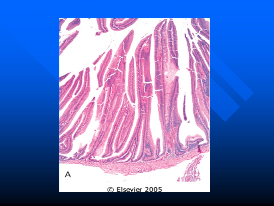
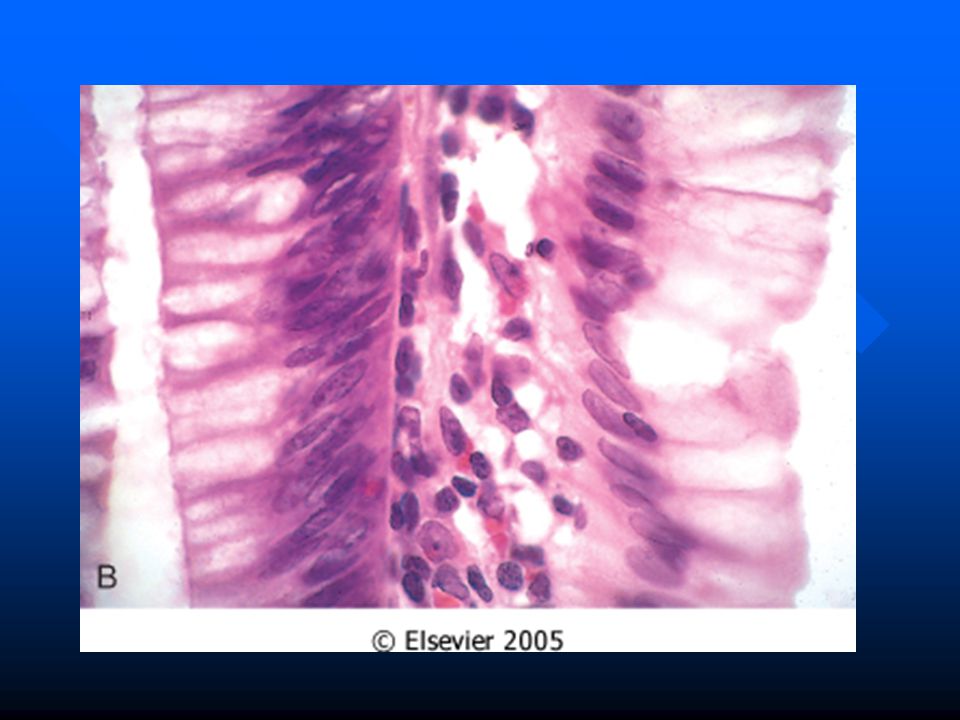
Polyp= a tumorous mass protruding into the lumen of the gut; may be pedunculated or sessile Major types: Non-neoplastic polyps: about 90% of polyps; formed as a result of abnormal mucosal maturation, inflammation or architecture Hyperplastic polyps Hamartomatous polyps Inflammatory polyps Neoplastic polyps: formed as a result of epithelial proliferation and dysplasia, i.e. pre-cancerous Adenomatous polyps

26
INTESTINAL POLYPS HYPERPLASTIC POLYPS
Majority of polyps Site: mostly in colon, >50% in rectosigmoid Patients: increase in frequency with age Appearance: Most are small <5 mm, nipple-like protrusion; multiple or single Pathology: histologically consist of abundant crypts lined by well-differentiated goblet or absorptive epithelial cells, seperated by scant lamina propria Asymptomatic or blood &/or mucus per rectum No malignant potential

28
INTESTINAL POLYPS HAMARTOMATOUS POLYPS
Juvenile polyps: Hamartomatous proliferation of lamina propria surrounding cystically dilated glands Patients: usually <5 yrs; in adults called “retention polyp” Appearance:1-3 cm in children; smaller in adults Single rounded or lobulated, +/- stalk Bleeding per rectum; stalk may twist and undergo painful infarction No malignant potential

29
INTESTINAL POLYPS HAMARTOMATOUS POLYPS
Peutz-Jeghers polyps: Rare autosomal dominant syndrome, characterized by GIT polyps, melanotic mucosal and cutaneous pigmentation around lips, oral mucosa, face, genitalia & palmar surfaces of hands Site: polyps may occur in stomach (25%), colon (30%) and small intestine (100%) Appearance: large peduculated lobulated polyps Pathology: branching framework of connective tissue & smooth muscle Polyps have no malignant potential, but PJS patients have increased risk of developing carcinoma of pancreas, breast, lung, ovary & uterus
, colon (30%) and small intestine (100%) Appearance: large peduculated lobulated polyps. Pathology: branching framework of connective tissue & smooth muscle. Polyps have no malignant potential, but PJS patients have increased risk of developing carcinoma of pancreas, breast, lung, ovary & uterus.")
31
INTESTINAL POLYPS ADENOMATOUS POLYPS
Vast majority of adenomas arise in colon Variable in size; pedunculated or sessile Results from epithelial proliferation and dysplasia, which may range from mild to carcinoma in situ Three architectural types: 1) Tubular adenoma 2) Villous adenoma 3) Tubulovillous adenoma Risk of malignant transformation depends on: Polyp size: <1 cm (v. rare); 1-2 cm (10%); >2 cm (45%) Histologic architecture: proportion of villous component Severity of dysplasia
 Tubular adenoma. 2) Villous adenoma. 3) Tubulovillous adenoma. Risk of malignant transformation depends on: Polyp size: <1 cm (v. rare); 1-2 cm (10%); >2 cm (45%) Histologic architecture: proportion of villous component. Severity of dysplasia.")
36
INTESTINAL POLYPS ADENOMATOUS POLYPS
Tubulovillous adenoma: 5-10% of adenomatous polyps; mixture of tubular & villous (20-50%) areas; intermediate in frequency of having a stalk, size, degree of dysplasia & risk of carcinoma Clinical features: 20-30% are <40 yrs; 50% after 60 Small polyps: asymptomatic Occult bleeding, anemia, hypoproteinemia & hypokalemia Intestinal obstruction or biliary obstruction Rx: Most are cured by adequate endoscopic excision Px: Annual endoscopic follow-up
 areas; intermediate in frequency of having a stalk, size, degree of dysplasia & risk of carcinoma. Clinical features: 20-30% are <40 yrs; 50% after 60. Small polyps: asymptomatic. Occult bleeding, anemia, hypoproteinemia & hypokalemia. Intestinal obstruction or biliary obstruction. Rx: Most are cured by adequate endoscopic excision. Px: Annual endoscopic follow-up.")
37
ADENOMATOUS POLYPS TUBULAR ADENOMA VILLOUS ADENOMA
>85% of adenomas Majority in distal colon; 50% in rectosigmoid Most are small (few mm-2 cm) with a stalk (-2 cm) Histology shows neoplastic epithelium forming glands & tubules Variable degrees of dysplasia to CIS Invasive carcinoma rare 1% of adenomas Rectum & rectosigmoid 75%, followed by cecum & ascending colon Sessile ranging from cm (most are 1-3 cm) Histology shows neoplastic cells forming villi (>50% villous) Carcinoma in situ 10% Invasive carcinoma 30%
 with a stalk (-2 cm) Histology shows neoplastic epithelium forming glands & tubules. Variable degrees of dysplasia to CIS. Invasive carcinoma rare. 1% of adenomas. Rectum & rectosigmoid 75%, followed by cecum & ascending colon. Sessile ranging from 1-10 cm (most are 1-3 cm) Histology shows neoplastic cells forming villi (>50% villous) Carcinoma in situ 10% Invasive carcinoma 30%")
38
INTESTINAL POLYPS FAMILIAL POLYPOSIS SYNDROMES
Familial adenomatous polyposis (FAP): Rare autosomal dominant trait Dx: at least 100 polyps (average 1000) Most are tubular adenomatous polyps Genetic defect of APC gene localized to chromosome 5q21 Risk of colon cancer: 100% at 30 years Rx: Prophylactic colectomy Gardner’s syndrome: + osteomas, epidermal cysts, fibromatosis, thyroid Ca.. Turcot’s syndrome: + CNS tumors (gliomas)
: Rare autosomal dominant trait. Dx: at least 100 polyps (average 1000) Most are tubular adenomatous polyps. Genetic defect of APC gene localized to chromosome 5q21. Risk of colon cancer: 100% at 30 years. Rx: Prophylactic colectomy. Gardner’s syndrome: + osteomas, epidermal cysts, fibromatosis, thyroid Ca.. Turcot’s syndrome: + CNS tumors (gliomas)")
40
INTESTINAL POLYPS FAMILIAL POLYPOSIS SYNDROMES
Lynch’s syndrome: aka: Hereditary nopolyposis colorectal cancer (HNPCC) Autosomal dominant syndrome characterized by an increased risk of colorectal cancer and extra-intestinal cancer, particularly endometrial carcinoma Multiple recurrent colorectal adenomatous polyps which present ar an early age Mutations in any of 4 genes (MSH2, MLH1, PMS1 & PMS2) involved in DNA repair, located in chromo-somes 2, 3 & 7, resulting in microsatellite instability Colonic carcinoma is typically located proximal to splenic flexure, often multiple, and not arising in preexisting adenomas
 Autosomal dominant syndrome characterized by an increased risk of colorectal cancer and extra-intestinal cancer, particularly endometrial carcinoma. Multiple recurrent colorectal adenomatous polyps which present ar an early age. Mutations in any of 4 genes (MSH2, MLH1, PMS1 & PMS2) involved in DNA repair, located in chromo-somes 2, 3 & 7, resulting in microsatellite instability. Colonic carcinoma is typically located proximal to splenic flexure, often multiple, and not arising in preexisting adenomas.")
41
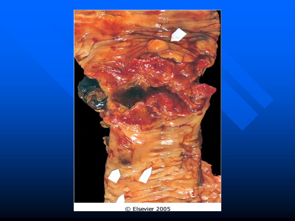
TUMORS OF LARGE INTESTINE COLORECTAL ADENOCARCINOMA
More than 98% of colonic malignancies Second most common cancer Peak incidence yrs; <20% before age of 50 yrs Wide geographic variation in incidence; more frequent in Western industrialized nations Environmental dietary factors have been implicated: Low unabsorbable vegetable fiber content High refined crabohydrate content High fat content Low protective micronutrients content (vit. A, C & E) Almost always arise in adenomatous polyps
 Almost always arise in adenomatous polyps.")
42
COLORECTAL ADENOCARCINOMA THE ADENOMA-CARCINOMA SEQUENCE
Cumulative alterations in the genome lead to progressive increase size, dysplasia & invasive potential “Multi-hit” concept for colon cancer carcniognesis: First hit: germline or somatic mutations of cancer suppressor genes i.e. APC & mismatch repair genes Second hit: Methylation abnormalities & inactivation of of normal alleles of APC & mismatch repair genes Protooncogene mutation, e.g. K-ras at 12p12 Homozygous loss of addtional cancer suppressor genes, e.g. DCC at 18q21 and p53 at 17p13 Additional mutations of many genes will occur in carcinoma

44
PATHOLOGY & CLINICAL FEATURES OF COLORECTAL ADENOCARCINOMA
Majority are sporadic; 1-3% in patients with FAP or IBD Mostly single; location is shifting to the right colon Proximal colon: fungating polypoid tumors Distal colon: annular encircling tumors (napkin-ring) Tumors penetrate colonic wall layer into serosal surface Histology range from well differentiated to undifferentiated; may be mucin-secreting Asymptomatic for year Right: fatigue, weakness, iron-deficiency anemia Left: occult bleeding, change in bowel habit, discomfort Dx: PE, lab, X-ray, endoscopy and biopsy
 Tumors penetrate colonic wall layer into serosal surface. Histology range from well differentiated to undifferentiated; may be mucin-secreting. Asymptomatic for year. Right: fatigue, weakness, iron-deficiency anemia. Left: occult bleeding, change in bowel habit, discomfort. Dx: PE, lab, X-ray, endoscopy and biopsy.")
49
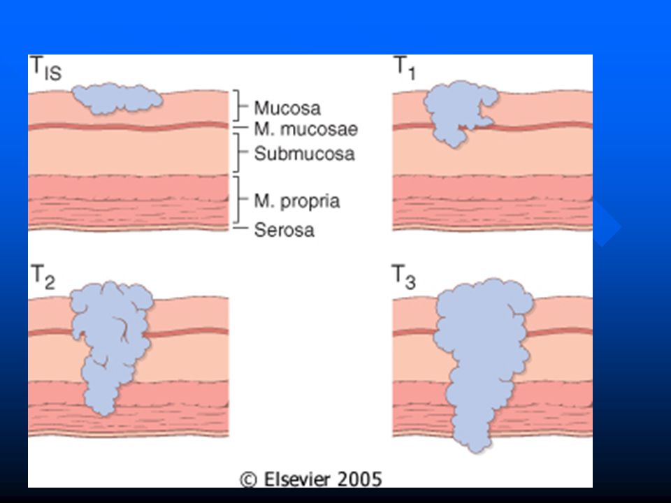
COLORECTAL ADENOCARCINOMA Modified Dukes’ (Astler-Coller) Staging System
 Staging System")
50
TUMORS OF SMALL & LARGE INTESTINE INTESTINAL LYMPHOMA
The GIT is the most common extranodal location of lymphomas May be primary or secondary Primary GI lymphoma: no evidence of liver, spleen or bone marrow involvement at diagnosis Adults, M=F, stomach>small intestine>colon Classification similar to nodal lymphomas: MALTomas, large cell lymphoma, … etc. Nonspecific symptoms Rx: surgery, chemo- and radiotherapy Px: better than nodal lymphomas

Similar presentations


 bowel habit change (-) bearing down sensation PMHx. hemorrhoidectomy,>")


>")