Download presentation
Presentation is loading. Please wait.

1
Malassezia Dermatitis: The Host-Pathogen Relationship Daniel O. Morris, DVM Diplomate, ACVD University of Pennsylvania Philadelphia, PA USA

2
D Morris ACVD Resident Review 2003 The genus Malassezia The genus Malassezia : Background Lipophilic: Able to utilize lipids as a source of carbon Able to utilize lipids as a source of carbon Lipid-dependent species lack the ability to synthesize long-chain (C12-C24 series) fatty acids- hence their requirement for an exogenous lipid source Lipid-dependent species lack the ability to synthesize long-chain (C12-C24 series) fatty acids- hence their requirement for an exogenous lipid source Only M. pachydermatis is truly non-lipid dependent, but its growth is enhanced by exogenous lipid Only M. pachydermatis is truly non-lipid dependent, but its growth is enhanced by exogenous lipid

3
D Morris ACVD Resident Review 2003 Ecology Opportunistic pathogens Commensal microflora in nearly all mammals (and some birds) studied Keratinophilic Dwell primarily within the stratum corneum Dwell primarily within the stratum corneum Isolated from the canine distal hair shaft Isolated from the canine distal hair shaft Rarely found in the follicular infundibulum Rarely found in the follicular infundibulum
 studied Keratinophilic Dwell primarily within the stratum corneum Dwell primarily within the stratum corneum Isolated from the canine distal hair shaft Isolated from the canine distal hair shaft Rarely found in the follicular infundibulum Rarely found in the follicular infundibulum")
4
D Morris ACVD Resident Review 2003 Species classification Lipid-dependent species M. furfur (the primary human pathogen) M. furfur (the primary human pathogen) M. sympodialis M. sympodialis M. globosa M. globosa M. obtusa M. obtusa M. restricta M. restricta M. slooffiae M. slooffiae M. equi M. equi Non-lipid dependent species M. pachydermatis (the primary animal pathogen) M. pachydermatis (the primary animal pathogen)
 M. furfur (the primary human pathogen) M. sympodialis M. sympodialis M. globosa M. globosa M. obtusa M. obtusa M. restricta M. restricta M. slooffiae M. slooffiae M. equi M. equi Non-lipid dependent species M. pachydermatis (the primary animal pathogen) M. pachydermatis (the primary animal pathogen).")
5
M. pachydermatis

6
M. globosa

7
M. restricta

8
M. sympodialis

9
-Occur in yeast form in-vivo (non-mycelial), but hyphae may occur in culture M. furfur
, but hyphae may occur in culture M. furfur")
11
-Thick, multilayered cell wall -Repetitive uni-polar (sympodial) budding to produce blastoconidia
 budding to produce blastoconidia")
12
D Morris ACVD Resident Review 2003 Microbiology of M. pachydermatis Optimum growth at 37 o C (range 32-40) Lipid-enhanced SDA or Dixon’s agar Microaerophilic conditions (5% CO 2 ) Colonies mature at 72 hours
 Lipid-enhanced SDA or Dixon’s agar Microaerophilic conditions (5% CO 2 ) Colonies mature at 72 hours.")
15
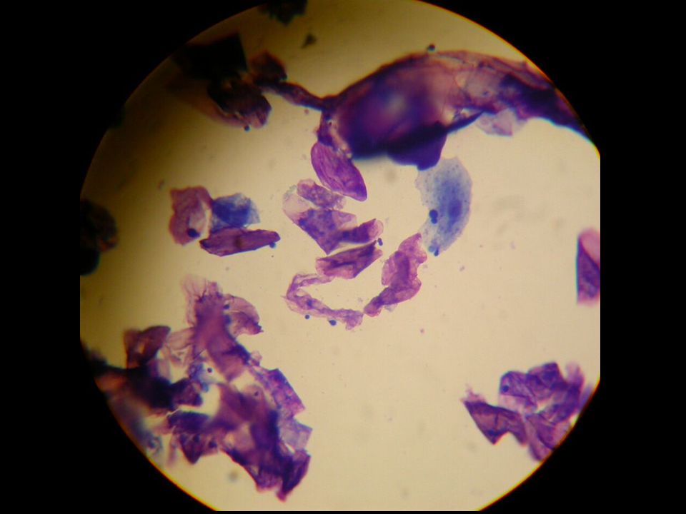
Candida species

16
D Morris ACVD Resident Review 2003 Microbiology: Isolation and I.D. Cytology Quantitative: direct impression smear, tape stripping Quantitative: direct impression smear, tape stripping Non-quantitative: cotton-tip swab, dry skin scraping Non-quantitative: cotton-tip swab, dry skin scrapingCulture Quantitative methods: tape-stripping, detergent scrub, contact plate Quantitative methods: tape-stripping, detergent scrub, contact plate Non-quantitative: sterile swab Non-quantitative: sterile swab

17
D Morris ACVD Resident Review 2003 Microbiology: Isolation and I.D. Molecular techniques Nested PCR: Species-specific primers for ribosomal RNA Nested PCR: Species-specific primers for ribosomal RNA DNA fingerprinting by PFGE: Allows testing for concordance of identity between strains (allows tracking of epizootics) DNA fingerprinting by PFGE: Allows testing for concordance of identity between strains (allows tracking of epizootics)
 DNA fingerprinting by PFGE: Allows testing for concordance of identity between strains (allows tracking of epizootics).")
18
D Morris ACVD Resident Review 2003 Correlation with pathogenic effect From the literature: Quantitative cytology Quantitative cytology >10 organisms per ½ inch 2 (evidence-based)* >1 organism/oil-immersion field (non-evidence based) Ear exudate: >5 organisms/oil-immersion field Quantitative culture Quantitative culture Colony counts from contact-plate cultures significantly correlate with cytological (tape-strip) counts † *Morris, et al. Type-1 hypersensitivity reactions to M. pachydermatis extracts in atopic dogs. AJVR 1998. † Nuttal, et al. Serum antibodies to Malassezia yeasts in canine atopic dermatitis. Vet Dermatol 2001

19
D Morris ACVD Resident Review 2003 Clinical manifestations Primary MD is rare: chronic wetting/ high humidity? Primary Malassezia otitis: much more common - “swimmer’s ear” Secondary MD: Atopy (and other allergic diseases) Seborrheic disorders Endocrine/metabolic diseases Systemic neoplasia (cats) Virulence differences between strains??
 Seborrheic disorders Endocrine/metabolic diseases Systemic neoplasia (cats) Virulence differences between strains .")
23
D Morris ACVD Resident Review 2003 Non-atopic disease associations Parakeratotic diseases Zinc-responsive dermatosis Generic dog food dermatosis Superficial necrolytic dermatitis Cutaneous T-cell lymphoma Canine Cushing’s disease

26
D Morris ACVD Resident Review 2003 Occurrence in the oral cavity Considered to be part of the normal oral microflora of dogs Implicated in a case of severe, ulcerative stomatitis/pharyngitis/ tonsillitis

27
D Morris ACVD Resident Review 2003 Malassezia dermatitis - feline Feline paraneoplastic alopecia

28
D Morris ACVD Resident Review 2003 Malassezia in avian species?? Role in feather picking?

30
D Morris ACVD Resident Review 2003 Malassezia in domestic farm animals Isolated from normal ruminants, horses, and swine. Role in otic or cutaneous diseases remains to be determined.

32
D Morris ACVD Resident Review 2003 Pathogen-dependent immunological factors (M. pachydermatis/M. furfur) Production of allergens relevant to dogs and human beings, respectively Fixation of complement (alternative pathway) anaphylatoxin production (C3a, C4a, C5a) inflammation Does not require sensitization or allergen- specific recognition Role in non-atopic inflammatory diseases?
 Production of allergens relevant to dogs and human beings, respectively Fixation of complement (alternative pathway) anaphylatoxin production (C3a, C4a, C5a) inflammation Does not require sensitization or allergen- specific recognition Role in non-atopic inflammatory diseases .")
33
D Morris ACVD Resident Review 2003 Immunological factors, cont… Directed effect to human keratinocytes leading to cytokine production; net effect is pro-inflammatory This effect varies according to yeast species M. furfur does not induce cytokine release in human keratinocytes M. pachydermatis does

34
D Morris ACVD Resident Review 2003 Pathogen virulence factors Adherence to corneocytes Increased at higher temperatures Increased at higher temperatures No influence by concurrent Staph. colonization No influence by concurrent Staph. colonization Mediated by trypsin-sensitive proteins/glyco- proteins and mannosyl-bearing carbohydrate ligands on the yeast cell wall, or by lipid ligands in otic infections Mediated by trypsin-sensitive proteins/glyco- proteins and mannosyl-bearing carbohydrate ligands on the yeast cell wall, or by lipid ligands in otic infections Marked variability amongst strains Marked variability amongst strains Implication: identification of an universal ligand inhibitor is unlikely Implication: identification of an universal ligand inhibitor is unlikely

35
D Morris ACVD Resident Review 2003 Pathogen virulence factors II Lipid hydrolysis FFA’s inhibition of competing microorganisms Production of catabolic enzymes (role in pruritus?) Proteinase – role in keratin penetration? Proteinase – role in keratin penetration? Chondroiton-sulphatase Chondroiton-sulphatase Phospholipase Phospholipase Hyaluronidase Hyaluronidase

36
Epidermal hyperplasia – relevance?

37
D Morris ACVD Resident Review 2003 Host factors: colonization of skin surface Dogs, cats, human beings: colonization occurs within hours of birth Sparsely haired skin Peri-orificial, periungual Peri-orificial, periungual Seborrheic “zones” (head and neck in human beings) Seborrheic “zones” (head and neck in human beings) External ear canals in dogs and cats External ear canals in dogs and catsMucosae Mucosal disease due to Malassezia has been reported but appears to be rare as opposed to Candidiasis. Anal sacs a reservoir for inoculation? Mucosal disease due to Malassezia has been reported but appears to be rare as opposed to Candidiasis. Anal sacs a reservoir for inoculation?

38
D Morris ACVD Resident Review 2003 Host factors: Microclimate Areas with increased moisture and/or sebum are favored Ear canals express a higher temp. and humidity than the skin surface Ear canals express a higher temp. and humidity than the skin surface Pinnal shape is unrelated to occurrence rates of MO Sterile saline added to the ear canal is sufficient to induce Malassezia otitis Sterile saline added to the ear canal is sufficient to induce Malassezia otitis Anecdotal reports of increased rate of primary Malassezia otitis and pododermatitis in humid climates Anecdotal reports of increased rate of primary Malassezia otitis and pododermatitis in humid climates

39
D Morris ACVD Resident Review 2003 Immunological responses Host factors II: Immunological responses Type-1 hypersensitivity IgE-mediated: IgE-mediated: Documented via IDAT, SPT, ELISA, RAST, and APT in atopic human beings. Major allergens characterized. Documented via IDAT, ELISA, and PCA testing in dogs with atopic dermatitis. Major allergens characterized. Cell-mediated response: Lymphocyte-mediated (Th 2 ) Lymphocyte-mediated (Th 2 ) Documented via in-vivo and in-vitro LT testing and cytokine profiling in human beings with AD Documented via in-vitro LT testing in dogs with AD
 Lymphocyte-mediated (Th 2 ) Documented via in-vivo and in-vitro LT testing and cytokine profiling in human beings with AD Documented via in-vitro LT testing in dogs with AD.")
40
D Morris ACVD Resident Review 2003 Evidence for Type-1 hypersensitivity in dogs Intradermal testing: Dogs with increased M. pachydermatis colonization, in association with atopic dermatitis, have stronger IDAT reactions to crude extracts of M. pachydermatis than do atopic dogs without MD. Dogs with increased M. pachydermatis colonization, in association with atopic dermatitis, have stronger IDAT reactions to crude extracts of M. pachydermatis than do atopic dogs without MD. Normal dogs do not react to the extract. Normal dogs do not react to the extract.

41
D Morris ACVD Resident Review 2003 Evidence for Type-1 hypersensitivity in dogs, II Testing for passive cutaneous anaphylaxis (P-K) Pooled serum from atopic dogs with MD that are IDAT positive for M. pachydermatis is injected ID in serial dilutions into normal dogs Pooled serum from atopic dogs with MD that are IDAT positive for M. pachydermatis is injected ID in serial dilutions into normal dogs At 24, 48 and 72 hours, the M. pachydermatis extract is injected ID at the former serum injection sites At 24, 48 and 72 hours, the M. pachydermatis extract is injected ID at the former serum injection sites M S H 1:2 1:4 1:8 1:161:321:64

42
D Morris ACVD Resident Review 2003 P-K testing, cont…. Formation of a wheal/flare reaction indicates transfer of anti-Malassezia antibody from the donor serum – persistence to 72 hours indicates IgE is present (vs. IgG) Heating (56 o c for 4 hours) and adsorption with anti- canine IgE removes the IgE
 Heating (56 o c for 4 hours) and adsorption with anti- canine IgE removes the IgE.")
43
D Morris ACVD Resident Review 2003 Evidence for Type-1 hypersensitivity in dogs, III ELISA testing: Atopic dogs (both with or without MD) have significantly higher levels of anti-Malassezia IgE than normal dogs, or non-atopic dogs with Malassezia overgrowth. Atopic dogs (both with or without MD) have significantly higher levels of anti-Malassezia IgE than normal dogs, or non-atopic dogs with Malassezia overgrowth. Normal dogs do not have high levels of circulating antibody toward the normal (commensal) population of yeast Normal dogs do not have high levels of circulating antibody toward the normal (commensal) population of yeast
 have significantly higher levels of anti-Malassezia IgE than normal dogs, or non-atopic dogs with Malassezia overgrowth. Normal dogs do not have high levels of circulating antibody toward the normal (commensal) population of yeast Normal dogs do not have high levels of circulating antibody toward the normal (commensal) population of yeast.")
46
D Morris ACVD Resident Review 2003 Evidence for Type-1 hypersensitivity in dogs, IV Clinical evidence Specific antifungal therapy of atopic dogs with MD will often ameliorate the majority of pruritus within several days. Rapid relapse of pruritus occurs when yeast counts rise again. Specific antifungal therapy of atopic dogs with MD will often ameliorate the majority of pruritus within several days. Rapid relapse of pruritus occurs when yeast counts rise again.

47
D Morris ACVD Resident Review 2003 Evidence for T-cell Dysregulation in atopic dogs with MD Lymphocyte blastogenesis testing (LT) methods: Peripheral blood mononuclear cells isolated by hypaque-ficoll density gradient Peripheral blood mononuclear cells isolated by hypaque-ficoll density gradient PBMC incubated for 7 days with M. pachydermatis extract at serial concentrations PBMC incubated for 7 days with M. pachydermatis extract at serial concentrations PHA used as pos. control; untreated media used as neg. control PHA used as pos. control; untreated media used as neg. control

48
D Morris ACVD Resident Review 2003 T-cell responses II Results: Atopic dogs with MD have increased LT response to M. pachydermatis compared to normal dogs and atopic dogs with Malassezia otitis. Atopic dogs without MD or MO are intermediate Atopic dogs with MD have increased LT response to M. pachydermatis compared to normal dogs and atopic dogs with Malassezia otitis. Atopic dogs without MD or MO are intermediate [Bond, et al: Basset hounds with idiopathic seborrhea have a blunted LT response to M. pachydermatis when compared to normal Bassets] [Bond, et al: Basset hounds with idiopathic seborrhea have a blunted LT response to M. pachydermatis when compared to normal Bassets]

49
D Morris ACVD Resident Review 2003 T-cell responses III Conclusions: T-cell dysregulation contributes to the pathogenesis of MD (as in human beings with AD) T-cell dysregulation contributes to the pathogenesis of MD (as in human beings with AD) Like human beings with Seb Derm, Basset hounds with seborrhea do not have a normal (inadequate?) T-cell response Like human beings with Seb Derm, Basset hounds with seborrhea do not have a normal (inadequate?) T-cell response Cerumen within the ear canal may be protective against penetration by (and recognition of) Malassezia allergens Cerumen within the ear canal may be protective against penetration by (and recognition of) Malassezia allergens
 T-cell dysregulation contributes to the pathogenesis of MD (as in human beings with AD) Like human beings with Seb Derm, Basset hounds with seborrhea do not have a normal (inadequate ) T-cell response Like human beings with Seb Derm, Basset hounds with seborrhea do not have a normal (inadequate ) T-cell response Cerumen within the ear canal may be protective against penetration by (and recognition of) Malassezia allergens Cerumen within the ear canal may be protective against penetration by (and recognition of) Malassezia allergens")
50
D Morris ACVD Resident Review 2003 A role for superantigens? What are superantigens? Microbial products that bind directly to the V region of the TCR without regard for antigen specificity Microbial products that bind directly to the V region of the TCR without regard for antigen specificity Result: activation of large “families” of T-cells that express a common V region Result: activation of large “families” of T-cells that express a common V region Activation of 5-30% of the T-cell population simultaneously with production of inflammatory cytokines and binding to “innocent” antigens = “innocent bystander” damage Activation of 5-30% of the T-cell population simultaneously with production of inflammatory cytokines and binding to “innocent” antigens = “innocent bystander” damage Role in Malassezia dermatitis: Ruled out in human beings Ruled out in human beings Studies in atopic dogs planned with newly developed monoclonal antibodies for V -region gene products of TCR Studies in atopic dogs planned with newly developed monoclonal antibodies for V -region gene products of TCR

51
D Morris ACVD Resident Review 2003 Future therapies for Malassezia dermatitis Currently, treatment continues to depend upon topical and systemic anti-fungal agents Allergen immunotherapy? Crude extracts vs. recombinant allergens? Crude extracts vs. recombinant allergens? Double-masked, placebo-controlled trials will be required Double-masked, placebo-controlled trials will be required 12 to 18 months in duration Anti-IgE therapies Trials on-going in human beings Trials on-going in human beings

Similar presentations


 Immune system.>")
 Or to precipitate.>")


 Lecture 18 Bacterial Pathogenesis (Based on other textbooks such as Madigan’s)>")


