Download presentation
Presentation is loading. Please wait.

1
Tissues and Membranes

2
Tissues… The human body consists of trillion of cells, but they do not work independently The cell must work together to perform various tasks to keep the body in homeostasis In order for cells to work together, they form tissues To help study the various cells and the tissues they make, anatomists have subdivided the tissues into four major groups and those groups are further subdivided according to their cellular makeup

3
General Information… Some tissues, such as epithelial, connective, muscle or nervous, may or may not have surface modifications Microvilli – contain microtubules which help increase the surface area of the cell Cilia – contain microfilaments which help move materials along the outer surface of the cell Cell junctions attach the cells of the tissues together forming an excellent barrier Three types of cell junctions 1.Tight junction – prevents movement of materials between cells and are usually found in epithelial cells 2.Adhering junction – holds the cells together and are found in skin and cardiac tissue 3.Gap junction

4
Three Types of Cell Junctions 1.Tight junction – prevents movement of materials between cells and are usually found in epithelial cells 2.Adhering junction – holds the cells together and are found in skin and cardiac tissue Desmosomes – the junction is a “spot” between the cells Intermediate junctions – the junction appears as a “band” around the cells 3.Gap junction – transmits impulse and small ions from cell to cell and are found in heart, smooth, and some nerve cells

5
The Four Tissue Groups… Tissue TypeCharacteristicType of cell within the tissues Epithelial (cover) Consists of cells that make up the inside or outside lining of organs Squamous, cuboidal, columnar Muscular (movement) Consists of cells that have the ability to contract and relax Skeletal muscle, smooth muscle, cardiac muscle Neural (control) Consists of cells that conduct impulses or cells that protect the nervous system Neuron, glial Connective (support) Consists of cells that have a matrix and typically fill internal spaces within the body Adipose, areolar, blood, bone, cartilage, dense, reticular
 Consists of cells that make up the inside or outside lining of organs Squamous, cuboidal, columnar Muscular (movement) Consists of cells that have the ability to contract and relax Skeletal muscle, smooth muscle, cardiac muscle Neural (control) Consists of cells that conduct impulses or cells that protect the nervous system Neuron, glial Connective (support) Consists of cells that have a matrix and typically fill internal spaces within the body Adipose, areolar, blood, bone, cartilage, dense, reticular")
6
Epithelia Characteristics… The cells fit closely together to form continuous sheets They are bound together at many points called cell junctions They always have one free surface called the apical surface, which is exposed to the body’s exterior or to the cavity of an internal organ it is lining

7
Epithelia Characteristics con’t… They rest on a basement membrane, which is a structureless material secreted by the cells They have no blood supply (avascular), therefore they depend on diffusion from capillaries under connective tissue for food and oxygen When well nourished, they can regenerate
, therefore they depend on diffusion from capillaries under connective tissue for food and oxygen When well nourished, they can regenerate")
8
Classification of Epithelia… Epithelia are classified according to: 1.Cell arrangement Simple – one layer of cells Stratified – two or more layers of cells All attached to a “basement” membrane 2.Shape If the cells are stratified, the free surface determines the name There are four main types i.Squamous ii.Cuboidal iii.Columnar iv.pseudostratified

9
Drawing Tissue… Draw each of the different types of tissue

10
Bell Ringer… What makes tissue? What are the 4 types of tissue and what is each of their main functions? Draw a simple squamous epithelial tissue – label the basement membrane, the apical surface, and cell junctions

11
The cells of Epithelial Tissue… Squamous cells Flat and irregular in appearance Found in the lining of the skin Our first line of defense Cuboidal cells Shaped like little squares Found in the lining of the urinary tubes Secrete and absorb material

13
The cells of Epithelial Tissue con’t… Columnar cells Shaped like columns Found in the lining of the trachea Secrete and absorb material Glandular cells Obtain needed materials from the blood and use them to make their secretion, which they discharge Two types of glands develop from epithelial sheets i.Endocrine glands ii.Exocrine glands

15
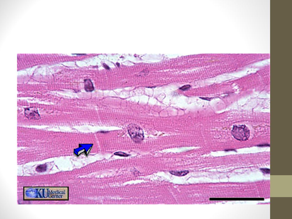
The cells of Muscular Tissue… Skeletal muscle cells Are elongated cells with striations Found making up the muscles associated with the skeletal system Contract and relax under voluntary control Smooth muscle cells Are elongated cells without striations Found making up the uterus and blood vessels Contract and relax under involuntary control

17
The cells of Muscular Tissue con’t… Cardiac muscle cells Consists of intercalated discs Found making up only the heart Contract and relax in a non-voluntary pulsating manner

19
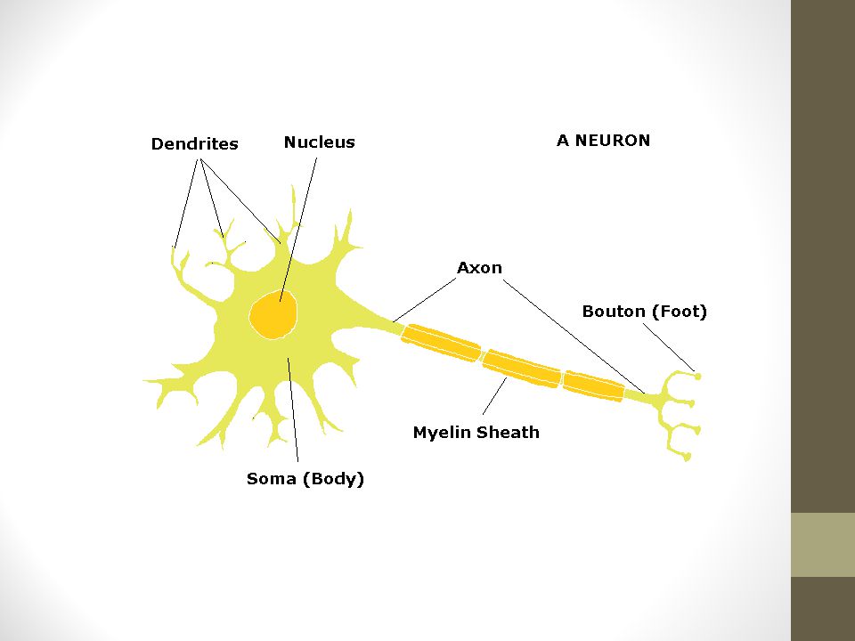
The cells of Neural Tissue… Neurons make up neural tissue Consists of dendrites, soma and an axon Found making up the nervous system such as the brain and spinal cord Conduct impulses Glial cells Have a variety of shapes and sizes Found either nearby or surrounding the neurons Provide protection for neurons

21
Connective Tissue Connective tissue represents the most diverse tissue group of the human body Even though cells are quite different from each other, they do have some commonality, which is they all have matrix of some sort The matrix is the material that surrounds the cells

22
The Major Matrix Types… There are 4 major matrix materials a.Fiber – the fibers of the matrix can be long, slender fibers or short, thick fibers b.Liquid – liquids such as plasma c.Solid – creates a tough, strong type of tissue d.Gel – creates a tough but yet very flexible type of tissue

23
Bell Ringer… Identify each type of tissue and explain why you choose it

24
Bell ringer… What is the one common thing that all connective tissue has in common? What type of cells protect nerve tissue?

25
The cells of Connective Tissue… Adipose cells Are round and appear empty, but they are full of fat Found surrounding various organs of the body Provide insulation Have a fiber matrix Areolar cells Are small and have long, thin fibers running between them making up the matrix Found between our skin and muscle and provide attachment of our skin to the muscle

27
The cells of Connective Tissue con’t… Blood cells Are small anucleated with a plasma matrix Can be found in our circulatory system Transport oxygen and carbon dioxide to and from other cells in the body Dense cells Are parallel fibers in a fiber matrix Make up tendons and ligaments Tendons provide attachment of muscles to bone Ligaments provide attachment of one bone to another

29
The cells of Connective Tissue con’t… Bone cell Form concentric rings around a central canal Can be found in our bones Provide strength In a solid matrix made of calcium phosphate Cartilage cell Are small and sit in a rather large lacuna Can be found within our joints Have a gel matrix, which allows for flexibility

31
The cells of Connective Tissue con’t… Reticular cell Are small and have short, thick fibers running between them Can be found making up the framework of the liver, spleen, tonsils, appendix, and thymus gland Have a fiber matrix that consists of short, thick fibers

33
Glandular tissue… Consists of one or more cells that make AND secrete a particular product called a secretion The secretion typically contains protein molecules in an aqueous fluid

34
2 Major Types of Glands 1.Endocrine glands Do not have ducts (ductless) Their secretions (all hormones) diffuse directly into the blood vessels found with in the gland Ex – thyroid, adrenal, pituitary 2.Exocrine glands Have ducts in which their secretions empty through to get to the epithelial surface Ex – sweat, oil, liver and pancreas (can be both internal and external)
 Their secretions (all hormones) diffuse directly into the blood vessels found with in the gland Ex – thyroid, adrenal, pituitary 2.Exocrine glands Have ducts in which their secretions empty through to get to the epithelial surface Ex – sweat, oil, liver and pancreas (can be both internal and external)")
35
Cartilage types… 1.Hyaline cartilage Most widespread type Supporting structures of the larynx, voice box, attaches the ribs to the sternum Covers the ends of bones where they form joints 2.Elastic cartilage Found where a structure with elasticity is desired – external ear 3.Fibrocartilage Forms the cushion-like discs between the vertebrae of the spinal cord

36
Body Membranes… There are 4 major types of membranes in the body a.Cutaneous membranes b.Mucous membranes c.Serous membranes d.Synovial membranes

37
Cutaneous membrane… The cutaneous membrane is actually the skin It covers the entire body and is made up of squamous epithelial cells

38
Mucous membranes… Typically consist of columnar epithelial cells Produce a mucus, which provides protection to the tissue they line, such as the cavities that open to the outside of the body The digestive tract – opens via the mouth and anus The respiratory tract – opens to the outside via the mouth and nose The reproductive tract – opens to the outside via the vaginal opening The urinary tract – open to the outside via the urethral opening

39
Serous membranes… Produce serous fluid, which provides protection for the tissue they cover Are made of epithelial cells that are supported by connective tissue Line internal cavities of the body that are not open to the outside The pleural membranes – cover the lungs The peritoneal membranes – cover organs such as the stomach and liver The pericardial membrane – covers the heart

40
Synovial membranes Produce synovial fluid This fluid helps to reduce abrasion at the joint site They are made of connective tissue They line the joints of the body

41
Tissue Injury… The body has many techniques for protecting itself from uninvited guests or injury at the tissue level: Intact physical barriers (skin and mucous membranes) Cilia Strong acid produced by glands in the stomach lining
 Cilia Strong acid produced by glands in the stomach lining")
42
Tissue injury… When tissue injury does occur, it stimulates both the body’s inflammatory and immune responses Inflammatory – a non-specific response that attempts to prevent further injury Immune response – extremely specific and attacks recognized invaders – bacteria, viruses, toxins

43
Tissue Repair… a.k.a wound healing Occurs in two major ways: 1.Regeneration- the replacement of destroyed cells by the same kind of cells 2.Fibrosis – repair involves dense connective tissue, which forms scar tissue

44
Determining the type of repair… The type of repair that occurs depends on: 1.The type of tissue damaged 2.The severity of the injury In general, incisions heal much more successfully than lacerations: Incision – a clean cut Laceration – ragged tear

45
Steps in tissue repair… 1.The broken blood vessels bleed, causing inflammatory chemicals to be released 2.The local blood vessels dilate and become more permeable, allowing white blood cells, fluid and plasma/clotting proteins to enter the injured area 3.The clotting proteins construct a clot, which stops the loss of blood and holds the wound together preventing harmful bacteria from spreading to surrounding tissue

46
Steps… 4.The surface dries and forms a scab 5.Granulation tissue forms It is a pink delicate tissue composed mainly of capillary buds that grow into the damaged area from the undamaged blood vessels The capillaries are fragile and bleed easily (pick a scab – what happens) Contains phagocytes the eventually dispose of the blood clot and fibroblast that synthesis collagen fibers (scar tissue) to permanently bridge the gap
 Contains phagocytes the eventually dispose of the blood clot and fibroblast that synthesis collagen fibers (scar tissue) to permanently bridge the gap")
47
Steps… 6.The surface epithelium begins to regenerate and makes its way across the granulation tissue just beneath the scab, which will then soon detach 7.The final result is a fully regenerated surface epithelium that covers an underlying area of fibrosis (the scar) The scar is either visible or invisible depending on the severity of the wound
 The scar is either visible or invisible depending on the severity of the wound")
48
Each type of tissue is different… Epithelial tissue – skin tissue and mucous membranes – regenerate beautifully Most fibrous connective tissue and bone tissue also regenerate well Skeletal muscle regenerates poorly, if at all Cardiac muscle and nervous tissue within the brain and spinal cord are only replaced by scar tissue

49
Why scar tissue is not beneficial… Scar tissue is strong, but is not as flexible as most normal tissue It also does not have the ability to perform the normal functions of the tissue it replaces This is why when scar tissue forms on any organ, it usually hampers the function of that organ

50
Developmental Aspects of Cells and Tissues… Growth through cell division (mitosis) occurs through puberty – why we grow Cells/tissue exposed to fiction (epithelium) replace lost cells throughout our lives Connective tissues remain mitotic and forms repair (scar) tissue Muscle tissue becomes amitotic by the end of puberty Nervous tissue becomes amitotic shortly after birth Amitotic tissues are severely handicapped by injury
 occurs through puberty – why we grow Cells/tissue exposed to fiction (epithelium) replace lost cells throughout our lives Connective tissues remain mitotic and forms repair (scar) tissue Muscle tissue becomes amitotic by the end of puberty Nervous tissue becomes amitotic shortly after birth Amitotic tissues are severely handicapped by injury")
51
Developmental aspects continued… Neoplasms, both benign and malignant, represent abnormal cell masses in which normal controls on cell division are not working Hyperplasia is the increase in size of tissues or organs that occurs when a tissue is strongly stimulated or irritated Atrophy is the decrease in size of tissues or organs that occurs when the organ is no longer stimulated normally

Similar presentations


 Connective tissue Muscle tissue.>")


