Download presentation
Presentation is loading. Please wait.

2
TMJ Radiography Jon K. Park, D.D.S., M.S. University of Maryland School of Dentistry Department of Oral Medicine Baltimore, MD 21201

3
TMJ Radiography Radiographs Used Panoramic Lateral Transcranial Transmaxillary PA Towne Tomogram Lateral PA Tomo CT MRI

4
TMJ Radiography Panoramic Used as a “scout” film

5
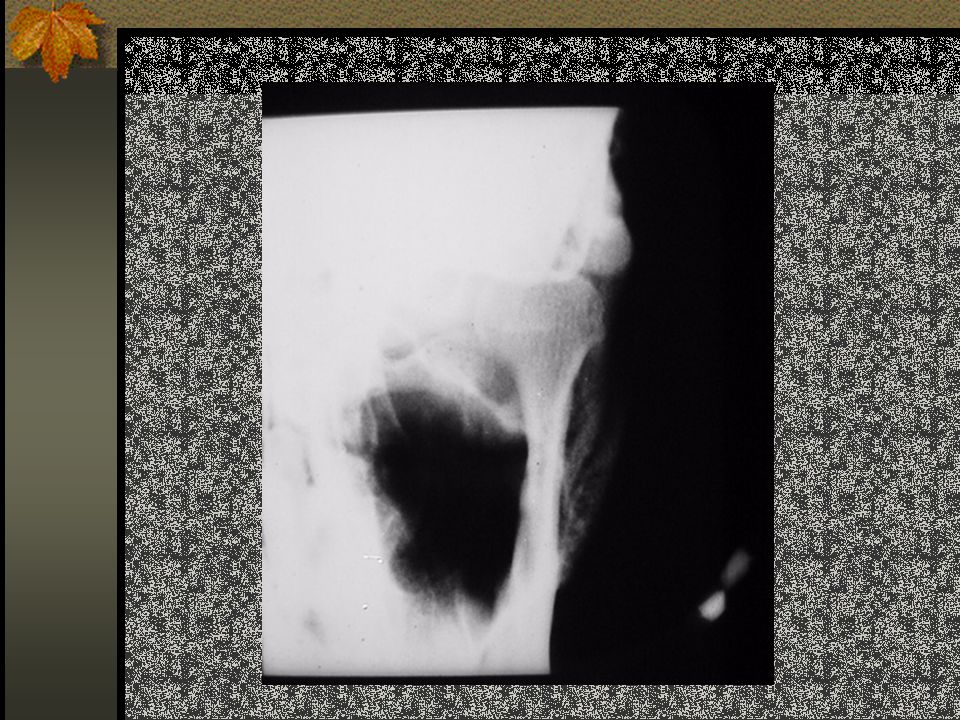
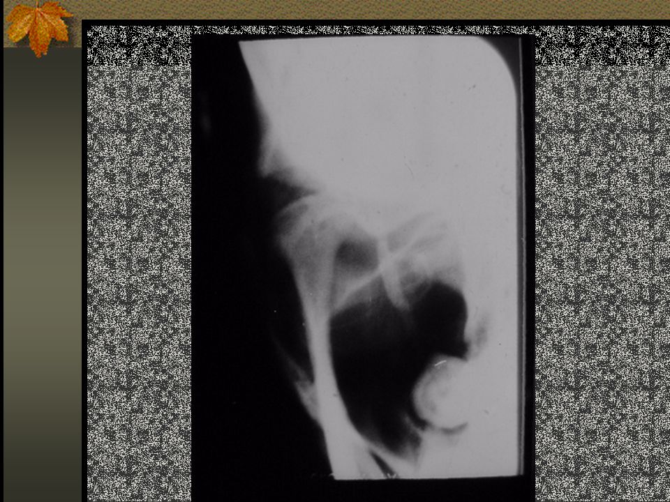
TMJ radiographs Lateral Transcranial Generally taken in “rest, closed, and open” positions Shows condylar morphology, lateral aspect Not good for evaluation of condylar position

6
TMJ radiography Transmaxillary Taken in open position Evaluates the superior aspect of the condyles from medial to lateral Taken through orbit, AP view

7
Lateral Transcranial Use TMJ Board X-ray tube position 11 deg. Down 1 in above and 1 inch behind EAM Align with opposite condyle

8
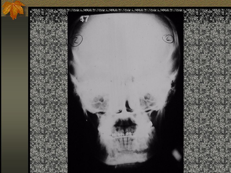
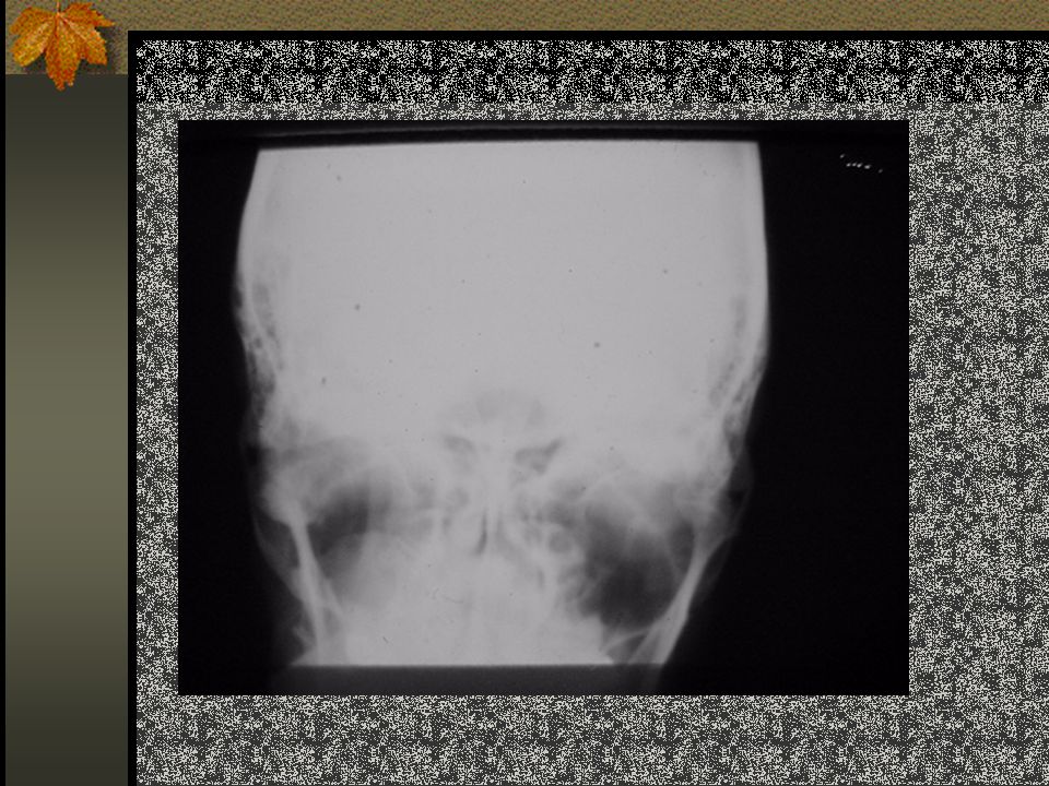
TMJ radiography PA Towne Taken in the “open” position Evaluates medial and lateral poles of condyle Patient must be able to open adequately for this film to be of any use

9
TMJ Tomogram Lateral Evaluates condylar position and condylar morphology Lateral, Open view Evaluates condylar translation PA Tomogram

10
TMJ CT and MRI CT Evaluates osseous and soft tissue components Generally does not show the meniscus MRI Good for assessing the meniscus and soft tissue

11
TMJ Arthrography Demonstrates meniscus Must be done under fluoroscopy Radiopaque dye injection Contraindicated with iodine sensitive patients

12
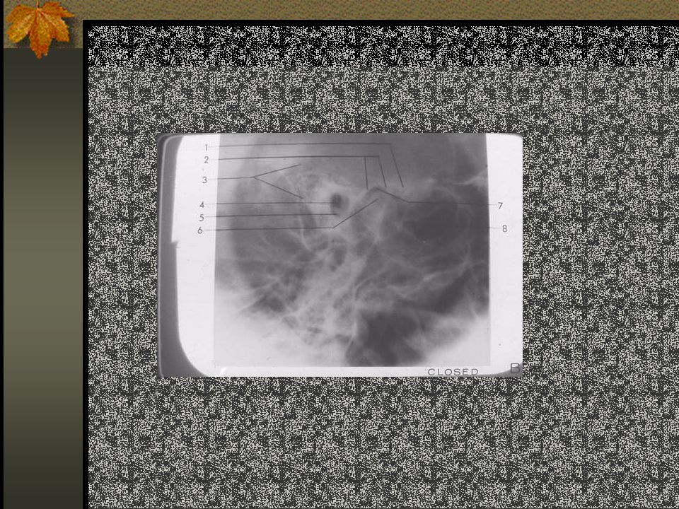
Lateral transcranial radiograph Closed Open Rest

13
Lateral Transcranial – closed view

41
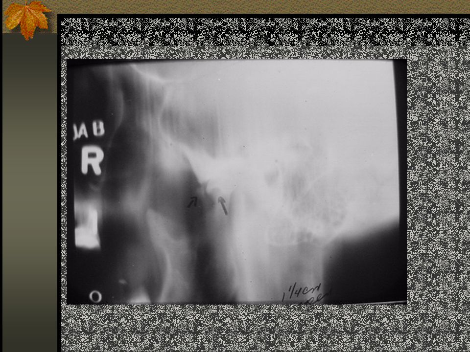
Transpharyngeal (Parma) view
 view")
42
MRI

43
MRI - TMJ

48
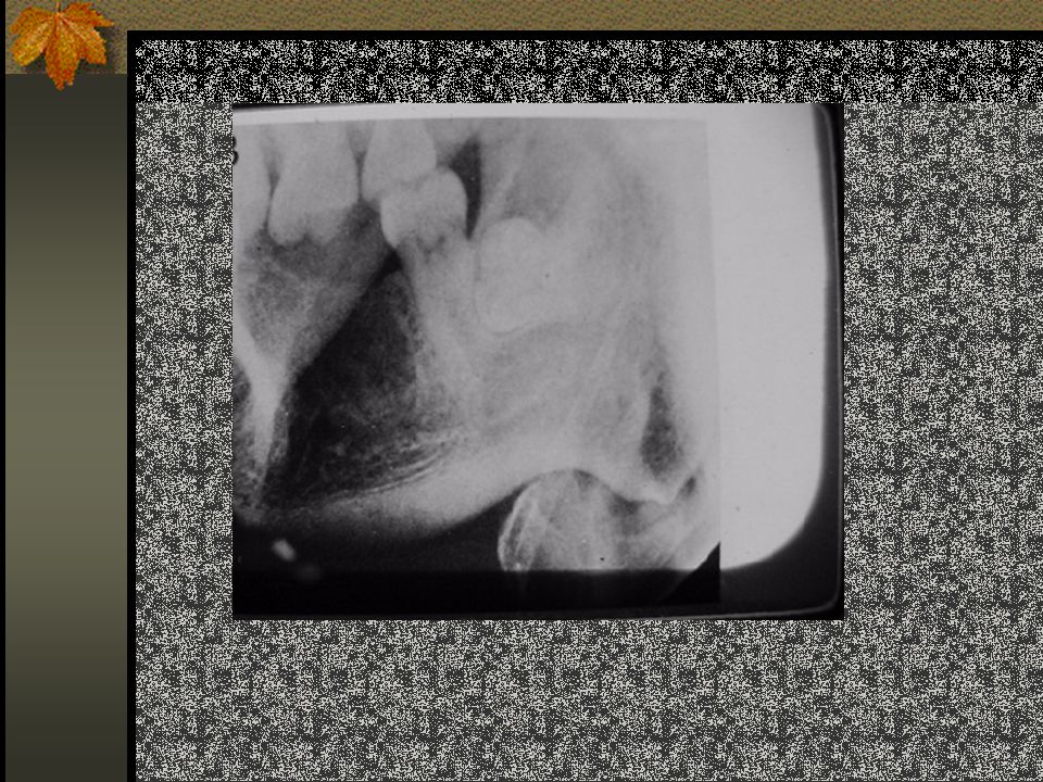
Flattening of the condyle

49
TMJ Radiography Jon K. Park, D.D.S., M.S. University of Maryland School of Dentistry Department of Oral Medicine Baltimore, MD 21201

Similar presentations