Download presentation
Presentation is loading. Please wait.

1
Slackers Guide to Blood and Urine Mike Ori

2
Disclaimer These represent my understanding of the subject and have not been vetted or reviewed by faculty. Use at your own peril. I can’t type so below are common missing letters you may need to supply e r l I didn’t use greek letters because they are a pain to cut and paste in.

3
List the conceptual classifications of anemia

4
Production Destruction (hemolysis) Loss (bleeding)
 Loss (bleeding)")
5
What is haptoglobin and what does a decrease in levels imply

6
A serum protein that binds free hemoglobin. Decrease is typically due excess hemoglobin in the blood as would be found in hemolytic anemia

7
What are the two categories of hemolytic anemia

8
Intrinsic – An issue with the RBC itself such as structural defects, enzyme defects, hemoglobin synthesis Extrinsic – A problem that arises external to the RBC but that impacts its integrity. – Antibodies – Mechanical damage

9
Differentiate the morphology of target cells, spherocytes, and schistocytes

10
Target cells are RBC’s with a small peak in the center of the central pallor Spherocytes are RBC’s that have lost cell membrane and thus loose their biconcave disk structure and its associated central pallor Schistocytes are RBC’s that have been fragmented

11
Distinguish heinz bodies, bite cells

12
Heinz bodies are condensations of hemoglobin

13
Describe hereditary spherocytosis

14
A disease of the spectrin substructure of RBC membranes due to mutations in ankyrin or spectrin itself. Splenomegaly Hemolytic anemia Reticulocytosis

15
Describe Glucose-6-phosphate dehydrogenase deficiency’s impact on RBC’s

16
Recall that G6PDH is important for oxidative repair of tissues. G6PDH defects occur in which the enzyme is labile as opposed to absent. In nucleated tissues, the enzyme is replaced but in the RBC it cannot be replaced. Therefore, enzyme concentration decreases more rapidly than in in wild-type individuals leading to more rapid accumulation of oxidative damage and premature RBC failure. Hemolysis occurs after an oxidative stress such as during illness or with fava bean ingestion

17
Describe sickle cell disease

18
Sickle cell disease occurs due to a single protein substitution in the beta globin chain. This mutation causes the affected B chains to polymerizes under oxidative stress. This leads to cell damage and the characteristic sickled shape. Membrane damage leads to shortened half-life. Sickle cell trait = heterozygous Sickle cell disease = homozygous

19
List the crisis types that can occur with sickle cell disease

20
Painful – due to occlusion of the microvasculature. Autosplenectomy and priapism Aplastic – sudden drop in RBC’s due to loss of RBC production as occurs with parvovirus infection in the marrow Sequestration – sludging in the spleen leads to accumulation of RBC’s

21
Why is sickle cell disease not typically apparent until late infancy?

22
Fetal hemoglobin production continues to occur giving a2g2 hemoglobin that is not susceptible to sickling. After about 6 months, a2B2 production increases and a2g2 decreases.

23
What is are cold and warm antibodies?

24
Cold antibodies are those that are active at temperatures less than that of the body. In contrast, warm antibodies are active at body temperature. IgG is typically a warm antibody while IgM is typically cold

25
Why do you care about cold and warm antibodies?

26
Cold antibodies typically do not cause severe hemolytic disease while warm antibodies can.

27
Distinguish megaloblastic from microcytic

28
Megaloblastic = MCV > 100 fl Microcytic = MCV < 75 fl

29
Define Reticulocyte distribution width

30
A number expressing the range in sizes of RBC’s found in a CBC. Megaloblastic, microcytic, schistocytes, spherocytes, reticulocytes all affect the RDW.

31
Define reticulocyte index

32
Measures the number of reticulocytes in blood corrected for HCT values and for maturation. Values > 2 indicate normal marrow response in anemia.

33
Distinguish anemias seen in chronic disease and in the following deficiencies: B12, Folate, Iron

34
B12 and folate deficiency cause a normochromic megaloblastic anemia. – B12 Neurologic defects Methylmalonic acid in urine – Folate No neuro deficits FIGlu in urine Iron deficiency causes a microcytic hypochromic anemia – Increased Total iron binding capacity (TIBC) – Decreased transferrin saturation – Decreased serum iron – Decreased stores (ferritin?) Anemia of chronic disease produces a normochromic normocytic anemia in response to increased IL-1, TNF-a, and IFN-g caused by chronic disease processes. – Normocytic normochromic to mild microcytic microchromic anemia – Decreased TIBC – Low serum iron – Normal stores (ferritin?)
 – Decreased transferrin saturation – Decreased serum iron – Decreased stores (ferritin ) Anemia of chronic disease produces a normochromic normocytic anemia in response to increased IL-1, TNF-a, and IFN-g caused by chronic disease processes. – Normocytic normochromic to mild microcytic microchromic anemia – Decreased TIBC – Low serum iron – Normal stores (ferritin ).")
35
Discriminate the causes of B12 deficiency

36
Inadequate amounts in diet Achloridia (failure to be freed in stomach) Lack of intrinsic factor (gastric parietal cells) Exocrine pancreas deficiency Ileal disease (leading to decreased absorption) In contrast to folate the body stores up to 5 years of B12
 Lack of intrinsic factor (gastric parietal cells) Exocrine pancreas deficiency Ileal disease (leading to decreased absorption) In contrast to folate the body stores up to 5 years of B12")
37
Describe aplastic anemia

38
Pancytopenia due to failure of the marrow. Most cases (65%) are idiopathic but any of the following are known causes – Viruses – Idiosyncratic drug reactions Chloramphenicol – Pesticides – Nuclear war Try slipping this one to the attending – Radiation Presumably from sources other than warheads – Fanconi’s anemia Microscopically the marrow will by hypocellular with fat infiltration
 are idiopathic but any of the following are known causes – Viruses – Idiosyncratic drug reactions Chloramphenicol – Pesticides – Nuclear war Try slipping this one to the attending – Radiation Presumably from sources other than warheads – Fanconi’s anemia Microscopically the marrow will by hypocellular with fat infiltration.")
39
Define red cell aplasia

40
Failure of the marrow to produce only RBC’s. Primary = idiopathic Secondary – Parvovirus – Chronic kidney disease with decreased EPO

41
What are the clinical manifestations of anemia

42
Acute – Anxiety and agitation – Headaches – Resting or orthostatic hypertension Light headedness Syncope – Diaphoresis – Systolic flow murmur (why?) Both – Pallor – Jaundice – Tachycardia, palpitations – Dyspnea
 Both – Pallor – Jaundice – Tachycardia, palpitations – Dyspnea")
43
List the blood transfusion products

44
IndicationAdult Packed RBCRaising hemoglobin1u = 1g/dl FFPAll clotting factors2 units PlateletsThrombocytopenia Platelet dysfunction 1 Single Donor 5-6 pooled 30-60k increase CryoprecipitateRestore fibrinogen Factor VIII or vWF 1 dose per 10kg Children dose at 10ml/kg

45
List the common transfusion reactions

46
Febrile – /\ T > 1C Allergic – Hives, itching, etc Hemolytic – Premature destruction of RBC’s Bacterial contamination – Especially platelets which are stored at room temp Transfusion Related Acute Lung Injury – Respiratory failure Fluid overload

47
What are the two types of hemostasis

48
Primary – Platelet aggregation – Early staunching of flow Secondary – Activation of coagulation cascade – Intrinsic, extrinsic, and common pathway

49
List the common coagulation tests

50
Prothrombin Time (PT) – Extrinsic pathway and common pathway Activated Partial Thromboplastin Time (aPTT) – Intrinsic pathway and common pathway D-Dimer – Fibrin split product specific for clots – Fibrin degradation products are less specific Mixing studies – Indicate whether a factor is missing or whether an inhibitor is present
 – Extrinsic pathway and common pathway Activated Partial Thromboplastin Time (aPTT) – Intrinsic pathway and common pathway D-Dimer – Fibrin split product specific for clots – Fibrin degradation products are less specific Mixing studies – Indicate whether a factor is missing or whether an inhibitor is present")
51
What is the presentation of inherited thrombophilias

52
First thrombosis < 50 yo Recurrent thrombosis Thrombosis at unusual site Life-threatening thromboembolism Family history

53
What is virchow’s triad

54
Stasis Endothelial injury Hypercoagulable state

55
List common genetic thrombophilia causes

56
Protein abnormalities – Factor V leiden Most common APC resistance due to mutation in the factor V – Protein C/S deficiencies Prothrombin Gene Mutation – Point mutation in gene at 20210 causes increased levels prothrombin Antithrombin Deficiency – Antithrombin inhibits several factors including thrombin (IIa).
.")
57
List the Acquired Thrombotic Disorders

58
Describe clotting effects of antiphospholipid antibodies

59
Common false positive with syphilis screening Paradoxical prolongation of aPTT – In vivo prothrombotic but in vitro anti-thrombotic Must have clinical disease in combination with antibody

60
Differentiate acute and chronic DIC

61
Acute – Widespread clotting results in decrease in clotting factors – Paradoxical bleeding, ischemia, multiorgan failure Chronic – Intermittent exposure to tissue factor – Compensated increase in clotting factors Treatment – Treat precipitating condition – Replace clotting factors or platelets as needed

62
What platelet levels are worrisome

63
Varies depending on condition. – 100k for surgery in closed spaces like eyes and brain – 50k for general surgery – 10k for general life

64
Differentiate the clinical sx of platelet v clotting factor bleeding

65
Platelet factors – generally present with petechiae, purpura, and mucocutaneous bleeding Clotting factor – bleeding presents with ecchymosis, hemarthrosis – Delayed bleeding when platelet plug disaggregates

66
What are the common groupings of thrombocytopenia

67
Decreased production Destruction Sequestration Dilution Artifactual

68
What is artifactual thrombocytopenia

69
Antiplatelet antibodies interact with platelets and EDTA preservatives to cause clumping of platelets that are not properly read by the CBC machines

70
What is HIT

71
Heparin induced thrombocytopenia

72
What is HIT Type I

73
HIT resulting from direct aggregation of platelets by heparin Rapid onset Often clinically benign – in contrast to type II – Consider prior type II HIT

74
What is type II HIT

75
Thrombocytopenia beginning 5-14 days after heparin exposure IgG reacts to heparin factor IV complex to cause platelet aggregation

76
What are the clinical manifestations, lab findings, and treatment of HIT

77
DVT and PE – Occasionally sever bleeding Lab findings – Thrombocytopenia – Platelet count 40-50% of pre-heparin level Treatment – DC heparin – Start anti-coagulant Do not start warfarin

78
Describe ITP, what are the types, what is the representative patient, treatment, etc

79
Idiopathic Thrombocytopenic Purpura – Isolated thrombocytopenia with no other explanation – IgG coat platelets which are then removed by spleen Acute – Children status post viral illness – Resolves spontaneously Restrict activity to avoid bleeding – Steroid therapy – IVIG Chronic – Adult females < 40 yo with no preceding illness – TX with steroids, IVIG, splenectomy

80
Describe TTP/HUS

81
TTP – Adult females – Deficiency in ADAMS13 prevents cleavage of long vWF multimers. – Long vWF multimers promote platelet aggregation – Neurologic issues HUS – Children following bacterial or viral infection with bloody diarrhea – Renal failure TX – Plasmaphoresis – Dialysis as necessary

82
What is the TTP/HUS pentad

83
Fever Microangiopathic hemolytic anemia Thrombocytopenia Neurologic deficits (TTP) Renal failure (HUS)
 Renal failure (HUS)")
84
What is the difference between qualitative and quantitative platelet disorders?

85
Qualitative – Normal quantity but deficient in function Quantitative – Abnormal number of platelets

86
Describe the role of glycoprotein Ib and IIb in platelet function

87
GPIb – Attaches platelet to vWF GPIIb – Attaches platelets to each other I before II

88
Describe the qualitative platelet disorders

89
Uremia – Mechanism unknown Bernard-soulier – Glycoprotein Ib defect Glanzmanns Thrombasthenia – GP IIb defect B before G

90
Describe Factor VIII deficiency (aka hemophilia A)
")
91
Defect in factor VIII X-linked Presentation/severity depends on residual activity – Severe < 1% – Moderate 2-5% – Mild 6-30% Before recombinant products, infectious disease was common If severe deficiency, immune response to FVIII can occur

92
Describes Factor IX deficiency

93
X-linked defect in FIX Clinically identical to Hemophilia A – 1/6 incidence of Hemophilia A

94
Describe vWF defects

95
Autosomal dominant Most common bleeding disorder vWF carries FVIII – Defect in platelet function predominates – Defect in coagulation cascade due to FVIII leads to aPTT increase

96
Describe vitamin K defects

97
Vitamin K is primary target for warfarin Vitamin Carboxylates II, VII, IX, X, C and S – Protein C has highest turnover so is affected first leading to hypercoagulation state in very early phase. Coagulation type bleeding

98
Where are most clotting factors synthesized

99
In the liver

100
What is the major zone of urinary continence in men and women

101
The external urethral sphincter

102
What additional factor is responsible for continence in females

103
Anterior vaginal wall and fascia Apposition of the urethra

104
Describe the role of the sympathetic and parasympathetic in urination

105
Sympathetic = storage Parasympathetic = pissing

106
Describe the neurotransmitter locations in the bladder

107
Acetylcholine = detrusor B2 = Bladder dome A1 = bladder neck

108
What are the fundamental classifications of urinary dysfunction

109
Wein classification – Failure to store urine – Failure to empty urine Due to… – Outlet – Bladder – Both

110
Describe detrusor overactivity

111
Involuntary bladder contractions – Failure to store urine due to bladder – Urge incontinence – Often idiopathic – Increases with age – M:F = 1:1

112
Describe urethral incompetence

113
Loss of function of the bladder outlet or urethra – Failure to store urine due to outlet – AKA sphincter incompetence Stress incontinence Causes – Radiation – Child birh – \/ estrogen

114
Describe overflow incontinence

115
Failure of the bladder to empty causes incontinence – AKA paradoxical incontinence – Wein classification = failure of the bladder to empty – Urine backs up in bladder until pressure overcomes urethral resistance. Continuous urinary leakage Causes – Neurogenic Areflexic – Obstructive Prostate

116
Describe mixed incontinence

117
Stress + urge incontinence History helps distinguish

118
List the conditions that result in continuous incontinence

119
Severe stress incontinence Overflow incontinence Urinary fistula Ectopic ureter

120
List the steps for evaluating Urinary Incontinence

121
Identify and treat reversible causes Identify complicating factors – Spinal cord injury – Urologic surgery Exclude overflow incontinence Classify type (stress, urge, continuous, etc) Trial of therapy
 Trial of therapy")
122
Describe Behavior Therapy of Incontinence

123
Behavior modification is first line therapy for both urge and stress incontinence Reduce fluid intake Eliminate bladder irritants like caffeine and alcohol Timed voiding Kegel exercises – Seriously there is a song for this and yes, it’s a bit lacivious – Start here http://www.youtube.com/watch?v=an3nheDsBXI http://www.youtube.com/watch?v=an3nheDsBXI – Then here http://video.google.com/videoplay?docid=- 8707040384334661843#http://video.google.com/videoplay?docid=- 8707040384334661843#

124
List the non-behavioral therapy for urge incontinence (and neurologic dysfunction)
")
125
Since urge incontinence is caused by detrusor over activity treatment should focus on reducing activity. Anti-muscarinincs – Not for areflexia Neuromodulation – Sacral – pacemaker – Tibial – weekly electro-acupuncture Botox – I still don’t get how it get there from the eye brows.

126
List the surgical strategies for managing neurologic urinary dysfunction

127
Catheters – Clean intermittent catheterization Not for Dysynergia as leakage would still occur – Suprapubic catheter – Sphincterotomy with condom catheter AKA Texas catheter Males only – Foley Augmentation cystoplasty – Making a bladder extension with a loop of bowel Urinary

128
List the non-behavioral therapy for stress incontinence

129
Female – Mid-urethral sling – gold standard – Urethral bulking agents – in-patient Male – Artificial sphincter – gold standard – Male urethral sling – Cunningham clamp – not loved by pts Meds – None

130
List the treatments for overflow incontinence

131
Bladder drainage Treatment of underlying cause – Neurologic – Obstructive

132
What are the general classifications of voiding dysfunction in neurologic damage

133
Detrusor overactivity Detrusor areflexia – Lack of contraction – Overflow incontinence Detrusor sphincter dyssnergia – Loss of coordination results in incomplete emptying – High post void residuals Urgency, frequency

134
List the likely symptoms for neurologic lesions above the brianstem

135
Detrusor over activity due to loss of control over PMC

136
List the urinary symptoms with brainstem lesions

137
Complex and variable…so not likely tested

138
List the symptoms of urinary dysfunction for suprasacral (above s2) vs sacral injury
 vs sacral injury")
139
Suprsacral (/\ s2) – Detrusor Sphincter dysynergia – Loss of PMC control Sphincter won’t relax Loss of coordination – Detrusor overactivity Detrusor controlled by primitive spinal reflexes Urgency, frequency Sacral (\/ s2) – Detrusor areflexia – Loss of PMC control Sphincter won’t relax – Loss of sacral control Detrusor won’t relax
 – Detrusor Sphincter dysynergia – Loss of PMC control Sphincter won’t relax Loss of coordination – Detrusor overactivity Detrusor controlled by primitive spinal reflexes Urgency, frequency Sacral (\/ s2) – Detrusor areflexia – Loss of PMC control Sphincter won’t relax – Loss of sacral control Detrusor won’t relax")
140
What are the symptoms of cystitis

141
Irritative voiding sx – Dysuria – Frequency – Urgency – Incomplete emptying Suprapubic pain Hematuria Malodorous urine Rarely fever/systemic sx

142
What is a colony forming unit and how is it applied in cystitis dx

143
CFU’s represent the number of viable bacterial cells in a sample. It is used in the diagnosis of UTI but standards are evolving – Old – 10 5 – Now – 10 2 – 10 4 if symptomatic Gram +/- may also be factored

144
What are the predisposing factors for cystitis in women

145
Short urethra Peri-urethral and vaginal flora Epithelial factors – Interact with bacterial pili to promote or retard colonization – Vaginal – Urethral Glucosamino-glycans NOTE: I recall this as a factor in both males and females

146
What are predisposing factors to cystitis in males

147
Obstruction – Prostate – Strictures Bladder dysfunction – Neurogenic – Why /\ males? Prostate surgery? Foreign body – Stones

148
What bacteria is associated with most UTI infections

149
E. coli = 80% (ambulatory) Fimbriae (mannose binding) P-pili (GAG binding)
 Fimbriae (mannose binding) P-pili (GAG binding)")
150
Describe the treatment of UTI

151
Uncomplicated – Antibiotics for 3 days – Fluoroquinolones preferred Recurrent – 2+ in six months or 3+ in one year – Longer ABX course – Preventative measures Post coital voiding Hydration Complete voiding

152
Differentiate chronic from acute prostatitis

153
Aside from chronic abacterial there is little difference between acute and chronic. SX same – LUTS – Fever – +/- enlarged prostate TX same – 4-6 weeks abx – Chronic add anti-inflammatory (why not in acute?)
.")
154
Describe the two glass test

155
Asks patient to provide urine sample The physician takes two double whiskeys Physician performs prostate massage Repeat urine collection Test 1Test 2Result +-Inconclusive? -+Bacterial prostatitis ++Cystitis --Abacterial prostatitis

156
Describe chronic non-bacterial prostatitis

157
Chronic LUTS and pelvic pain Multiple courses of abx with no relief WBC on prostate massage Treatment – Alpha blocker – NSAIDS – Hot Sitz bath – Muscle relaxants

158
What is the presentation of epididymitis

159
Adult males Frequently hemiscrotal discomfort with radiating flank pain Erythematous scrotum Prehns sign – Relief on scrotal elevation in supine patient

160
What are the likely pathogens of epididymitis and orchitis

161
Epididymitis – Chlamydia if < 35 yo – E. coli otherwise Orchitis – Gonorrhea – Chlamydia – mumps

162
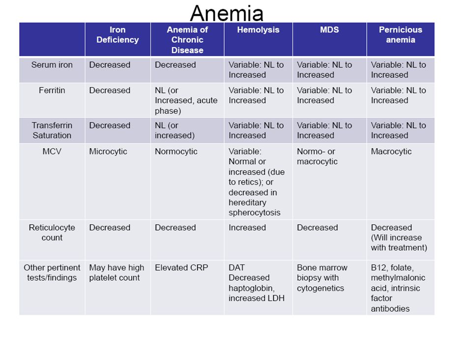
Summarize clinical anemias as presented in CPC 1

164
Differentiate Acute intermittent porphyria, porphyria cutanea tarda, congenital porphyria, and pseudoporphyria

165
Acute intermittent porphyria – Neurologic and GI involvement – Pain – Red urine due to excreted porphobilin – Autosomal dominant porphyria cutanea tarda – Defect in Uroporphyrinogen decarboxylase – Skin involvement with blisters – Autosomal dominant Congenital porphyria – Early childhood – Red teeth – Hypertrichosis (excessive hair) – Autosomal recessive Pseudoporphyria – Mimics tarda but without defects in genes – Drugs and UV exposure
 – Autosomal recessive Pseudoporphyria – Mimics tarda but without defects in genes – Drugs and UV exposure")
166
What are porphyrins

167
Precursors of heme The most important precursors for our purposes are – D-aminolevulinic acid step 1 product Urine test – Porphobilinogen step 2 product Urine test Acute intermitttent – Uroporphyrinogen III Step 4 product Tarda – FYI, all accumulate due to failure of step +1 enzyme

168
How do you treat acute intermittent porphyria

169
Hemin – Heme precursor that feeds back to stop d-ALA synthesis Glucose drip – Not sure why Stop triggers – Alcohol – Estrogen supplements

170
What is thalassemia

171
Autosomal recessive diseases that result in reduced rates (unbalanced rates) of hemoglobin synthesis. The subunits produced are fully functional but they are in short supply. This leads to an imbalance in the quantities of a/B and aberrant assembly of the subunits into tetramers. Abnormal tetramers predominate as defects in production become more pronounced.

172
Describe the layout and quantity of hemoglobin genes

173
Alpha – Two per chromosome for four total (epsilon excluded) – CR16 Beta – Multiple subclasses but only one per subclass per chromosome – CR11
 – CR16 Beta – Multiple subclasses but only one per subclass per chromosome – CR11")
174
Describe the defect in alpha thalassemia

175
Loss of transcription of alpha subunits. Most commonly this occurs due to frank deletion. Severity of disease increases with the number of alpha genes lost.

176
Describe the alpha thalassemia categories

177
Genotype (loci deleted) NamePhenotype 0Normal 1Asymptomatic carrierNormal 2Minor -Mild hypochromic microcytic anemia 3Intermedia AKA HbH disease Microcytic hypochromic, Heinz body precipitates, HbH( B 4 ), Hb Barts (g 4 ), occasional transfusions 4MajorHydrops fetalis
 NamePhenotype 0Normal 1Asymptomatic carrierNormal 2Minor -Mild hypochromic microcytic anemia 3Intermedia AKA HbH disease Microcytic hypochromic, Heinz body precipitates, HbH( B 4 ), Hb Barts (g 4 ), occasional transfusions 4MajorHydrops fetalis")
178
Describe the defect in beta thalassemia

179
Loss of transcription of beta subunits due to deletion or promoter defects. The disease increases in severity based on the extent of loss.

180
List the hemoglobin forms that appear in B thalassemia

181
HbA1a2B2a2B2 Normal decreases HbA2a2d2a2d2 Delta increase HbFa2g2a2g2 Fetal increase Mechanisms leading to increases in delta and fetal forms are not understood.

182
Describe the genotypes and phenotypes in B thalassemia

183
GenotypeNamePhenotype B/BNormal B + /B to B + /B 0 MinorMild Microcytic usually asymptomatic B + /B + to B + /B 0 IntermediaMicrocytic with occasional transfusions B + /B 0 to B 0 /B 0 MajorMicrocytic hypochromic anemia with regular transfusions. Hemochromatosis The important point here is to understand that the relative function is important. One gene functioning at 90% is better than two at 10%.

184
What is the treatment for hemochromatosis

185
iron chelators – Deferoximine – IV – Deferasirox – oral Fer = iron De = reverse/opposite Defer = un iron (wrinkle?)
")
186
What nutritional supplementation needs to be given to thalassemia patients?

187
Folate (b12 was not mentioned but seems sensible)
")
188
How prevalent are burns and who gets them

189
2-3 million/year Young, old, unlucky, careless

190
What is a 1 st, 2 nd, 3 rd, and 4 th degree burn?

191
First – Epidermis – Sunburns – No scars Second – Partial thickness of dermis Third – Full dermal thickness Fourth – Subcutaneous tissue involvement

192
Describe the zones of a burn

193
Coagulation – the central zone of dead tissue – Not rescuable Stasis – Damaged tissue but salvagable – Cytokines can cause loss of viability Hyperemia – Viable tissue (undamaged?) – Responding to cytokines but not likely killed by them ??
 – Responding to cytokines but not likely killed by them")
194
Describe the relation of burns and trauma

195
Many burns have underlying trauma due either to the mechanism of the burn or due to the victims actions while alight – leaping from a balcony – explosion, plane crash Look for and treat trauma

196
Describe fluid management in burns

197
Burn patients have significant insensible loss and edema. Massive fluid infusions are give in the first 24 hours to offset these losses.

198
When would you refer a patient to a specialized burn center

199
Age – 10% – Adult TBSA > 20% – > 50 yo and TBSA > 10% Location – Genitals and perinerum – Face – Across joints – Hands, feet Causative agent – Chemical – Electrical – Inhalation Other – PT comorbidities

200
What is the major determinant for grafting

201
Wound will not be healed by 3 weeks. Early excision \/ mortality

202
Why won’t systemic abx help with burns

203
Burns are devascularized so abx are unable to effectively penetrate the wound Topical abx are the way to go Early bacterial late fungal

204
Why is inhalation injury bad

205
Increases fluid requirement Increases mortality

206
What are the Bad things caused by burns

207
Decreased immune function Anemia – RBC t ½ = 40 days Edema Catabolism – Massive calorie requirement

208
What are the types of insults that can cause burns

209
Thermal – Heat – Cold Chemicals – Acids – Bases Electrical

210
How do electrical burns differ from thermal burns

211
Electrical burns often have small entry and exit wounds the belie the devastating injury that occurs below the surface as a result of the dissipation of electrical energy within the tissue. Technetium tests are useful for locating muscle damage. Thermal burns, aside from inhalation ones, are generally obvious.

Similar presentations