Download presentation
Presentation is loading. Please wait.

1
Dr.T.M.J.K.Tennakoon

2
Red cell indicies RBC number Hb% PCV- Red cell Mass MCV-Volume of a red cell MCH-Amount of haemoglobin in a red cell MCHC-Amount of haemoglobin in unit of red cell mass RDW-Indicates the different sizes of red cells

3
Total WBC count Neutrophils Lymphocytes Eosinophils Monocytes Basophils

4
Platelet count MPV PDW

5
Definition : Low haemoglobin level compare to the age and sex of the normal population. In Sri Lanka Adult Male – 12g% Adult Female- 11g% Children – 11g% 2 nd and 3 rd trimesters of the pregnancy- 10.5g%

6
Depends on the red cell size (MCV) Low MCV- Hypochromic microcytic anaemia. Normal MCV- Normocytic Normochromic Anaemia. High MCV- Macrocytic Anaemia.

7
Iron deficiency Anaemia Thalassemia Trait Sideroblastic Anaemia (Including Lead poisoning) Long standing Anaemia of Chronic disorder
 Long standing Anaemia of Chronic disorder")
8
Acute blood loss / Haemolysis Anaemia of chronic disorder Chronic Renal Failure Endocrine Disorders

9
Vit.B12 /Folate Deficiency Alcohol Excess Liver Disease Retculocytosis with blood loss or Haemolysis Cytoreductive treatment MDS /Aplastic Anaemia Hypothyroidism Anti Folate treatment

10
IDA Thal Trait RBC MCV MCH MCHC RDW or

11
In real life, both co-exisists Place for blood picture and iron studies S.ferritin level S.Iron TIBC Iron saturation% For confirmation of thalasemia- HPLC

12
ML is a 64-year old male who has not had any primary care for several years. When he tried to donate blood last week, he was told that he was anemic. He presents to your clinic for evaluation. What would you do??

13
HISTORY ◦ Is the patient bleeding? Actively? In past? ◦ Is there evidence for increased RBC destruction? ◦ Is the bone marrow suppressed? ◦ Is the patient nutritionally deficient? Pica? ◦ PMH including medication review, toxin exposure

14
REVIW OF SYMPTOMS Decreased oxygen delivery to tissues ◦ Exertional dyspnea ◦ Dyspnea at rest ◦ Fatigue ◦ Signs and symptoms of hyperdynamic state Bounding pulses Palpitations ◦ Life threatening: heart failure, angina, myocardial infarction Hypovolemia ◦ Fatiguablitiy, postural dizziness, lethargy, hypotension, shock and death

15
PHYSICAL EXAM Stable or Unstable? -ABCs -Vitals Pallor Jaundice -hemolysis Lymphadenopathy Hepatosplenomegally Bone Pain Petechiae Rectal-? Occult blood

16
Initial Testing ◦ FBC (includes RBC indices) ◦ Reticulocyte count ◦ Peripheral blood smear
 ◦ Reticulocyte count ◦ Peripheral blood smear")
17
Iron Studies ◦ Ferritin ◦ TIBC ◦ % Saturation Urinalysis Upper and lower GI endoscopy referral

18
Blood loss? ◦ Age places him at risk for colon CA Decreased Production? ◦ Alcohol use, Iron deficiency Increased Destruction? ◦ “Darker urine” lately

19
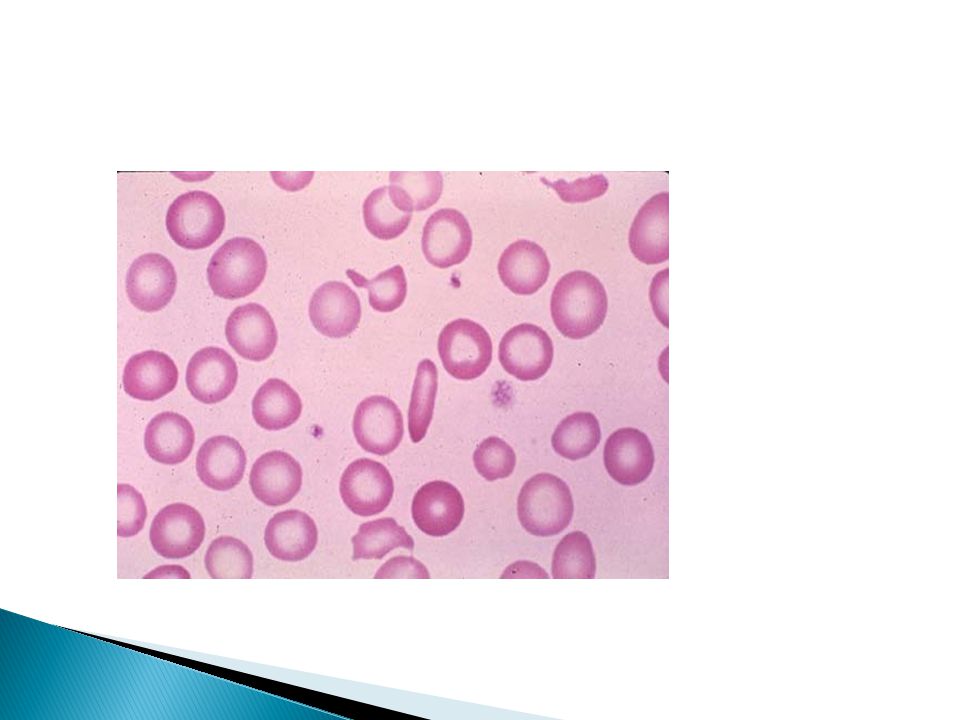
FBC, Hb-9.5g%,MCV-72fl, MCH-25pg WBC-8200/ml, Plt- 221000/ml Smear reveals microcytic, hypocrochromic RBCs Retic count is normal Urinalysis -normal Iron Studies ◦ Ferritin: 10 ◦ TIBC: 350 ◦ % Sat: 15

22
Rule out Sources of Bleeding ◦ FOB ◦ Upper and lower GI endoscopies Consider oral iron therapy Dietary counseling (iron sources, limiting alcohole, etc)
")
23
Colonoscopy revealed small suspicious lesion in sigmoid colon. Histo- adenocarcinoma. Excised surgically, no mets.

24
Routine FBC, one year later, reveal Hb of 12g%. He feels “better than ever”!

25
Anemia is a sign, not a disease. Anemias are a dynamic process. Its never normal to be anemic. The diagnosis of iron deficiency anemia mandates further work-up.

26
A 21-year-old married woman was referred for evaluation of anemia. History- recently married and wants to have a family. Feels lethargic. No history of melena or bleeding PR. Menstrual history- normal. Family history - anaemia O/E – pale. No icterus. CVS and RS are normal. Spleen-palpable. No edema. FBC: WBC= 4600, platelets 421,000/ul, Hb- 8.1g%, RBC- 5.2M/ul, MCV 59, (MCH) 17, MCHC- 32. Retic 3.1%, Ferritin 482 ng/ml, serum iron 149, transferrin 193, Iron sat 77%.
 17, MCHC- 32. Retic 3.1%, Ferritin 482 ng/ml, serum iron 149, transferrin 193, Iron sat 77%..")
28
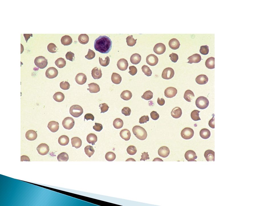
Ix: ◦ Smear: microcytic/hypochromic, misshapen RBCs HPLC- elevated HbA2. Dx- Thalassemia trait Tx: ◦ Mild: None ◦ Severe: RBC transfusions + Fe chelation, Stem cell transplants

29
Genetic defect in hemoglobin synthesis ◦ synthesis of one of the 2 globin chains ( or ) ◦ Imbalance of globin chain synthesis leads to depression of hemoglobin production and precipitation of excess globin (toxic) ◦ “Ineffective erythropoiesis” ◦ Ranges in severity from asymptomatic to incompatible with life (hydrops fetalis)
 ◦ Imbalance of globin chain synthesis leads to depression of hemoglobin production and precipitation of excess globin (toxic) ◦ Ineffective erythropoiesis ◦ Ranges in severity from asymptomatic to incompatible with life (hydrops fetalis)")
30
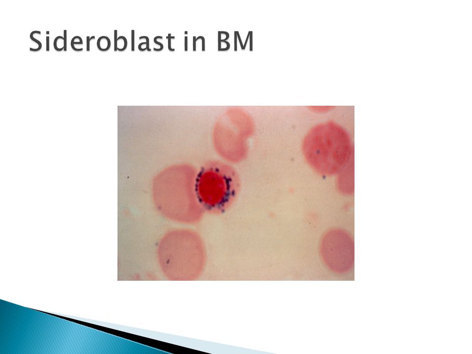
Heterogenous group of anemias Defined by presence of ringed sideroblasts in the BM Etiologies: ◦ Hereditary (rare), ◦ Some types of porphyria ◦ Myelodysplasia ◦ Ethanol ◦ Drugs (INH, Chloramphenicol) Tx: ◦ Trial of pyridoxine for hereditary or INH induced SA
, ◦ Some types of porphyria ◦ Myelodysplasia ◦ Ethanol ◦ Drugs (INH, Chloramphenicol) Tx: ◦ Trial of pyridoxine for hereditary or INH induced SA")
32
55 yr, Female with moderately severe Rheumatoid Arthritis on Prednisolone, Celecoxib and MTX with FA, is referred to you for evaluation of anemia. FBC: Hb = 9g%, MCV = 82, WBC = 5400/ l, plt = 345 000/ l Smear – Normochromic normocytic anaemia Retic count = 2 % Ferritin = 330 g/dL (20-160)
.")
33
A-- Marrow Failure B-- Iron Deficiency C-- Thalassemia D– Anaemia of chronic disorder.

36
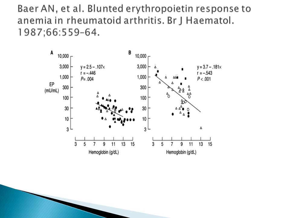
erythropoietin Utility of supraphysiologic doses of erythropoietin in the setting of Anaemia of chronic disorder.

39
A 69 yr old woman with progressive anemia with macrocytosis. FBC- leukopenia and thrombocytopenia. BP- hypersegmented granulocytes. Neurologic examination – normal Serum folate - normal. Erythrocyte folate level - below the normal range. Which statement is true? The patient has folic acid deficiency The serum follate is normal because the patient has taken a green salad in the past 24 hrs These findings are not specific to folate def. and a B12 level should be checked A bone marrow test would be helpful A serum epo level should be checked

40
Etiology: ◦ Anemia-- Vitamin B12 and folate are needed for DNA synthesis deoxyuridate to thymidylate, including RBC precursors ◦ Causes for B12 deficiency - Dietary intake, acid-pepsin in the stomach, pancreatic proteases, gastric secretion of intrinsic factor, an ileum with Cbl-IF receptors Causes for Foliate deficiency - Poor dietary intake alcohol, malabsorption, increased demand (pregnancy, hemolytic anemias),
,")
41
Ix : ◦ Smear: Macrocytic (High MCV) RBCs, +/- hypersegmented neutrophils, +/- modest neutropenia, but… ◦ B12 Low serum B12, elevated serum methylmalonic acid levels Anti-IF Abs, Schilling test (?), PA accounts for 75% ◦ Folate Serum folate level-- can normalize with a single good meal
 RBCs, +/- hypersegmented neutrophils, +/- modest neutropenia, but… ◦ B12 Low serum B12, elevated serum methylmalonic acid levels Anti-IF Abs, Schilling test ( ), PA accounts for 75% ◦ Folate Serum folate level-- can normalize with a single good meal")
42
Tx: ◦ B12 deficiency: B12 1 mg IM. EOD for 6 doses and repeat monthly. ◦ Folate deficiency: Improved diet, folate 1 mg/day ◦ Monitor the response. ◦ Pernicious Anemia – monitor for GI cancers.

43
A 32-year-old woman with Crohn’s disease for 15 years. A recent flare beginning 2 weeks ago was treated with sulfasalazine and corticosteroids. Despite improvement in diarrhea and abdominal pain, she continues to feel ill and experiences easy fatigability with dyspnea and palpitations on mild exertion. On physical examination, pallor, trace scleral icterus, and active bowel sounds are noted. Laboratory studies show: Hb =8g%; WBC= 14,000/ul. (90% neutrophils with left shift ) reticulocyte count= 7%; platelets =Normal
 reticulocyte count= 7%; platelets =Normal.")
45
Hemolytic anemias are either acquired or congenital. The laboratory signs of hemolytic anemias include: 1. Increased LDH (LDH1) - sensitive but not specific. 2. Increased indirect bilirubin - sensitive but not specific*. 3. Increased reticulocyte count - specific but not sensitive 4. Decreased haptoglobin - specific but not sensitive. 5. Urine hemosiderin - specific but not sensitive. *The indirect bilirubin is proportional to the hematocrit, so with a hematocrit of 45% the upper limit of normal is 1.00 mg/dl and with a hematocrit of 22.5% the upper limit of normal for the indirect bilirubin is 0.5mg/dl. Since tests for hemolysis suffer from a lack of sensitivity and specificity, one needs a high index of suspicion for this type of anemia
 - sensitive but not specific. 2. Increased indirect bilirubin - sensitive but not specific*. 3. Increased reticulocyte count - specific but not sensitive 4. Decreased haptoglobin - specific but not sensitive. 5. Urine hemosiderin - specific but not sensitive. *The indirect bilirubin is proportional to the hematocrit, so with a hematocrit of 45% the upper limit of normal is 1.00 mg/dl and with a hematocrit of 22.5% the upper limit of normal for the indirect bilirubin is 0.5mg/dl. Since tests for hemolysis suffer from a lack of sensitivity and specificity, one needs a high index of suspicion for this type of anemia.")
46
Thank you

Similar presentations