Download presentation
Presentation is loading. Please wait.

1
Histology

2
Body Tissues Cells are specialized for particular functions Tissues Groups of cells with similar structure and function Extracellular Matrix “cell glue” between cells Histology study of tissue

3
Body Tissues Four primary types (functional categories) Epithelium protection /secretion /absorption/ filtration Connective support & structure Nervous communication & control Muscle movement (internal & external)
 Epithelium protection /secretion /absorption/ filtration Connective support & structure Nervous communication & control Muscle movement (internal & external)")
4
Primary Germ Layers Endoderm (Epithelial) Mesoderm (Epithelial, Muscle, Connective) Ectoderm (Epithelial, Nervous) Digestive & respiratory epithelium MusclesEpidermis Urethra epitheliumSkeleton (bones & cartilage)Lining of mouth, anus, nostrils BladderBloodSweat & sebaceous glands Liver & pancreasBlood vessel epitheliumHair DermisBrain & spinal cord Excretory & reproductive organsEyes, nose, ear epithelium
 Mesoderm (Epithelial, Muscle, Connective) Ectoderm (Epithelial, Nervous) Digestive & respiratory epithelium MusclesEpidermis Urethra epitheliumSkeleton (bones & cartilage)Lining of mouth, anus, nostrils BladderBloodSweat & sebaceous glands Liver & pancreasBlood vessel epitheliumHair DermisBrain & spinal cord Excretory & reproductive organsEyes, nose, ear epithelium")
5
EPITHELIAL TISSUES

6
Epithelial Functions Protection Sensory Secretion Absorption Excretion

7
Epithelium Characteristics High cellularity cells fit closely together very little EC matrix Contains specialized contacts tight junctions & desmosomes Avascular no blood vessels within it diffusion provides nutrients & carries waste away lots of nerve fibers Reinforcement & connection Defines boundaries cancer causes a breach in these boundaries Regenerate easily if well nourished Found in areas of high friction

10
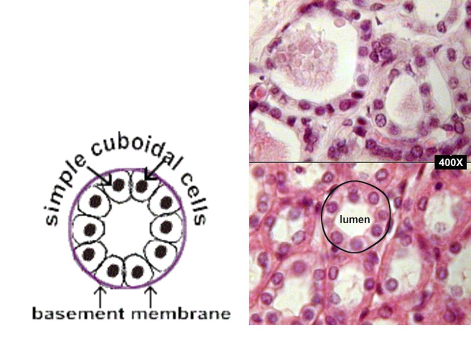
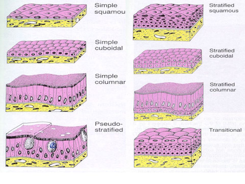
Classification of Epithelium Combination of shape & # of cells is used to name tissues Shape of cells Squamous – flattened Cuboidal – cube-shaped Columnar – column-like

11
Classification of Epithelium Number of cell layers Simple one layer found in areas of transport Stratified more than one layer High abrasion areas for protection Secretory membranes

12
Classification of Epithelium Pseudostratified “False” layers Ciliated (respiratory tract) Non-ciliated (male urethra) Transitional multiple layers of epithelial cells, “hodge-podge” Found in urinary tract can look cuboidal until bladder stretches, then looks squamos
 Non-ciliated (male urethra) Transitional multiple layers of epithelial cells, hodge-podge Found in urinary tract can look cuboidal until bladder stretches, then looks squamos")
14
Keratinized Epithelium Keratin tough waterproof material found in upper layers of some stratified squamos epithelium

15
Non-Keratinized Epithelium

16
Epithelial Membranes Mucous membranes Line body cavities OPEN to the exterior Example: digestive, respiratory, urogenital Mucus protects by trapping microorganisms, substances in mucus will destroy them Cutaneous Membranes Skin Helps waterproof & protect body First line of defense in immune system Serous Membranes Lines all CLOSED body cavities Serous fluid located between layers to reduce friction due to organ motion

17
Epithelial Membranes

18
Glandular Epithelium Gland one or more cells that secretes a particular product Two major gland types Endocrine gland Ductless – secretes product directly into blood or tissue Produces hormones Exocrine gland Empty through ducts to the epithelial surface Include sweat and oil glands

19
Glandular Epithelium Apocrine secrete their product from intact cells Ex: Mammary glands Holocrine entire cells and their secretions accumulate as the gland’s secretory product cells rupture Ex: Sebaceous (oil) glands Merocrine Most common accumulate their secretory product at the apical surface of each cell, which then separates from the remainder to form a secretion in the lumen of the gland. cells then repair themselves. Ex: Salivary glands

20
Glandular Epithelium

21
Connective Tissue Support & strengthen & provide structure

22
Connective Tissue Found everywhere in the body Includes the most abundant and widely distributed tissues Functions Binds body tissues together Supports the body Provides protection Insulates to maintain body temperature Transportation of other molecules

23
Origin of Connective Tissue Mesenchyme: embryonic tissue that differentiates into all forms of CT

24
Connective Tissue Characteristics Variations in blood supply Some tissue types are well vascularized Adipose – danger of hemorrhage with liposuction Some have poor blood supply or are avascular Blood supply is necessary for healing brings oxygen & “spare parts”

25
Extracellular Matrix Two main elements Ground substance proteins and polysaccharide molecules function as a molecular sieve to diffuse nutrients & other substances Fibers Produced by the cells Three types Collagen fibers – tensile strength Elastic fibers – stretch with recoil Reticular fibers - support

26
Connective Tissue Cells Prefixes Fibro Osteo Chondro Hemo(cyto) Suffixes Blast – build the cells Cyte – cell Clast – breakdown the cells
 Suffixes Blast – build the cells Cyte – cell Clast – breakdown the cells")
27
Connective Membranes Synovial Lines spaces between bone and joint Secrete synovial fluid to reduce friction

28
Connective Tissue Outline Bone Cartilage Hyaline Fibrocartilage Elastic Perichondrium Fibrous Areolar (Loose connective) Adipose Reticular Dense fibrous Regular Irregular Blood Plasma Cells Eryhtrocytes Leukocytes Thrombocytes
 Adipose Reticular Dense fibrous Regular Irregular Blood Plasma Cells Eryhtrocytes Leukocytes Thrombocytes")
29
Connective Tissue Types - BONE Bone (osseous tissue) Composed of: Bone cells in lacunae (cavities) Hard matrix of calcium salts Large numbers of collagen fibers Osteon – primary anatomical and functional unit of compact bone Used to protect and support the body Hematopoiesis – formation of blood cells
 Composed of: Bone cells in lacunae (cavities) Hard matrix of calcium salts Large numbers of collagen fibers Osteon – primary anatomical and functional unit of compact bone Used to protect and support the body Hematopoiesis – formation of blood cells")
30
Connective Tissue Types - CARTILAGE Hyaline cartilage Most common cartilage Composed of: Abundant collagen fibers Rubbery matrix Avascular Found in Entire fetal skeleton Ventral ends of ribs Articular surface of bones

31
Fibrocartilage Highly compressible Strongest & most durable forms cushion-like discs between vertebrae, pubic symphysis, meniscus Connective Tissue Types - CARTILAGE

32
Elastic cartilage Provides elasticity, very flexible Supports the external ear, larynx Connective Tissue Types - CARTILAGE

33
Perichondrium Surrounds cartilage Connective Tissue Types - CARTILAGE

34
Connective Tissue Types - FIBROUS Areolar (Loose Connective) Most widely distributed connective tissue (surrounds blood vessels & nerves) Soft, pliable tissue Contains all fiber types Can soak up excess fluid Main function – to cushion and protect organs Types of fibers Collagenous - collagen Elastic – elastin, stretchable Reticular – very thin
 Most widely distributed connective tissue (surrounds blood vessels & nerves) Soft, pliable tissue Contains all fiber types Can soak up excess fluid Main function – to cushion and protect organs Types of fibers Collagenous - collagen Elastic – elastin, stretchable Reticular – very thin")
35
Connective Tissue Types - FIBROUS Adipose Matrix is an areolar tissue in which fat globules predominate Many cells contain large lipid deposits Functions Insulates the body Protects some organs Serves as a site of fuel storage

36
Adipose Connective Tissue Types - FIBROUS

37
Reticular Delicate network of interwoven fibers Holds together adipose tissue Forms stroma (internal supporting network) of lymphoid organs; soft skeleton Lymph nodes Spleen Bone marrow Connective Tissue Types - FIBROUS
 of lymphoid organs; soft skeleton Lymph nodes Spleen Bone marrow Connective Tissue Types - FIBROUS")
38
Dense fibrous Main matrix element is collagen fibers Cells are fibroblasts Irregular – not parallel dermis Regular - parallel Tendons – attach muscle to bone Ligaments – attach bone to bone Connective Tissue Types - FIBROUS Regular Irregular

39
Connective Tissue Types - FIBROUS

40
Connective Tissue Types - BLOOD Fibers are visible during clotting Functions as the transport vehicle for materials 55% Liquid component plasma 45% Blood cells Erythrocytes - RBC Leukocytes - WBC Thrombocytes - platelets

41
Connective Tissue Outline Bone Cartilage Hyaline Fibrocartilage Elastic Perichondrium Fibrous Areolar (Loose connective) Adipose Reticular Dense fibrous Regular Irregular Blood Plasma Cells Eryhtrocytes Leukocytes Thrombocytes
 Adipose Reticular Dense fibrous Regular Irregular Blood Plasma Cells Eryhtrocytes Leukocytes Thrombocytes")
42
Muscle Tissue Highly vascular & highly cellular Less matrix = more flexibility More blood flow = more ATP made Actin & myosin – contractile myofilaments Function is to produce movement 3 types Skeletal Smooth Cardiac

43
Muscle Tissue Types Skeletal muscle Can be controlled voluntarily Cells attach to connective tissue Cells are striated Cells have more than one nucleus attached to bone Smooth muscle Involuntary muscle Surrounds hollow organs Attached to other smooth muscle cells No visible striations One nucleus per cell Viscera of hollow internal organs

44
Cardiac muscle Found only in the heart Function is to pump blood (involuntary) Cells attached to other cardiac muscle cells at intercalated disks Cells are striated One nucleus per cell Muscle Tissue Types
 Cells attached to other cardiac muscle cells at intercalated disks Cells are striated One nucleus per cell Muscle Tissue Types")
45
Nervous Tissue Consists of brain, spinal cord, nerves Carry electrical signals Neurons: generate & conduct electricity Usually nonregenerative Neuroglia: support neurons

46
Tissue Repair Determination of type of repair Type of tissue damaged Severity of the injury Regeneration Replacement of destroyed tissue by the same kind of cells Fibrosis Repair by dense fibrous connective tissue (scar tissue)
")
47
Regeneration of Tissues Regenerate easily Epithelial tissue Bone Regenerate poorly Skeletal muscle – replaced with connective tissue, not muscle Cartilage Nervous Replaced largely with scar tissue Cardiac muscle Nervous tissue within the brain and spinal cord Connective - keloids

48
Steps of Tissue Repair 1. capillaries dilate – brings blood to supply clotting factors 2. clot forms – seal off injury 3. scab forms – protect injury 4. debris is cleaned out – macrophages eat away damaged tissue to leave room for repair 5. Organization of tissue parts – granulation occurs (a type of intermediate tissue) 6. Macrophages digest & remove original clot 7. Surface epithelium regenerates – scab usually falls off at this time End result: Healed injury!
 6. Macrophages digest & remove original clot 7. Surface epithelium regenerates – scab usually falls off at this time End result: Healed injury!.")
49
Tissue Disease

Similar presentations


 Connective tissue Muscle tissue.>")


