Download presentation
Presentation is loading. Please wait.

1
Skin & Body Membranes Chapter 4

2
FACTS ABOUT THE SKIN Called the Integument: means covering.
Covers entire body. Wt. Approximately 9 pounds. 7% of total body wt. 4

3
Facts Cont. Every square centimeter of skin contains:
-70cm of Blood Vessels -55cm of nerves (230 sensory receptors) -100 sweat glands -15 oil glands -1/2 million cells dying and are constantly being replaced.
 -100 sweat glands. -15 oil glands. -1/2 million cells dying and are. constantly being replaced.")
4
I. Classification of Body Membranes

5
A. Epithelial Membranes
Epithelial sheet Underlying layer of connective tissue

6
1. Cutaneous Membrane Skin Keratinizing stratified squamous epithelium
Anchored by dense fibrous CT

7
2. Mucous Membranes Epithelium varies Underlying loose CT
Lines all cavities open to exterior

8
3. Serous Membranes Layer of simple squamous Layer of areolar CT
Lines body cavities closed to exterior Made of two layers Parietal: lines cavity Visceral: cover outside of organs in cavity Serous fluid in between layers Decrease friction

9
Figure 4.1 Classes of epithelial membranes

10
B. Connective Tissue Membranes
Synovial Membranes: soft areolar CT No epithelial cells Line fibrous capsules surrounding joints Provide smooth surface & secrete lubricating fluid

11
II. Integumentary System

12
A. Basic Skin Functions Mechanical Damage: keratin toughens cells
Pressure receptors alert nervous system to possible damage Chemical damage: keratinized cells relatively impermeable Pain receptors alert nervous system

13
Bacterial Damage: secretions are acidic & inhibit bacteria
Phagocytes ingest foreign substances & pathogens Ultraviolet radiation: melanin produced by melanocytes offer protection Thermal Damage: heat, cold, & pain receptors Desiccation: keratin in cells reduce evaporation

14
Heat loss: activating sweat glands
Allowing blood to flush into skin capillary beds Heat retention: reducing flow of blood into skin capillary beds Excretion of urea & uric acid in sweat Modified cholesterol molecules in skin converted to vitamin D by sunlight

15
B. Types of Nerve Endings
Cutaneous sensory receptors - (exteroceptors) - respond to stimuli outside the body. Meissners Corpuscles in dermal papillae - sense gentle touch and feel in the skin
 - respond to stimuli outside the body. Meissners Corpuscles in dermal papillae - sense gentle touch and feel in the skin.")
16
Pacinian receptors - deeper dermis - deeper pressure
Root Hair Plexuses - wind in the hair Bare nerve endings - sense cold, heat etc.

17
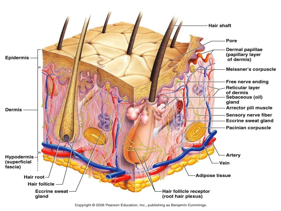
C. Structure of the Skin Most superficial: Epidermis
Second Layer: Dermis Hypodermis: not actual skin but is known as the fat layer of the skin. Underneath the dermis Mostly fat, insulate and absorb shock Anchors skin to underlying structures 5

19
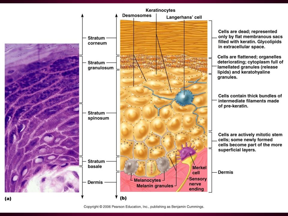
Epidermis: 4 Cell types 6

20
1. Keratinocytes: main structural cell…new epidermis every 35-45 days
Produce Keratin: fibrous protein used for protection. 2. Melanocytes: give skin color, accumulate on superficial side of keratinocytes. 3. Langerhans cells: macrophages of the immune system. 4. Merkel Cells: Combines with nerve receptors to form Merkel disc which is sensitive to touch. 7

22
5 layers of Epidermis (pg. 100 fig. 4.3)
1. Stratum Basale (Basal Layer) -Mostly young Keratinocytes, -One cell layer thick 8
 -Mostly young Keratinocytes, -One cell layer thick. 8.")
23
2. Stratum Spinosum (Spiny Layer) - Mostly Langerhans cells that
surround keratinocytes that are flat and prickly. -Keratinocytes in this layer contain tonofilaments: -Thick bundles of tension fibers 9

24
3. Stratum Granulosum (Granular Layer) -3-5 Layers of Keratinocytes
-Tough Layer -Water resistant, to slow water loss from the body 10

25
4. Stratum Lucidum (Clear Layer) -Only present in thick skin
-Mainly 2-3 rows of Keratinocytes 11

26
5. Stratum Corneum (Horny Layer)
-Outermost layer, mostly dead keratinocytes or keratin filled cells layers thick, thickest layer - prevent abrasion and penetration -Waterproofing from environment -Protects deeper cells 12

27
Dermis: 2nd Layer of Skin
Hide of our skin Richly supplied with nerves, blood & lymph vessels. Cell types are mostly macrophages & fibroblasts. House major portion of hair follicles, oil and sweat glands. 13

28
The dermis has 2 major layers
1. Papillary Layer: connect epidermis to dermis - Contain the majority of blood vessels. - Form identifying finger and foot prints. 14

29
2. Reticular Layer - 80% of the Dermis
- Dense irregular connective tissue - Form cleavage lines: skin heals better when an incision is made along these lines. 15

30
- If overstretched such as in pregnancy, dermal tears form striae or sretchmarks.
- Blister: Separation of Epidermis from Dermis, fluid filled.

31
D. SKIN COLOR 3 PIGMENTS CONTRIBUTE TO SKIN COLOR 16

32
1. MELANIN -Color ranges: yellow - reddish brown - black
-More melanin the darker the color. Freckles and moles are local accumulation of melanin. -Sun exacerbates melanin buildup 17

33
2. CAROTENE Color range: yellow - orange.
Found in certain plants: carrots, rich sources of Vitamin A. Most found in the palms and soles. 18

34
3. HEMOGLOBIN Color range: - Pale: caused by lack of blood.
Yellow: caused by build up of bile in the blood (Jaundice). Blue: cyanosis, lack of oxygen Pink: Normal hue 19
. Blue: cyanosis, lack of oxygen. Pink: Normal hue. 19.")
35
Bronze: Addisons disease, metallic appearance
Blue: Lack of Oxygen (cyanosis). Redness: Erythema, blushing, inflammation, or hypertension. Bruises: blood has escaped & clotted in tissue spaces 20
. Redness: Erythema, blushing, inflammation, or hypertension. Bruises: blood has escaped & clotted in tissue spaces. 20.")
36
E. APPENDAGES OF THE SKIN
21

37
1. HAIR AND HAIR FOLLICLES
- Hair (Pili): made of keratin Shaft: projects from the skin Root: embedded in skin, depending on shape signifies, straight or curly hair. - Hair pigment depends on melanocytes located in follicle 22
: made of keratin. Shaft: projects from the skin. Root: embedded in skin, depending on shape signifies, straight or curly hair. - Hair pigment depends on melanocytes located in follicle. 22.")
38
- Hair Follicles (pg. 105 fig. 4.7) - contains hair root
- Nerve endings - Knot of capillaries: papilla, to supply nutrients - Bundle of smooth muscle: arrector pili: raiser of hair. 23

40
2. Distribution of Hair growth
Fine Vellus: fine body hair of children or females Terminal hair: course scalp hair and eyebrows - grow in response to sex hormones, the more testosterone the more terminal hair 24

41
Male patterned baldness: genetic and sex-influenced condition
Alopecia: hair loss, as we get older new hairs do not replace old hairs as quickly. Male patterned baldness: genetic and sex-influenced condition 25

42
3. Nails Hard keratin 26

43
4. Sweat Glands sudoriferous glands
-Cover entire skin surface except for nipples, and parts of external genitalia. 2.5 million per person 27

44
Types of Sweat Glands Eccrine: most numerous
location: palms, soles of feet, forehead secretion: sweat 99% water, salts, nitrogenous wastes acidic pH 4-6 purpose: temperature regulation emotion induced sweating, we have no control 08/02/98 28

46
Apocrine: Location: axillary, genital areas
Size: Larger than eccrine glands Secretions: same as eccrine plus fatty deposits and protein. - Has foul odor when fats and proteins are decomposed (body odor) -Begin to function at puberty 31
 -Begin to function at puberty. 31.")
47
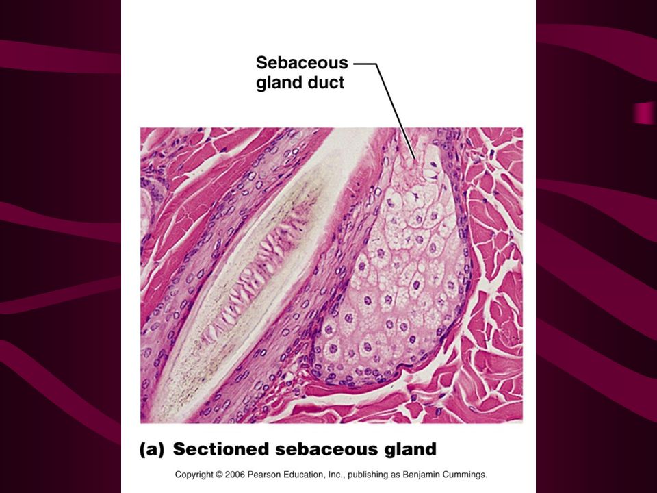
5. Sebaceous (oil) glands
Location: all over the body except for palms and soles of feet. Secretion: Sebum, oily substance Function: smooth and soften hair and skin and slows water loss during dry weather. Acne: active inflammation of gland Bacteria 34

49
Blocked duct: Whitehead is formed
if this oxidizes and dries it becomes a blackhead Seborrhea: (Cradle Cap) in infants is over secretion of sebaceous glands 35
 in infants is over secretion of sebaceous glands. 35.")
50
III. Homeostatic Imbalances
39

51
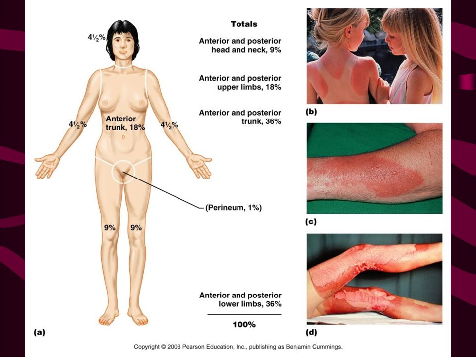
A. Burns Partial thickness burns - 1st and 2nd degree
Full thickness burns - 3rd degree 40

52
1. Problem with burns Fluid and electrolyte imbalance Shock Infection
41

53
2. Treatment Dependent on percentage of burn calculated by the Rule of nines. pg. 108 fig. 4.11 Fluid and electrolyte replacement Antibiotics Supportive care Debridment of eschar(burned skin) Grafting
 Grafting.")
55
B. Skin Cancer 1. Basal Cell Carcinoma
most common, least malignant, slow growth Pearly edge 99% cure rate with early excision

56
2. Squamous Cell Carcinoma
In keratinocytes of the stratum spinosum Scaly red papule (round elevated) rapid growth, meets to lymph Good cure rate if caught early and radiation is followed through.
 rapid growth, meets to lymph. Good cure rate if caught early and radiation is followed through.")
57
3. Malignant Melanoma CA of Melanocytes Most dangerous
Accounts for about 5% of Skin CAs Little chance of survival, better if caught early Tx is surgical excision with chemotherapy

59
4. American Cancer Society
ABCD rule for examination of skin CA A - Asymmetry B - Border irregularity C - Colors different D - Diameter is larger than 6mm (pencil eraser)
")
Similar presentations


 Largest organ of the body (15% of body weight) Skin thickness variable, normally 1-2 mm Protection –chemical barrier (waterproof)>")

 Weight = 4-5 kg (8-9lbs) 7% of body weight Thickness – 1.5-4.0 mm Millions rub off each day- New epidermis.>")