Download presentation
Presentation is loading. Please wait.

1
Cardiovascular Examination
Deling Zou

2
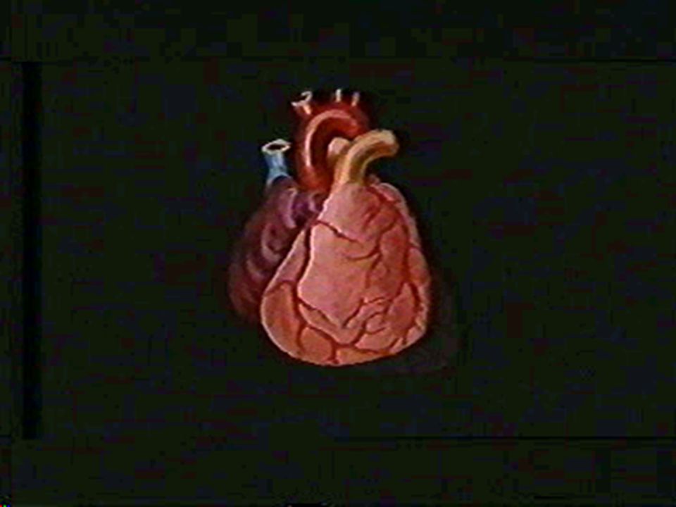
Anatomy

4
Inspection 1 Precardial projection and excavation 2 Apical impulse
3 Abnormal pulsations of precardium

5
Inspection 1 Precardial projection and excavation
congenital heart disease: tetralogy of Fallot Valvular heart disease MS,PS pericardial effusion (large , childhood)
")
6
The second right intercostal space(2nd ICS-RS)
aneurysm of aortic arch dilatation of ascending aorta 2) flat chest 3) pigeon chest/funnel chest
 flat chest. 3) pigeon chest/funnel chest.")
7
Inspection 2 Apical impulse *Normal:
position—the fifth left intercostal space cm medial to the midclavicular line range— cm in diameter

8
*Abnormal 1) Location #diaphragm:
“transverse position” upper,outward obesity ,child, pregnacy; ascites; tumor of abdominal cavity “vertical position” (thin, high, emphysema) inferior,inner
 inferior,inner.")
10
#mediastinum: one side pleural effusion or pneumothorax—to the healthy side one side atelectesis or pleural adhesion—to the affected

11
#enlargement of the heart
right ventricular dilatation –left or slightly upper left ventricular dilatation—left inferior LV &RV dilatation –left inferior (both side dilatation)
")
13
#Posture: recumbent position—upper
left lateral position—to the left 2-3cm right lateral position—to the right cm Dextrocardia: 5-ICS—RS

14
Inspection- apical impulse - abnormal 2)Intensity and extent changes
Intensity and extent changes")
15
-apical impulse - abnormal
Inspection -apical impulse - abnormal 3)Inward impulse: apex excavation in the systole seen: adhensive pericarditis prominent RV hypertrophy
Inward impulse: apex excavation in the systole. seen: adhensive pericarditis prominent RV hypertrophy.")
16
Inspection Abnomal pulsations of percardium
1)left third-forth intercostal space lateral to the sternum(3,4ICS-LS) seen: RV hypertrophy
left third-forth intercostal space lateral to the sternum(3,4ICS-LS) seen: RV hypertrophy.")
17
2)hypoxiphoid process seen: difference deep inspiration RV hypertrophy ↑ abdominal aorta (aneurysm) ↓
 ↓ .")
18
3)basal part of the heart
2 ICS-LS: dilatation of the pulmonary artery or pulmonary hypertensin, occasionally healthy young man 2 ICS-RS: aneurysm of aortic arch or dilatation of ascending aorta

19
Palpation 1 Apical impulse and pulsation of precardium 2 Thrill
3 Pericardial friction rub

20
1 Apical impulse and pulsation of precardium
Palpation 1 Apical impulse and pulsation of precardium Exact position of apex The beginning of systole of ventricle first sound Heaving apex impulse: reliable of LV hypertrophy

21
2 Thrill One of characteristic signs of organic heart disease.
Mechanism : the flow of blood→narrowed orifice→vortices→ vibration→chest wall thrill-high frequency murmurs-low frequency Method:position,phase of cardiac cycle,clinical significance seen: CHD or valvular stenosis , occasionally insurficiency

22
CHD:congenital heart disease

23
3 Pericardil friction rub
1)Precardium-4th ICS-LS 2) both phases of the cardiac cycle 3) systolic period, sitting erect and leaning forward, the end of expiration 4)mechanism: rub of the visceral and parietal layers of pleura 5)seen:acute pericarditis
Precardium-4th ICS-LS. 2) both phases of the cardiac cycle. 3) systolic period, sitting erect and leaning forward, the end of expiration. 4)mechanism: rub of the visceral and parietal layers of pleura. 5)seen:acute pericarditis.")
24
Percussion Aim:to determine the size and shape of the heart .
Absolute dullness: contain no gas Relative dullness : real size

25
1 murneuver of percussion
patient in erect position –the pleximeter is vertical with the intercostal space patient in the recumbent position –the pleximeter is parallel with the intercostal space

26
2 order : left—right ; upwards ; inward
left margin : from 2-3 cm lateral to the apex beat up to the 2nd ICS right margin : one intercostal space higher than the border of liver dullness up to the 2nd ICS size: vertical distance from margin to the anterior midline

28
Percussion

29
Percussion

30
(2)The upper border –the lower border of the anterior end of the third rib↑
(3)The basal part —the second intercostal space upward left: aortic node and PA (4)Concave part –between the aorta and the left ventricle
The basal part —the second intercostal space upward. left: aortic node and PA. (4)Concave part –between the aorta and the left ventricle.")
31
5 Changes in the area of cardiac dullness and its significance
Percussion 5 Changes in the area of cardiac dullness and its significance Cardiac factors : 1)LV enlargement: “boot shape” Seen:aortic valvular disease , hypertension heart disease
LV enlargement: boot shape Seen:aortic valvular disease , hypertension heart disease.")
32
2)RV enlargement : slightly↑--absolute dullness↑ Prominent↑--relative dullness↑ to the left side prominently Seen:PHD, MS 3)Two ventricle ↑: “generally enlarged heart” seen:DCM , Kashan cardiomyopathy
Two ventricle ↑: generally enlarged heart seen:DCM , Kashan cardiomyopathy.")
33
4)LA and/or pulmonary artery: LA:concave part disappear LA+PA:2,3 ICS-LS outwards
“pear shape” Seen: MS--- “mitrial type”

34
5)pericardial effusion: enlargement of both sides of the border
body’s position: recumbent position:widening of base of the heart erect position:“triangular shape”

35
6)dilatation of the aorta /ascending aortic aneurysm:
widening if the dull area of first and second intercostal space (with systolic pulsation)
")
36
Extacardial factors : 1)large pleural effusions and pneumothorax → to the healthy side 2)atelectasis /pleural pachynsis →to the affected 3)a large amount of ascites or big abdominal tumor: diaphragm elevated→transverse position →left side enlargement
large pleural effusions and pneumothorax → to the healthy side 2)atelectasis /pleural pachynsis →to the affected 3)a large amount of ascites or big abdominal tumor: diaphragm elevated→transverse position →left side enlargement")
37
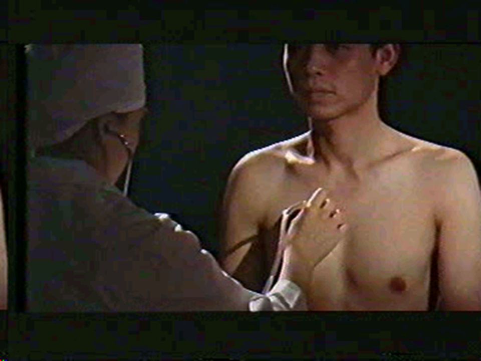
Ausclutation

38
1 Ausclutatoty valve areas
1)ausclutatory mitral area: apical area 2)ausclutatory pulmonary area:2 ICS-LS 3)ausclutatory aortic area: 2 ICS-RS 4)second ausclutatory aortic area: 3rd ICS-LS—Erb area 5)tricuspid area :4,5 ICS-LS
ausclutatory mitral area: apical area 2)ausclutatory pulmonary area:2 ICS-LS 3)ausclutatory aortic area: 2 ICS-RS 4)second ausclutatory aortic area: 3rd ICS-LS—Erb area 5)tricuspid area :4,5 ICS-LS.")
40
2 Order: MV---PV---AV1---AV2---TV 3 Contents : 1) rate 2)rhythm 3)heart sound 4)extra heart sound 5)murmurs 6)pericardial friction sound
 rate 2)rhythm 3)heart sound 4)extra heart sound 5)murmurs 6)pericardial friction sound.")
41
1)heart rate: 60~100bpm F>M child (<3 years) > 100bpm tachycardia: normal adult >100bpm child(<3 years) >150bpm bradycardia: HR <60 bpm

42
Ausclutation heart rate:60-100bmp

43
2)cardiac rhythm:. sinus arrythmia—affected by breath
2)cardiac rhythm: *sinus arrythmia—affected by breath *premature beat: classification:atrial~ ventricular ~ junctional ~ frequently:>6 bpm occasionally: <6 bpm bigeminy trigeminy
cardiac rhythm: *sinus arrythmia—affected by breath *premature beat: classification:atrial~ ventricular ~ junctional ~ frequently:>6 bpm. occasionally: <6 bpm. bigeminy trigeminy.")
44
*atrial fibrillation: absolute irregular rhythm S1 intensity inequality Pulse deficit seen:MS,CHD,hyperthyroidism, PHD,DCM

45
Ausclutation atrial fibrillation

46
3) cardiac sound
 cardiac sound")
47
Ausclutation content cardiac sound S1: S2:

49
4)Abnormal cardiac sound *Intensity:
position of the atrioventricular valve Ventricular contractility and output Valvular integrity and activity

50
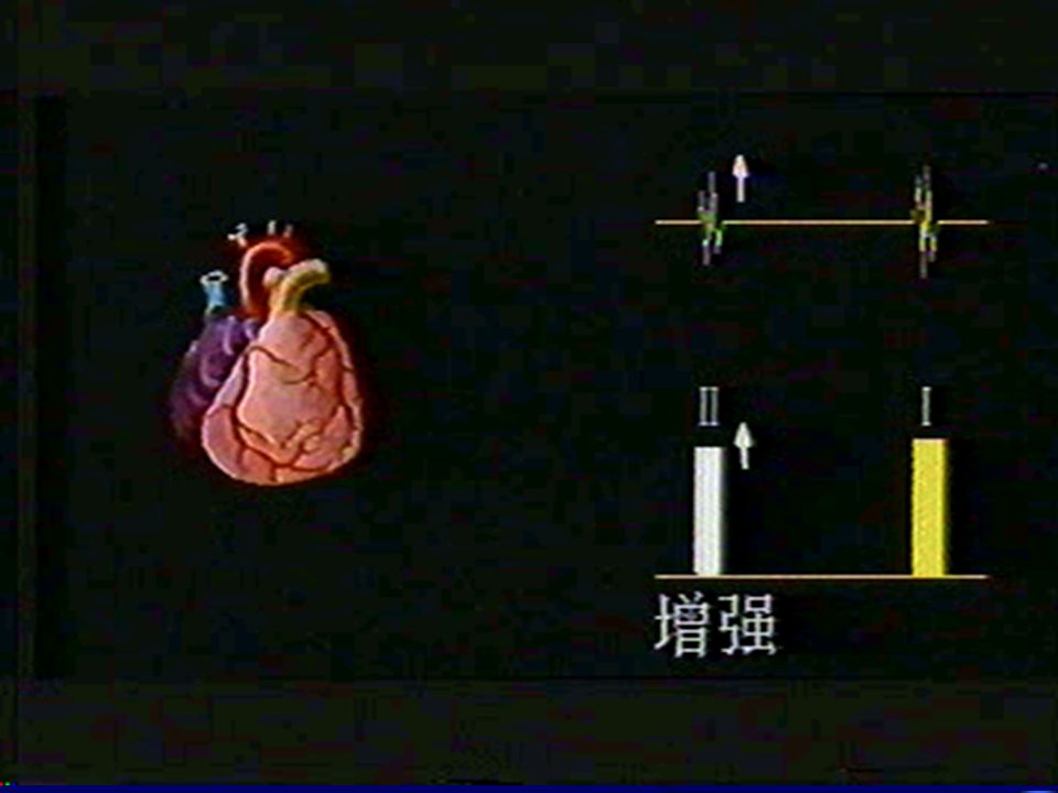
S1: Accentuation: MS HR↑contractility↑ fever,anemia,hyperthyroidism complete AVB →cannon sound

52
S1 attenuation : MI P-R interval enlong AI myocarditis,myopathy,MI,HF inequality: af, III°AVB

54
S2---A2,P2 S2 ↑ ---pressure and flow of blood ↑ A2 : hypertensin, arterisclerosis P2 : PHD,CoHD(L--R),LVF S2 ↓ ---pressure↓ flow ↓ Seen:hypotension,AS/AL,PS/PI

56
*Quality mono rhythm pendular rhythm---embryocardia *Splitting of heart sound S1 splitting: seen—RBBB, right heart failure Ebetein malformation ,MS LA myxoma

58
S2 splitting: (1)physiological splitting :end of inspiration (2)general splitting : most commonly seen: CRBBB, PS, MS,MI ,VSD (3)fixed splitting :ASD (4)paradoxical splitting(reversed splitting) :pathological seen: CLBBB ,AS, hypertension
physiological splitting :end of inspiration (2)general splitting : most commonly seen: CRBBB, PS, MS,MI ,VSD (3)fixed splitting :ASD (4)paradoxical splitting(reversed splitting) :pathological seen: CLBBB ,AS, hypertension")
60
5)extra cardiac sound Diastolic period 1)gallop rhythm: --protodiastolic gallop: S1+S2+S3 the third sound gallop (sign of organic heart disease) seen : HF(AMI, severe myocarditis , myopathy etc.) -- late diastolic gallop: atrial gallop S1+S2+S4 seen : HBP ,HCM ,AS ,CHD -- summation gallop: quadruple rhythm seen:HF,cardiomyopathy

63
5) extra cardiac sound Diastolic period
2)opening snap:MS 3)pericardial knock: constrictive pericarditis 4)tumor plop: LA myxoma
opening snap:MS 3)pericardial knock: constrictive pericarditis 4)tumor plop: LA myxoma.")
65
Ausclutation CONTENT Tumor plop

66
S1----mid<0.08″ late>0.08″ seen: mitral prolapse
Systolic period (1)early systolic ejection sound(click) pulmonary :pulmonary hypertension; pulmonary artery dilatation PS, ASD, VSD Aortic: hypertension, aneurysm , AS, AI ,aorta constriction (2)mid and late systolic click: S1----mid<0.08″ late>0.08″ seen: mitral prolapse
early systolic ejection sound(click) pulmonary :pulmonary hypertension; pulmonary artery dilatation PS, ASD, VSD Aortic: hypertension, aneurysm , AS, AI ,aorta constriction (2)mid and late systolic click: S1----mid<0.08″ late>0.08″ seen: mitral prolapse.")
69
iatrogenic (1)prosthetic valvular sound (2)pacemaker
prosthetic valvular sound (2)pacemaker")
70
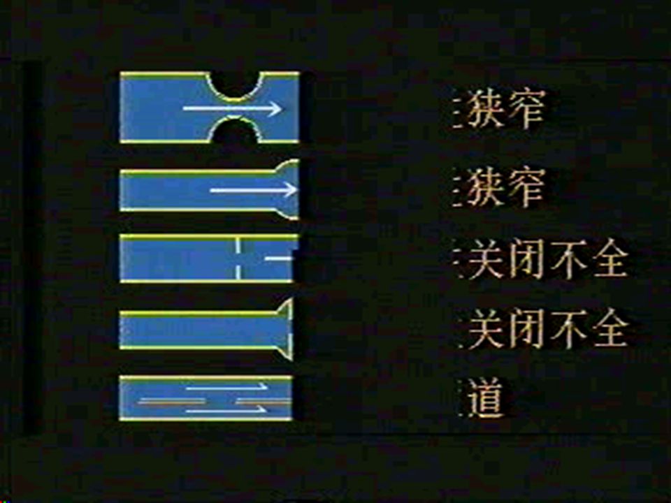
6)cardiac murmurs
cardiac murmurs")
72
*characterization of murmur and ausclutatory key points (1)location:L3,4 –VSD L2,3—PDA (2)transmission: MI ---left axilla AS---neck (3)phase: systolic murmurs diastolic ~ continuous ~ biphasic ~ early,mid,late,whole murmurs
phase: systolic murmurs diastolic ~ continuous ~ biphasic ~ early,mid,late,whole murmurs.")
73
(4)quality: blowing—MI rumbling—MS sighing--AI machinery--PDA
(5)intensity :Levine 6 grade classification shape: crescendo---MS decrescendo---AI crescendo-decrescendo---AS continuous---PDA regular---MI murmurs
intensity :Levine 6 grade classification shape: crescendo---MS decrescendo---AI crescendo-decrescendo---AS continuous---PDA regular---MI. murmurs.")
74
(6) others: body position: MS--left lateral position AI--sitting erected and forward MI,TI,PVS--lie on one’ back Lie → stand: HCM breath:expiration--LV murmurs inspiration --RV murmurs valsalva--HCM exercise: HR↑--murmurs ↑ murmurs

75
clinical significance murmurs: functional and organic 7)pericardial friction sound:
both phases , unaffected by respiration . seen: pericarditis , RHD ,AMI ,renal failure, SLE

76
* clinical significance of cardiac murmurs
systolic murmurs MV:functional:exercise,fever,anemia,pregnancy, hyperthyroidism relative:HBP,CHD,DCM,anemia organic:MI(RHD),mitral prolapse
,mitral prolapse.")
78
* clinical significance of cardiac murmurs
systolic murmurs Aortic area:organic:AS relative:dilatation of ascending aorta

80
* clinical significance of cardiac murmurs systolic murmurs
pulmonary :physiology relative:MS、ASD organic:PS TV:relative :RV enlarged organic :rare

81
* clinical significance of cardiac murmurs
Diastolic murmurs MV:organic:RHD(MS) relative:AI(severe) Austin Flint murmur AV:AI
 relative:AI(severe) Austin Flint murmur. AV:AI.")
84
* clinical significance of cardiac murmurs
Diastolic murmurs PV:organic murmur is rare PI(dilatation of pulmonary artery) MS+P Graham Steell murmur TV:rare
 MS+P Graham Steell murmur. TV:rare.")
85
* clinical significance of cardiac murmurs
continuous murmurs PDA innocent murmur

86
Vascular examination The second clinical hospital of CMU

87
pulse pulse rhythm tensions and state of arterial wall intensity
pulse rate pulse rhythm tensions and state of arterial wall intensity pulse wave

88
pulse pulse rate Atrial fibrillation and frequent premature beat stroke volume peripheral artery no pulse pulse rate less than HR(pulse deficit)
")
89
pulse pulse deficit; pulse rhythm bigeminal pulse,trigeminal pulse;
dropped pulse

90
pulse tensions and state of arterial wall
Artery tension depending on blood pressure (mainly SBP). Judge state of artery wall
. Judge state of artery wall.")
91
pulse intensity Bownding pulse seen:high fever, hyperthyroidism, AI
Microsphygmia seen:HF,AS and shock

92
pulse pulse wave normal pulse wave
composed of upstroke(knocking wave)、peak (tide wave)and downstroke(dicrotic wave)
、peak (tide wave)and downstroke(dicrotic wave)")
93
pulse pulse wave water hammer pulse seen:AI,hyperthyroidism,PDA, severe anemia pulse tardus seen:AS dicrotic pulse seen:HCM pulsus alternans seen:HBP,AMI,AI paradoxical pulse seen:cardiac tamponade,constrictive pericarditis Pulseless seen:serious shock, arteritis

94
blood pressure method of measurement direct measurement method
indirect measurement method

95
blood pressure standard
definition of Bp level and classification(older than 18 years old) classification SBP(mmHg) DBP(mmHg) Ideal BP 80 Normal BP 85 High limit of BP Grade 1(mild) subgroup: boundline hypertension Grade 2(moderate) Grade 3(severe) ≥ ≥110 Simple systolic hypertension 90 boundline systolic hypertension 90
 classification SBP(mmHg) DBP(mmHg) Ideal BP 120 80. Normal BP 130 85. High limit of BP Grade 1(mild) subgroup: boundline hypertension Grade 2(moderate) Grade 3(severe) ≥ 180 ≥110. Simple systolic hypertension 140 90. boundline systolic hypertension 90.")
96
blood pressure clinical significance of BP changes
hypertension:higher than 140/90mmHg for 3 times not in the same day hypotension:lower than 90/60-50mmHg Shock,,MI,acute cardiac tamponade obvious difference between bilateral upper limbs:more than 10mmHg---arteritis,congenital artery malformation difference between upper and lower limbs:lower limb BP is 20-40mmHg higher than upper one normally pathological:constrictive aorta ,arteritis(chest-abdominal aorta) change of pulse BP: 40mmHg,wide pulse BP---hyperthyroidism,AI 30mmHg,narrow pulse BP---AS,pericardial effusion
 change of pulse BP: 40mmHg,wide pulse BP---hyperthyroidism,AI. 30mmHg,narrow pulse BP---AS,pericardial effusion.")
97
blood pressure dynamic BP monitoring Average BP for 24h 130/80mmHg;
bright day 135/85mmHg; night: 125/75mmHg Peak:6am—10am,4pm—6pm

98
Vessel murmur and peripheral vessel sign
venous murmur jungular murmur:is caused by the rapid flow of jungular vein into SVC (superior vena cava)
")
99
Vessel murmur and peripheral vessel sign
artery murmur Continuous murmur in the lateral lobe of thyroid in the patient with hyperthyroidism Systolic murmur in the upper abnormal region or lumber region caused by stenosis of renal artery. Arterio-venous fistula

100
Vessel murmur and peripheral vessel sign
pistol shot sound Seen:AI,hyperthyroidism,severe anemia Durozier’s murmur capillary pulsation

101
The main symptoms and signs of common diseases of circulatory system

102
Mitrial stenosis Causes:
RHD:rheumatic heart disease CHD:congenital heart disease Other reasons: senile retrograde

103
Symptoms: hemoptysis;
cough; hemoptysis; dyspnea: dyspnea on exertion→ paroxysmal nocturnal dyspnea → pneumonedema

104
Palpation :diastolic thrill palpable over the apical area
Signs: Inspection : mitrial face Apex impulse may be displaced to the left Palpation :diastolic thrill palpable over the apical area Percussion : normal heart borders→pear shape heart

105
Auscultation : )the first sound (S1)↑ )diastolic murmur :apical area; localized; mild and late diastolic ;crescendo ;rumbling; more clearly when the patient is lying on his left side ) opening snap may be auscultatory 4)accentuation of second pulmonary sound (P2↑), splitting 5)Graham Steel’s murmur (PV diastolic) 6)Maybe atrial fibrillation(late stage)
 opening snap may be auscultatory 4)accentuation of second pulmonary sound (P2↑), splitting 5)Graham Steel’s murmur (PV diastolic) 6)Maybe atrial fibrillation(late stage).")
106
Mitral Insufficiency RHD / non-RHD ; acute/chronic Symptoms:
fatigue, palpitations, dyspnea on exertion, Left heart failure

107
Signs : Inspection : apex beat is displaced downwards and to the left

108
Palpitation : Percussion :
apical impulse forceful Heaving apex impulse Severe systolic thrill Percussion : the area of dullness to left and downwards

109
Auscultation : 1)S1 ↓(attenuation)
2)murmurs: harsh; pansystolic murmur; blowing; /6 grade ↑ wide spread-transmitted to left axilla left infrascapular angle
murmurs: harsh; pansystolic murmur; blowing; 3/6 grade ↑ wide spread-transmitted to left axilla left infrascapular angle.")
110
Aortic Stenosis Causes: RHD Congenital Senile retrograde Symptoms :
palpitation ,dizziness, angina pectoris, syncope, HF-dyspnea

111
Inspection : apical impulse increase Displaced to left and downwards
Signs : Inspection : apical impulse increase Displaced to left and downwards Palpation : apex beat is elevated and forceful systolic thrill can be palpated over aortic auscultatory valve area Pulse tardus

112
Percussion: Auscultation :
the area of dullness is normal or to left and downward Auscultation : 1)murmur: aortic auscultatory valve area systolic murmur harsh ,ejection sound , 3/6 grade ↑(thrill) transmitted to neck 2)A2 ↓,reversed splitting 3)S4
murmur: aortic auscultatory valve area systolic murmur. harsh ,ejection sound , 3/6 grade ↑(thrill) transmitted to neck 2)A2 ↓,reversed splitting 3)S4.")
113
Aortic Insufficiency Causes:
RHD Non-RHD:congenital prolapse syphilis aortitis arteriosclerosis endocarditis acute/chronic

114
Symptoms : Signs Inspection :
palpitation, dizziness, LHF Signs Inspection : apical impulse to left and downwards Palpation : apex impulse to left and downwards Heaving apex impulse

115
Percussion : the area of cardiac dullness is enlarged downwards and to the left; the concave part of the heart is not enlarged (boot shape)
")
116
Auscultation : 1)specific murmur:
diastolic ; sighing ; aortic area; heard clearly sitting erect and forward 2)Austin Flint murmur :relative MS (rumbling mid-diastolic murmur)
Austin Flint murmur :relative MS (rumbling mid-diastolic murmur)")
117
Peripheral vascular signs
*head bobbing (Musset’s sign):nodding motion of the head with each systole; *signs of capillary pulsation; *water hammer pulse; *pistol shot sounds : esp. Femoral arteries; *Duroziez’s murmur; *Visible pulsation of carotid arteries
:nodding motion of the head with each systole; *signs of capillary pulsation; *water hammer pulse; *pistol shot sounds : esp. Femoral arteries; *Duroziez’s murmur; *Visible pulsation of carotid arteries.")
118
Pericardial effusion Causes: Symptoms :
infective and non-infective pericarditis Symptoms : pain over the pericardial region Dyspnea, cough, fever, lassitude Shock

119
Signs : Inspection : diminution in strength of the apex beat or absence of the apex beat ; jugular venous enlargement

120
Palpation : *diminution in strength of the apex beat or the apex beat palpated uneasily *paradoxical pulse may be present

121
Percussion : enlargement of the cardiac dullness bilaterally, changed with posture

122
Auscultation : *pericardial friction sound
*HR↑,diminution of intensity of cardiac sound (S1/S2↓) *pericardial knock may be heard
 *pericardial knock may be heard.")
123
*Large effusion: Jugular varicosity Liver enlargement Paradoxical pulse Pulse pressure ↓

124
* Kussmaul sign: deep inspiration –jugular vein distension *Ewart sign: left infrascapular region vocal fremitus↑ dullness -- percussion bronchovesicular breath sound-- auscultation

125
Causes : Symptoms: Heart Failure myopathy ; ventricular load ↑
promote factors Symptoms: 1 LHF: fatigue, cough, frothy sputum dyspnea(on exertion → orthopnea → paroxysmal nocturnal ~) 2 RHF: abdominal distension, oliguria, nausea, vomiting
 2 RHF: abdominal distension, oliguria, nausea, vomiting.")
126
Signs : 1 LHF: *Inspection : tachypnea , cyanosis, semireclining/sitting position Acute pneumoedema:
frothy sputum, hyperhidrosis *Palpation :pulse alternans *Percussion : *Auscultation :diastolic gallop rhythm P2↑ Fine rales, rhonchi

127
2 RHF: *Inspection :Jugular distension Pericardial cyanosis Edema(pitting, pendulous) *Palpation : liver enlargement, tenderness Hepatojugular reflux(+) *Percussion : pleural effusion (right side) ascites *Auscultation : RV diastolic gallop rhythm TV systolic blowing murmurs
 ascites *Auscultation : RV diastolic gallop rhythm TV systolic blowing murmurs.")
Similar presentations


 Chapter 8 Are G. Talking, MD, FACC Instructor Patricia L. Thomas, MBA, RCIS.>")


