Download presentation
Presentation is loading. Please wait.

1
COMPARATIVE STUDY OF THE CLINICAL OUTCOME OF THREE TYPES OF DRESSINGS IN THE MANAGEMENT OF DIABETIC FOOT ULCERS Dr. Nafees Javed Qureshi Dr. M S Sridhar

2
Introduction Diabetic Foot Ulcers (DFU) – common complication. India leads the world with 40.9 million population with diabetes. Foot ulcers are single most important risk factor for non-traumatic amputations. Diabetic foot ulcers need standard care with multi- disciplinary approach.

3
OBJECTIVES To compare the clinical outcome of three types of dressings in the management of diabetic foot ulcers. - Alginate+Hydrocolloid dressings. - Povidone iodine +Hydrogen peroxide dressings. - Normal Saline soaked gauze dressings.

4
Materials and methods Randomised Prospective study. 3 group of patients - Group (A)- Alginate+hydrocolloid dressings. - Group (B)- Povidone iodine+hydrogen peroxide dressing - Group (C)- Normal saline soaked gauze dressings. For classification of wounds, S(AD)SAD classification system was used. Study period - September 2011 to November 2013.
- Alginate+hydrocolloid dressings. - Group (B)- Povidone iodine+hydrogen peroxide dressing - Group (C)- Normal saline soaked gauze dressings. For classification of wounds, S(AD)SAD classification system was used. Study period - September 2011 to November")
5
Criteria INCLUSION CRITERIA- (A). Type 1 or Type 2 diabetic patients with foot ulcers. (B). Age 18 years or above. (C). Able and willing to give informed consent. EXCLUSION CRITERIA- (A). Patients with peripheral vascular disease. (B). Patients with known allergy to iodine. (C). Patients on immunosuppressive/corticosteroid therapy
. Age 18 years or above. (C). Able and willing to give informed consent. EXCLUSION CRITERIA- (A). Patients with peripheral vascular disease. (B). Patients with known allergy to iodine. (C). Patients on immunosuppressive/corticosteroid therapy.")
6
S(AD) SAD Classification GradeAreaDeepSepsisArteriopathyDenervation 0Skin intact Intact 1Lesion <1cm2Superficial tissue No infectionPedal pulses reduced Reduced 2Lesion 1- 3cm2 Upto tendon & joint capsule Cellulitis associated lesions Absence of both pedal pulses Absent 3Lesions >3cm2 Lesion in bones & joints OsteomyelitisGangreneCharcot joint
 SAD Classification GradeAreaDeepSepsisArteriopathyDenervation 0Skin intact Intact 1Lesion <1cm2Superficial tissue No infectionPedal pulses reduced Reduced 2Lesion 1- 3cm2 Upto tendon & joint capsule Cellulitis associated lesions Absence of both pedal pulses Absent 3Lesions >3cm2 Lesion in bones & joints OsteomyelitisGangreneCharcot joint")
8
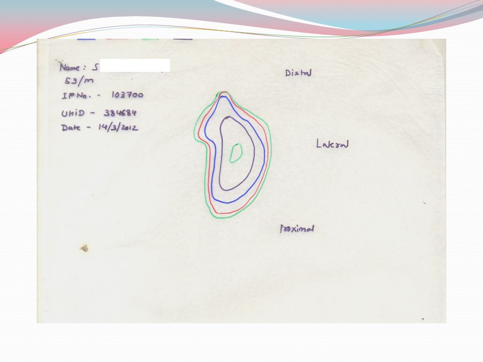
All patients were followed up personally after 1 week, 15 days, 1 month and 3 months. Size of the ulcer was measured on a transparent sheet using a marker pen at first visit and on subsequent follow up visits.

9
Follow Up Criteria Ulcer Size on transparency: Floor: Discharge: Surrounding Area: Signs of inflammation: GRBS :

10
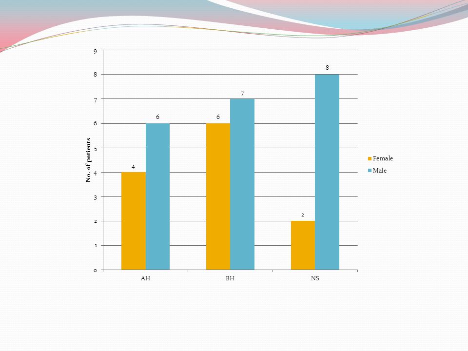
Follow Up All patients seen personally. Total 46 patients. Lost to follow up / excluded – 13 patients. Final Analysis – 33 Group A – 10 patients Group B – 13 patients Group C – 10 patients

11
Follow up Group A – 10 patients. Group B – 13 patients. Group C – 10 patients.

15
Results Descriptives Age NMean Std. Deviation Std. Error 95% Confidence Interval for Mean Minimu mMaximum Lower Bound Upper Bound 11062.808.8792.80856.4569.154472 21355.4612.1773.37748.1062.822974 31061.707.7752.45956.1467.264571 Total3359.5810.3111.79555.9263.232974 ANOVA F=1.826, P=0.179

19
Results FactorsGroup AGroup BGroup C ComorbiditiesHypertension – 5 IHD – 3 Hypertension – 6 Duration of Diabetes5 years Patient Insulin / OHAOHA – 6 Insulin – 4 OHA – 6 Insulin – 7 OHA – 7 Insulin – 4 History of Foot related complications 321 Smoking342 Total Leucocyte Count 10,000 11,000 GRBS at first visit230228221 X ray footNo osteomyelitisOsteomyelitis – 1 Arterial DopplerNot done2 – normal

20
First Visit Group AGroup BGroup C Ulcer Size10x15 – 3x3 cm12x10 – 3x3 cm10x8 – 3x4 cm Plantar Location687 FloorSlough – 6Slough – 7Slough - 6 Vascularity (ABI)Good Signs of Inflammation (+) 676 Debridement done535 SAD Score3-73-83-7
Good Signs of Inflammation (+) 676 Debridement done535 SAD Score")
21
Follow up Follow UpGroup AGroup BGroup C Reduction in Size on Transparency Max – 100% Min – 20% Max- 100% Min – 10% Max – 100% Min – 25% Complete Healing of Wound464 FloorHealthy Discharge234 Surrounding AreaNormal InflammationNo Diabetic status>200 – 4>200 – 5>200 - 5

22
Group A Complete Healing within 3 months – 4 50 % Size Reduction -3 < 50% Size reduction - 3 1 patient developed wound infection after 2 months follow up, but was managed with debridement.

23
Group B Complete healing within 3 months -6 Size reduction 50 % - 4 Size reduction 10-30 % 3

24
Group C Complete healing within 3 months - 4 Size reduction 50 % -4 Size reduction 25 % -2

25
SAD score & wound healing Odds ratio =0.769( 95% CI (0.301, 1.968), P=0.584 With every 1 unit increase in SAD score, healing of ulcers decreased upto 25 %. Variables in the Equation DressingBS.E.WalddfP value Odds ratio 95% C.I.for EXP(B) Lower Upp er AHStep 1 a SADScore-.262.479.2991.584.769.301 1.96 8 Constant.8392.343.1281.7202.314 BHStep 1 a SADScore-1.168.7262.5901.108.311.075 1.29 0 Constant5.7423.7122.3931.122311.702 NSStep 1 a SADScore-.128.401.1021.750.880.401 1.93 2 Constant.3192.353.0181.8921.376 a. Variable(s) entered on step 1: SADScore.
 Lower Upp er AHStep 1 a SADScore Constant BHStep 1 a SADScore Constant NSStep 1 a SADScore Constant a. Variable(s) entered on step 1: SADScore..")
26
Review of Literature

27
Comparison with Literature Foam dressings were associated with higher odds of ulcer healing compared with basic wound contact dressings. Estimate was considered to be of low quality. In general, estimates had large uncertainty due to low sample sizes. No significant difference between foam dressings v/s basic contact dressings. Dumville JC, Soares MO, O’Meara S, Cullum N (2012) Systematic review and mixed treatment comparison: dressings to heal diabetic foot ulcers. Diabetologia 55 : 1902–1910
 Systematic review and mixed treatment comparison: dressings to heal diabetic foot ulcers. Diabetologia 55 : 1902–1910.")
28
Review of Literature Foot ulcers occur in 12-25% patients with diabetes. Precedes 84% of all non-traumatic amputations. Amputations are 15 times more common in patients with Diabetes. Standard care heals 24% of ulcers in 24 weeks. - Removal of mechanical stress. - Debridement. - Moist wound environment. Brem H., Sheehan P., Rosenberg H.J., Schneider J S., Boulton A J. et al (2006) Evidence-Based Protocol for Diabetic Foot Ulcers. Plastic and Reconstructive Surgery. 117; 193-209
 Evidence-Based Protocol for Diabetic Foot Ulcers. Plastic and Reconstructive Surgery. 117;")
29
Alginate + Hydrocolloid Dressings Alginates are highly absorbent, gel-forming materials with haemostatic properties. Calcium alginate dressings inhibits growth of Staphylococcus aureus in vitro, with no increase in growth of Pseudomonas, Streptococcus pyogenes, or Bacteroides fragilis. Safe to use on infected foot ulcers, provided there are regular and thorough dressing changes. Hydrocolloids provide moist environment. Hilton J.R., Williams D.T., Beuker B., Miller D.R., Harding K.G (2004) Wound Dressings in Diabetic Foot Disease. Clinical Infectious Diseases 39; 100-103
 Wound Dressings in Diabetic Foot Disease. Clinical Infectious Diseases 39;")
30
Povidone-iodine + Hydrogen Peroxide Dressings Povidone iodine - decreases bacterial count of ulcers, - promote revascularization, - do not cause the emergence of drug-resistance - have broader antimicrobial spectrum. Hydrogen peroxide - wound antiseptic. - greatest activity is towards Gram-positive bacteria. - loosens debris and necrotic tissue of wound. In-vitro studies – Povidone iodine shown to be cytotoxic to cells essential to the wound healing process, such as fibroblasts, keratinocytes, leukocytes. Brem H., Sheehan P., Rosenberg H.J., Schneider J S., Boulton A J. et al (2006) Evidence-Based Protocol for Diabetic Foot Ulcers. Plastic and Reconstructive Surgery. 117; 193-209
 Evidence-Based Protocol for Diabetic Foot Ulcers. Plastic and Reconstructive Surgery. 117;")
31
Normal Saline Dressings Physiologically normal Isotonic with plasma Provide moist environment for healing of wounds. As the water in gauze evaporates, dressing becomes hypertonic, draws fluid from wound by osmosis. Wound fluid then dilutes - to reach dynamic equilibrium. Movement of wound fluid into sponge contribute to its effectiveness as a dressing. Lim J.K., Saliba L, Smith M.J., McTavish J., Raine C. et al. Normal saline wound dressing – is it really normal?: British Journal of Plastic Surgery: 2000; 53; 42–45.

32
Limitations Small sample size.

33
Conclusion No significant difference between clinical outcome of 3 types dressings in diabetic foot ulcers. SAD score helps in predicting healing of ulcers.

34
Thank You

Similar presentations