Download presentation
Presentation is loading. Please wait.

1
Anatomy of the coronary circulation & Angiographic VISUALIZATION
Dr Sandeep Mohanan Department of Cardiology Calicut Medical College 1/10/12

2
OUTLINE Coronary arterial anatomy Variations in coronary circulation
Coronary venous anatomy Angiographic views of coronary arteries

3
Coronary arterial anatomy
1st anatomical drawings- Leonardo da Vinci Oblique inverted crown

4
The coronary arteries and their major branches are sub-epicardially located

5
Pericardium (Epicardium)
Epicardial Vessel Subepicardium Myocardium Subendocardium

6
LCA ostium ~ 4mm RCA ostium~ 3.2mm

7
The LEIDEN convention 1R2LCx pattern
Each artery arises from respective aortic sinuses - Right coronary sinus(anterior) - Left coronary sinus(left posterior) - Non-coronary sinus(right posterior) 1R2LCx pattern
 - Left coronary sinus(left posterior) - Non-coronary sinus(right posterior) 1R2LCx pattern.")
10
Right coronary artery ~ 9.8cm
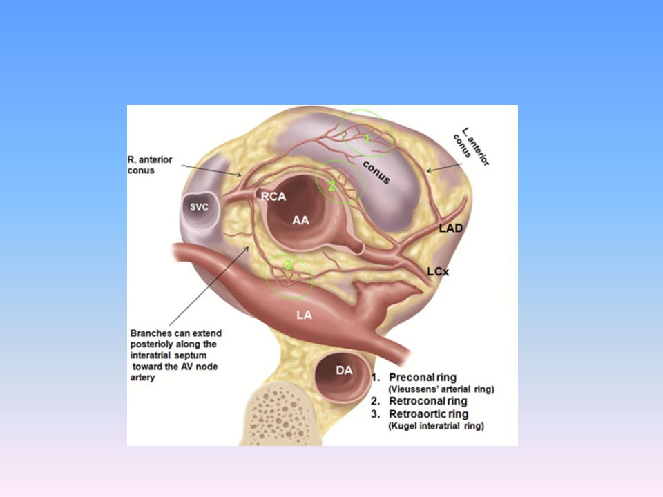
1)Conus artery/ Infundibular/ Third coronary/ Adipose /Arteria of Vieussens Separate ostium in 23% - 51% - Circle of Vieussens
Conus artery/ Infundibular/ Third coronary/ Adipose /Arteria of Vieussens. Separate ostium in 23% - 51% - Circle of Vieussens.")
12
Right coronary artery 2) Atrial branches of the RCA - < 1mm
SA nodal artery ( Ramus crista terminalis) – %
 – 55-65%")
13
Right coronary artery 3) Right ventricular branches
Acute right marginal artery Ramus crista supraterminalis (Superior septal artery) – % , males
 – % , males.")
14
Right coronary artery 4) Posterior descending artery Dominance
Posterior septal branches - < 15mm 5) AV nodal artery %
 AV nodal artery %")
15
Right coronary artery 6) Postero-lateral branches to the LV - Inferior wall of the LV
 Postero-lateral branches to the LV - Inferior wall of the LV")
16
Clinical division of the RCA
Proximal - Ostium to 1st main RV branch Mid st RV branch to acute marginal branch Distal acute margin to the crux

17
Left coronary artery LMCA
10-15mm(upto 30mm) length & 3-6mm(upto 10mm diameter) Trifurcates in 1/3rd : Ramus intermedius/ median artery/ left diagonal artery/straight LV artery Rare variations – absent LMCA/ pentafurcation
 length & 3-6mm(upto 10mm diameter) Trifurcates in 1/3rd : Ramus intermedius/ median artery/ left diagonal artery/straight LV artery. Rare variations – absent LMCA/ pentafurcation.")
18
Left anterior descending artery
- ~ cm ; Type I (22%) , Type II & Type III diagonal branches 90deg bend after turning around P. conus as it gives off 2nd diagonal branch Right ventricular branches( left conal/pre-infundibular A) ~ 10 septal perforating branches (40-80mm X mm) anchors the LAD LAD supplies 45-55% of LV
 , Type II & Type III diagonal branches. 90deg bend after turning around P. conus as it gives off 2nd diagonal branch. Right ventricular branches( left conal/pre-infundibular A) ~ 10 septal perforating branches (40-80mm X mm) anchors the LAD. LAD supplies 45-55% of LV.")
19
LAD(contd) 1st proximal septal A is prominent (His Bundle and LBB)
Myocardial bridging – % overall (28% in children) Rarely dual LADs
 Rarely dual LADs.")
20
Clinical division of the LAD
Proximal - Ostium to 1st major septal perforator Mid st perforator to D2 (90 degree angle) Distal D2 to end
 Distal - D2 to end.")
21
Left circumflex artery
~9.3 cm long ; mm Left atrial branches Kugel’s artery (Arteria anastomotica auricularis magna) LV branches are called the Obtuse marginal arteries LCx supplies 15-25% of LV (40-50% in dominant LV)
 LV branches are called the Obtuse marginal arteries. LCx supplies 15-25% of LV (40-50% in dominant LV)")
23
Clinical division of the LCX
Proximal - Ostium to 1st major obtuse marginal branch Mid OM1 to OM2 Distal OM2 to end

24
Coronary segment classification system
CASS investigators – 27 segments BARI – 29 segments ( ramus intermedius and 3rd diagonal branch) - Obstructive CAD : > 50% stenosis
 - Obstructive CAD : > 50% stenosis.")
25
Prognostication scores
Califf scoring system Gensini scoring system Candell-Riera scoring system CASS investigators: - no. of vessels - no. of proximal segments - Global LV function

26
“Dominance” A misnomer giving rise to PDA, at least 1 PLV & AV nodal A
(BARI classification) - 85% right dominant 8% left dominant 7% co-dominant (70%/ 10%/ 20% – Hurst’s THE HEART) Left dominance is 25-30% in Bi-AoV Gensini GG. Coronary Arteriography. Mount Kisco,NY: Futura Publishing Co; 1975:260–274.
 - 85% right dominant. 8% left dominant. 7% co-dominant. (70%/ 10%/ 20% – Hurst’s THE HEART) Left dominance is 25-30% in Bi-AoV. Gensini GG. Coronary Arteriography. Mount Kisco,NY: Futura Publishing Co; 1975:260–274.")
27
Nodal blood supply Studies on nodal blood supply principally by James (1961) and Hutchinson( 1978) - James : SA node - RCA 55% & LCA 45% AV node- RCA 90% & LCA 10% Hutchinson : SA node - 65% & 35% AV node- 80% & 20% AV node may have dual supply in 2% cases

28
Arterial anastomoses Seen at the intracoronary/inter-coronary levels in abundance– significant in development in collaterals in CAD Most abundant at the septum Intracoronary : cm X micm Inter-coronary: cm X micm

29
Coronary artery variations
2 coronary artery system is a recent evolutionary acquisition Fish and amphibia – 1 coronary artery Birds – ~ 40% have single coronary arteries. 1-5% of those undergoing CAG Angelini P – Coronary artery anomalies – current clinical issues. Definition, classifications, incidence, clinical relevance and treatment guidelines. Tex Heart Inst J 2002;29:

30
Coronary artery variations
Definition of a coronary artery is not based on its origin and proximal course, but by focusing on its intermediate and distal segments/ its dependent microvascular bed. Angelini P – Coronary artery anomalies – current clinical issues. Definition, classifications, incidence, clinical relevance and treatment guidelines. Tex Heart Inst J 2002;29:

31
? Coronary artery Variation vs Anomalies
A broad spectrum of variations of which some may cause adverse effects Most of the coronary variations may have no clinical implications as can be proven by myocardial perfusion studies. The regional distribution of a coronary artery, rather than its absolute origin and characteristics.

32
Level of variables 1) Ostium ) Size ) Proximal course 4) Mid-course 5) Intramyocardial ramifications 6) Termination Anomalies without a shunt: 1. Abnormal number : 1/ 3/ 4 ostia 2. Anomalous origin: a) Outside SOV b) Independent origin from same sinus c) Opposite sinus d) Other artery 3. Myocardial bridge 4. Segmental stenosis/hypoplasia Anomalies with shunt: 1) Fistula ) APOCA C. Aneurysms
 Mid-course 5) Intramyocardial ramifications 6) Termination. Anomalies without a shunt: 1. Abnormal number : 1/ 3/ 4 ostia. 2. Anomalous origin: a) Outside SOV. b) Independent origin from same sinus. c) Opposite sinus. d) Other artery. 3. Myocardial bridge. 4. Segmental stenosis/hypoplasia. Anomalies with shunt: 1) Fistula 2) APOCA. C. Aneurysms.")
33
A puzzling issue….. Proximal course of the LAD may be very different
LCx may run over atrial or ventricular surface. An RCA that terminates in the AV groove well before the crux may not always be an obstruction: 7 – 10% (Grossman) Double ostia from the RCS All 3 arteries from a single sinus One single artery……………..and so on……
 Double ostia from the RCS. All 3 arteries from a single sinus. One single artery……………..and so on……")
34
The most common coronary variation (Cleveland Clinic-1,26,000 patients) was separate ostia for LAD & LCX – 0.41% and 2nd commonest was LCX from RCS / RCA – 0.37% However, in another series of 1950 angiograms coronary anomalies were seen in 5.6% cases and split RCA (1.2%) was the commonest. 127,000 patients (Grossman) Angelina P. Coronary artery anomalies. Philadelphia, Lippincott Williams & Wilkins, 1999.
 was the commonest. 127,000 patients (Grossman) Angelina P. Coronary artery anomalies. Philadelphia, Lippincott Williams & Wilkins,")
35
1) Ostium 2) Size 3) Proximal course
Level of variables 1) Ostium ) Size ) Proximal course 4) Mid-course 5) Intra-myocardial ramifications 6) Termination MSCT with retrospective ECG gating is now considered the gold standard for characterization of coronary anomalies. Prompt a search for underlying CHDs 1) Shi H, Aschoff AJ, Brambs HJ. Multislice CT imaging of anomalous coronary arteries. Eur Radiol. 2004;14: 2) Memisoglu E, Hobikoglu G, Tepe MS. Congenital coronary anomalies in adults: Comparison of anatomic course visualized by catheter angiography and electron beam CT. Catheter Cardiovasc Interv. 2005;66:34-42.
 Ostium 2) Size 3) Proximal course. 4) Mid-course 5) Intra-myocardial ramifications. 6) Termination. MSCT with retrospective ECG gating is now considered the gold standard for characterization of coronary anomalies. Prompt a search for underlying CHDs. 1) Shi H, Aschoff AJ, Brambs HJ. Multislice CT imaging of anomalous coronary arteries. Eur Radiol. 2004;14: ) Memisoglu E, Hobikoglu G, Tepe MS. Congenital coronary anomalies in adults: Comparison of anatomic course visualized by catheter angiography and electron beam CT. Catheter Cardiovasc Interv. 2005;66:")
36
Abnormal position of ostia
Coronary orifice below the cuspal margin: - 10% RCS 15% LCS Coronaries above the sinotubular jn ~ 6% - leads to difficult cannulation, esp RCA with a high anterior ostium.

37
Abnormal number of coronary arteries
Single coronary artery %, usually benign D/d- 2 separate ostia from same sinus, atresia.. Course is important – in 25% a major branch crosses the infundibulum. 3 coronaries - 1) Separate origin of conus artery from RCS (36- 50%) 2) Absent LMCA with separate ostia for LAD & LCX 4 coronaries - case reports Dual LAD % (Morettin ,1976)
 Separate origin of conus artery from RCS (36- 50%) 2) Absent LMCA with separate ostia for LAD & LCX. 4 coronaries - case reports. Dual LAD % (Morettin ,1976)")
38
Absent LMCA ~0.4% - 1 ostia at the LCS/ 2 ostia in LCS/ 1 ostia in LCS & other RCS Increased incidence of Left dominance 6% incidence of bridging Not usually associated with CHDs Similar incidence of atherosclerosis Difficulty in selective cannulation Topaz et al. Absent left main coronary artery: angiographic findings in 83 patients with separate ostia of the left anterior descending and circumflex arteries at the left aortic sinus. Am Heart J.1991 Aug;122(2):
:")
39
Shepherd’s-crook RCA ~5%
Acute superiorly angled take-off of the RCA from the aorta. Difficult RCA lesion angioplasty Ethan Halpern. Cardiac CT . Functional anatomy.

40
Dual LAD (Duplication)
~ % of normal hearts Proximal LAD (LAD proper) bifurcates early into a short and long LAD -Type I : Short LAD in AIVS, Long LAD on prox AIVS, LV side, distal AIVS -Type II : Short LAD in AIVS, Long LAD on prox AIVS, RV side, distal AIVS -Type III: Short LAD in AIVS, Long LAD intra-myocardially in septum -Type IV: Very short LAD proper and short LAD, Long LAD from RCA Spindola-Franco H et al. Dual left anterior descending coronary artery: angiographic description of Important variants and surgical implications. Am Heart J 1983:105;445–55.
 bifurcates early into a short and long LAD. -Type I : Short LAD in AIVS, Long LAD on prox AIVS, LV side, distal AIVS. -Type II : Short LAD in AIVS, Long LAD on prox AIVS, RV side, distal AIVS. -Type III: Short LAD in AIVS, Long LAD intra-myocardially in septum. -Type IV: Very short LAD proper and short LAD, Long LAD from RCA. Spindola-Franco H et al. Dual left anterior descending coronary artery: angiographic description of Important variants and surgical implications. Am Heart J 1983:105;445–55.")
41
Coronary artery Ectasia
1 - 5% in angiographic series, more in males % are congenital Dialatation of a segment to at least 1.5times of the adjacent normal coronary artery.

42
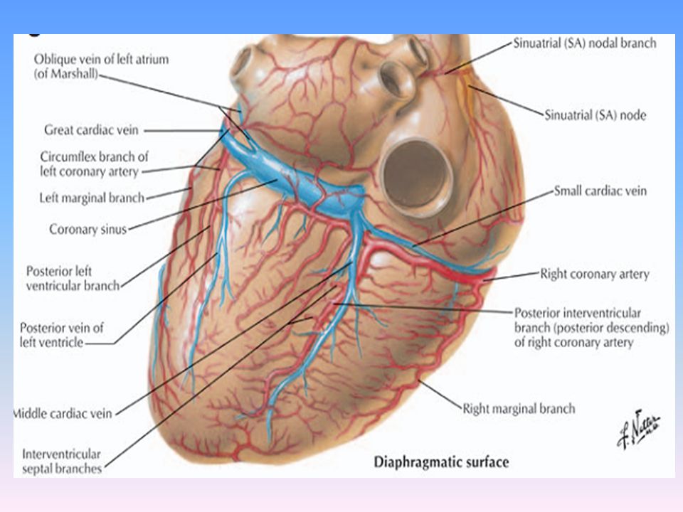
Coronary venous anatomy
Targeted drug delivery Retrograde cardioplegia administration Potential conduit to bypass cor. artery stenosis Stem cell delivery to the infarcted region Access to LA & LV myocardium for arrythmia mapping & ablation LV epicardial pacing in CRT

43
Coronary venous anatomy

44
THEBESIAN veins – Venae cordis minimae

45
Conventional coronary venous nomenclature
Coronary sinus - Thebasian valve Anterior IV vein(Great cardiac vein) - Vieussens valves - Left marginal vein of LV - Postero-lateral LV vein Middle cardiac vein Small cardiac veins SEGMENTAL CLASSIFICATION The highly variable existence of the conventional veins calls for segmental classification (ant, lat, post, base, mid and apex -9 segments of the LV) of coronay veins for better epicardial localization of veins for interventional electrophiography purposes.
 - Vieussens valves. - Left marginal vein of LV. - Postero-lateral LV vein. Middle cardiac vein. Small cardiac veins. SEGMENTAL CLASSIFICATION. The highly variable existence of the conventional veins calls for segmental classification (ant, lat, post, base, mid and apex -9 segments of the LV) of coronay veins for better epicardial localization of veins for interventional electrophiography purposes.")
46
Segmental venous classification
Thus 9 LV venous segments are derived which when added with the conventional classification gives the best comprehensive information to place the epicardial LV leads for CRT purposes

47
Retrograde coronary venography
Lateral LV wall venous branches can be profiled by individualizing the different radiological views- considering the anterior IV vein and middle cardiac vein as reference points.

48
MDCT angiogram delineating coronary veins along with arteries
Before venogram for better characterisation of coronary vein variations. However additional 60ml contrast and 9-11mSv exposure.

49
Coronary Angiographic Views
Cardiac Cath 1st by Werner Forssman in 1929 1st contrast angiography by Chavez in 1947 CART 1st performed by F. Mason Sones in 1958 a high-resolution image-intensifier television system with digital cineangiographic capabilities. - Radiograph tube below and Image intensifier above (Flouroscopic imaging system with C-arm) - Physiologic monitoring system, sterile supplies, resuscitation equipment, Contrast injector (3-8ml/sec) and contrast media Xray generator, Xray tube , Image intensifier and detector, digital angio imaging. A higher angulation increases the radiation scatter. Fluoroscopy has only 1/5th rad exposure of cine angiography NCRPM guideline: not >3 rem per 3months.. Advised safe limit is 100mrem/week for cath lab personnel. Skin and thyroid- 15rem/year, gonads, eyes, bonemarrow- 5rem/yr Cxray= 3 -5 mRoentgen ( 1 R = 1 rad for skin, 1R= 4rad for bone due to more absorption) R= radiation exposure, Rad = radiation absorptioon) Rem= radiation equivalent dose in man. 1 rem= 1rad. 1SV= 1J/kg=1Gy 1gy=100rad 1Sv= 100rem 1mrem=10micSv
 - Physiologic monitoring system, sterile supplies, resuscitation equipment, Contrast injector (3-8ml/sec) and contrast media. Xray generator, Xray tube , Image intensifier and detector, digital angio imaging. A higher angulation increases the radiation scatter. Fluoroscopy has only 1/5th rad exposure of cine angiography. NCRPM guideline: not >3 rem per 3months.. Advised safe limit is 100mrem/week for cath lab personnel. Skin and thyroid- 15rem/year, gonads, eyes, bonemarrow- 5rem/yr. Cxray= 3 -5 mRoentgen ( 1 R = 1 rad for skin, 1R= 4rad for bone due to more absorption) R= radiation exposure, Rad = radiation absorptioon) Rem= radiation equivalent dose in man. 1 rem= 1rad. 1SV= 1J/kg=1Gy. 1gy=100rad. 1Sv= 100rem. 1mrem=10micSv.")
50
Information from a CAG:
CAG helps visualization of the major epicardial arteries up to their 2nd and 3rd order branches - Coronary anatomy Characteristics and distribution of coronary stenosis Distal vessel size Intracoronary thrombus Index of coronary flow Mass of myocardium served Collateral vasculature Optimal injection rate: 7ml (2.1ml/s) for LCA and 4.8ml (1.7ml/s) for RCA
 for LCA and 4.8ml (1.7ml/s) for RCA.")
51
Pitfalls of CAG – A Lumenogram

52
Interpretation of the significance of a lumenogram
Multiple projections from different angles, preferably orthogonal Knowledge of the normal calibre of major coronaries: LMCA: 4.5 ± 0.5 mm LAD: 3.7 ± 0.4 mm LCX : 3.5 ± 0.5 mm ( 4.2 mm if dominant) RCA: 3.9 ± 0.6 mm ( 2.8 mm if non-dominant) IVUS Functional studies : FFR
 RCA: 3.9 ± 0.6 mm ( 2.8 mm if non-dominant) IVUS. Functional studies : FFR.")
53
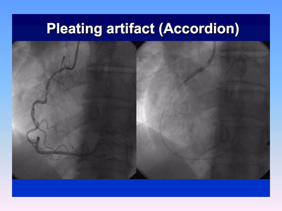
Mistakes in CAG interpretation
Inadequate number of projections used Improper/inadequate contrast injection Super-selective injection Catheter induced vasospasm Coronary artery variations Myocardial bridges Total ostial occlusions Wire induced spasm (ACCORDION EFFECT) Accordion effect: A mechanical alteration in the geometry and curvature of the vessel due to straightening and shortening of the artery due to wire advancement.
 Accordion effect: A mechanical alteration in the geometry and curvature of the vessel due to straightening and shortening of the artery due to wire advancement.")
55
Angiographic projections
LAO and RAO views help furnish the true PA and lateral views of the heart D/A s - foreshortening - superimposition Cranial view: Image-intensifier tilted towards head Caudal view: Image-intensifier tilted towards the feet -however the optimal angiographic view varies with coronary anatomy, body habitus and location of lesion

56
Angiographic projections
Kern MJ. Cardiac Catheterization Handbook. 5th edition,2011.

57
RAO and LAO projections

58
Optimal angiographic views for coronary segments
Carlo Di Mario, Nilesh Sutaria. CORONARY ANGIOGRAPHY IN THE ANGIOPLASTY ERA: PROJECTIONS WITH A MEANING Heart 2005;91:968–976.

59
RAO- LCA When the LMCA, LAD, LCX have an initial leftward course the long axis of these arterial segments are projected away frm the image intensifier and prevent optimal visualisation in the RAO view.

60
RAO- RCA

61
Optimal angiographic views for coronary segments
Carlo Di Mario, Nilesh Sutaria. CORONARY ANGIOGRAPHY IN THE ANGIOPLASTY ERA: PROJECTIONS WITH A MEANING Heart 2005;91:968–976.

62
Shallow RAO cranial - LCA

63
AP cranial - LCA

64
RAO cranial - RCA

65
Optimal angiographic views for coronary segments
Carlo Di Mario, Nilesh Sutaria.CORONARY ANGIOGRAPHY IN THE ANGIOPLASTY ERA: PROJECTIONS WITH A MEANING Heart 2005;91:968–976.

66
RAO caudal - LCA

67
Optimal angiographic views for coronary segments
Carlo Di Mario, Nilesh Sutaria.CORONARY ANGIOGRAPHY IN THE ANGIOPLASTY ERA: PROJECTIONS WITH A MEANING Heart 2005;91:968–976.

68
AP (Shallow RAO) caudal- LCA
 caudal- LCA")
69
Optimal angiographic views for coronary segments
Carlo Di Mario, Nilesh Sutaria.CORONARY ANGIOGRAPHY IN THE ANGIOPLASTY ERA: PROJECTIONS WITH A MEANING Heart 2005;91:968–976.

70
LAO - LCA

71
LAO - RCA

72
Optimal angiographic views for coronary segments
Carlo Di Mario, Nilesh Sutaria. CORONARY ANGIOGRAPHY IN THE ANGIOPLASTY ERA: PROJECTIONS WITH A MEANING Heart 2005;91:968–976.

73
LAO cranial - LCA Some overlap with LCX can be overcome by more 60 degree LAO tilt. However when the proximal LCA is superiorly directed it is not an optimal view- use LAO caudal

74
LAO cranial - RCA

75
Optimal angiographic views for coronary segments
Carlo Di Mario, Nilesh Sutaria.CORONARY ANGIOGRAPHY IN THE ANGIOPLASTY ERA: PROJECTIONS WITH A MEANING Heart 2005;91:968–976.

76
LAO caudal (Spider view) - LCA
Enhanced by maximal expiration as the heart becomes more horizontal

77
Optimal angiographic views for coronary segments
Carlo Di Mario, Nilesh Sutaria.CORONARY ANGIOGRAPHY IN THE ANGIOPLASTY ERA: PROJECTIONS WITH A MEANING Heart 2005;91:968–976.

78
Lateral view Mid & distal LAD Proximal LCX Mid RCA LIMA graft to LAD

79
Optimal angiographic views for coronary segments
There is no single magical projection that can be applied uniformly to all patients for visualizing a particular coronary atery According to Grossman: For LCA – RAO caudal and LAO caudal for LMCA and proc LAD in orthogonal & RAO cranial and LAO cranial for mid and distal LAD in orthogonal For RCA: LAO for proximal RCA and RAO cranial for distal, PDA, PLV and Lateral view for mid Carlo Di Mario, Nilesh Sutaria. CORONARY ANGIOGRAPHY IN THE ANGIOPLASTY ERA: PROJECTIONS WITH A MEANING Heart 2005;91:968–976.

80
Panoramic coronary angiography
GIORGIO TOMMASINI et al. Panoramic Coronary Angiography. JACC 31(4),March 15, 1998:871–7
,March 15, 1998:871–7.")
81
References Hurst’s The Heart 13th Edition
Braunwalds Heart Disease 9th edition Grey’s Anatomy Kern’s Handbook of Interventional Catheterization Kjell C Nikus. Coronary angiography. Grossman’s Textbook of Cardiac Catheterization Carlo Di Mario, Nilesh Sutaria. CORONARY ANGIOGRAPHY IN THE ANGIOPLASTY ERA: PROJECTIONS WITH A MEANING Heart 2005;91:968–976 David M Fiss. Normal coronary anatomy and anatomic variations. Applied Radiology, Jan 2007. Horia Muresian. Coronary arterial anomalies and variations. MAEDICA. A journal of clinical Medicine,1(1), 2006. Singh et al. The coronary venous anatomy. A segmental approach to aid CRT 2005, 46(1), Shilpa Bhimali et al. A STUDY OF VARIATIONS IN CORONARY ARTERIAL SYSTEM IN CADAVERIC HUMAN HEART. World Journal of Science and Technology 2011, 1(5): ISSN: 2231 – 2587.
, Singh et al. The coronary venous anatomy. A segmental approach to aid CRT 2005, 46(1), Shilpa Bhimali et al. A STUDY OF VARIATIONS IN CORONARY ARTERIAL SYSTEM IN CADAVERIC HUMAN HEART. World Journal of Science and Technology 2011, 1(5): ISSN: 2231 –")
82
Thank you

Similar presentations


 in thorax, in inferior mediastinum>")


