Download presentation
Presentation is loading. Please wait.

1
Kirstin Blackie Nima Mohan
Medical Abdomen Kirstin Blackie Nima Mohan

2
Objectives Be aware of common conditions presenting with abdominal symptoms Understand important factors in the history, examination, investigation and management of common abdominal pathologies.

3
Causes of Abdo Pain

4
What other symptoms would you want to ask about?
Case Study Mr C, 35 year old man, presents to his GP with mild abdominal pain and yellowing of the whites of his eyes (noticed by his girlfriend who is a nurse). What other symptoms would you want to ask about?
. What other symptoms would you want to ask about")
5
Signs and Symptoms of Liver pathology
Abdominal pain (RUQ) Jaundice Nausea, vomiting Weight loss Abdo distension Haematemesis and malaena Breast swelling, tesicular atrophy Confusion Spider naevi Palmar erythema Dupuytrens contracture Hepatomegaly, Spenomegaly
 Jaundice. Nausea, vomiting Weight loss. Abdo distension. Haematemesis and malaena. Breast swelling, tesicular atrophy. Confusion. Spider naevi. Palmar erythema. Dupuytrens contracture. Hepatomegaly, Spenomegaly.")
6
Case study cont: Has recently has ‘flu’ – has felt generally unwell, tired and vaguely nauseated. He is unsure but thinks he may have had a mild fever. What risk factors would you ask about?

7
Risk factors for liver disease
High Alcohol intake Blood-to-blood contact (IVDU, Tattoos, infected transfusions, needlestick injuries) Unprotected sex Drugs (prescribed, OTC, herbal) Travel Family history of liver disease (autoimmune hepatitis, Wilson’s disease) Mr C is in monogamous sexual relationship with girlfriend for 2 months– she is on OCP. No barrier contraception. Drinks approximately 30 units alcohol / week. Denies any other risk factors. What first line investigations would you like to do?
 Unprotected sex. Drugs (prescribed, OTC, herbal) Travel. Family history of liver disease (autoimmune hepatitis, Wilson’s disease) Mr C is in monogamous sexual relationship with girlfriend for 2 months– she is on OCP. No barrier contraception. Drinks approximately 30 units alcohol / week. Denies any other risk factors. What first line investigations would you like to do")
8
Liver Function Tests Viral hepatitis: Alcoholic hepatitis
ALT greatly raised (10-100x upper limit of normal) Alcoholic hepatitis ALT moderately raised (2-10x upper limit of normal) Drug induced hepatitis Mixed picture: raised hepatic (AST, ALT) and Cholestatic (Alk Phos and GGT) markers Abnormal clotting (prolonged PT or INR) may indicate acute liver failure
 Alcoholic hepatitis. ALT moderately raised (2-10x upper limit of normal) Drug induced hepatitis. Mixed picture: raised hepatic (AST, ALT) and Cholestatic (Alk Phos and GGT) markers. Abnormal clotting (prolonged PT or INR) may indicate acute liver failure.")
9
Drugs commonly associated with Hepatitis
Acute hepatocellular damage: Paracetamol (dose related) Alcohol (dose related) TB drugs Anticonvulsants Azathioprine Methotrexate Chronic active hepatitis Nitrofurantoin Isoniazide Intrahepatic cholestasis Oestrogens erythromycin
 Alcohol (dose related) TB drugs. Anticonvulsants. Azathioprine. Methotrexate. Chronic active hepatitis. Nitrofurantoin. Isoniazide. Intrahepatic cholestasis. Oestrogens. erythromycin.")
10
Other causes: EBV, CMV, paravirus B19, dengue, yellow fever.
Hep A Hep B Hep C Hep D Hep E Route of transmissio n Faecal-oral (contaminate d water/food, oro-anal sex) Blood products, body fluids, sexual contacts Blood products As for Hep B Contaminated water People at risk Childcare workers, MSM vertical transmission, sexual partners, healthcare workers, Tattoos, body piercings, blood transfusions, IVDU IVDU, tattoos Less commonly: vertical transmission, sexual transmission Those at risk of Hep B or with chronic Hep B Travel to endemic areas, and sporadic outbreaks associated with poor sanitation eg refugee camps Potential for chronic infection none Common in infants (90%), rarer in adults (10%) 80% develop chronic infection Co-infection with Hep B Incubation period 3 weeks (range 2-7) 10 weeks (range 4-26) 7 weeks (range 2-21) 5 weeks (range 3-7) (range 3-8) Other causes: EBV, CMV, paravirus B19, dengue, yellow fever.
 Blood products, body fluids, sexual contacts. Blood products. As for Hep B. Contaminated water. People at risk. Childcare workers, MSM. vertical transmission, sexual partners, healthcare workers, Tattoos, body piercings, blood transfusions, IVDU. IVDU, tattoos. Less commonly: vertical transmission, sexual transmission. Those at risk of Hep B or with chronic Hep B. Travel to endemic areas, and sporadic outbreaks associated with poor sanitation eg refugee camps. Potential for chronic infection. none. Common in infants (90%), rarer in adults (10%) 80% develop chronic infection. Co-infection with Hep B. Incubation period. 3 weeks (range 2-7) 10 weeks. (range 4-26) 7 weeks. (range 2-21) 5 weeks. (range 3-7) (range 3-8) Other causes: EBV, CMV, paravirus B19, dengue, yellow fever.")
11
Hepatitis E endemic areas

12
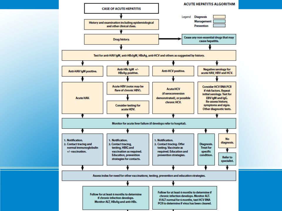
Mr C and his girlfriend are both tested for viral hepatitis

13
Hepatitis B Serological Markers

15
What are the differential diagnoses?
65 year old man who hasn’t been to his GP in years comes into A+E with an uncomfortable swollen abdomen What are the differential diagnoses?

16
5 Fs of distended abdomen
Fat Faeces Fluid Foetus Flatus How would you examine for fluid (ascites)?
")
17
Shifting Dullness Does the presence of ascites prove that this patient has liver disease?

18
What are the possible causes of ascites?

19
Cirrhosis: common end point of many disease processes
Alcohol excess Hepatitis B Hepatitis C Non-alcoholic Fatty Liver disease / Non-alcoholic Steatohepatitis Haemachromatosis Primary Biliary Cirrhosis Primary Scelosis Cholangitis Autoimmune hepatitis Wilson’s disease and other inherited metabolic disorders

21
How would you investigate decompensated liver disease?
Bloods: likely increased biliruben, AST, ALT, alk phos, GGT; Decreased albumin, increased PT/INR (reduced synthetic function); Decreased WCC and platelets (hypersplenism); Look for the cause: serology, autoantibodies, iron studies Imaging: liver US and doppler, MRI Ascitic tap: Biopsy: confirm clinical diagnosis
; Decreased WCC and platelets (hypersplenism); Look for the cause: serology, autoantibodies, iron studies. Imaging: liver US and doppler, MRI. Ascitic tap: Biopsy: confirm clinical diagnosis.")
22
US liver “fibrotic, structurally abnormal nodules in liver …. Compatible with cirrhosis. Doppler shows signs of portal hypertension.”

23
Complications of Cirrhosis
Anaemia (folate deficiency, hypersplenism) Thrombocytopenia (hypersplenism) Coagulopathy (reduced production of clotting factors) – can lead to DIC Oesophageal varices Spontaneous Bacterial Peritonitis Hepatic encephalopathy Hepatocellular carcinoma
 Thrombocytopenia (hypersplenism) Coagulopathy (reduced production of clotting factors) – can lead to DIC. Oesophageal varices. Spontaneous Bacterial Peritonitis. Hepatic encephalopathy. Hepatocellular carcinoma.")
24
How would you manage this patient?
Patient education and support Treat underlying cause Adequate nutrition (calorie and protein intake) Careful prescribing Therapeutic ascetic tap Alcohol abstinence (also important in non- alcohol induced cirrhosis) Alcohol dependent individuals will require: Chlordiazepoxide, Thiamine, Vitamin B Monitoring for further complications: oesophageal varicies or HCC ?transplant
 Careful prescribing. Therapeutic ascetic tap. Alcohol abstinence (also important in non- alcohol induced cirrhosis) Alcohol dependent individuals will require: Chlordiazepoxide, Thiamine, Vitamin B. Monitoring for further complications: oesophageal varicies or HCC. transplant.")
25
What other questions would you ask her???
Case Study “A 17 year old girl presents to the GP with a 8 week history of fatigue. She also reports frequent episodes of pyrexia and intermittent episodes of diarrhoea over this period. Over the last 48 hours she has had 14 episodes of watery diarrhoea” What other questions would you ask her???

26
She denies...... jaundice, dyspepsia, vomiting, malena, constipation, ulcers Changes in appetite Changes in mensustral cycle urinary symptoms No recent travel No changes / alterations to her diet She reports Fatigue – low energy levels SOBOE Palpitations Frequent Pyrexia Abdominal pain – generalised cramps Diarrhoea – no blood or mucus Weight Loss

27
What are you going to do next??
Clinical Examination Investigations Urine Dip and MSU Bloods : FBC, U&E's , CRP, ESR, LFT's, TFT's, Electrolytes, Anti -TTG, Blood Cultures?? Stool culture Imaging

28
What is your immediate management plan?
Clinic On examination Tachycardic – 101 regular, good volume. normotensive – 110/76 CPT > 3 sec Pale conjunctive Cardio- respiratory examination - NAD Diffuse tenderness in the abdomen normal PR What is your immediate management plan? WCC 15.9 HB 8.2 MCV 89 Platelets 289 ESR HIGH CRP 110 urea 17 creatnine K+ 4.6 na+ 135 LFT's NAD TFT's Cultures No growth

29
RADIOLOGICAL FEATURES
CROHN'S DISEASE ULCERATIVE COLITIS CLINICAL FEATURES Abdominal pain peri-anal disease Constitutuional Symptoms Gastro-inestinal bleeding Diahorrea < 6 episodes /day Rectal Spasm EXAMINATION FEATURES RIF mass Per-anal skin tags Fistulas Scars / Stomas – ileostomy / colostomy bags. RADIOLOGICAL FEATURES Fistula formation Asymetrical / skip lesions Rose thorn ulcers Ileal involvement Rectal involvement Superficial ulcers ENDOSCOPIC FEATURES Cobble stone mucosa Transmural disease Granluoma formation Stricture formation Superficial ulceration Stud ulcers Pseudopolyps. Crypt abscesses COMPLICATIONS & PROGNOSIS Fistula formation – perianal Toxic megacolon – perforation Small bowel obstruction Malignancy Large Bowel obstruction Toxic Megacolon Primary sclerosing cholangitis. Increased Risk of Malignancy

30
WHAT TYPE OF IMAGING. NAME OF SIGN. WHAT DISEASE
WHAT TYPE OF IMAGING? NAME OF SIGN? WHAT DISEASE? 5 OTHER EXTRA INTESTINAL MANIFESTATIONS OF THIS DISEASE?

31
EXTRA INTESTINAL MANIFESTATIONS
EYES : episcleritis, uveitis MOUTH: Apthous ulcers, angular stomatitis JOINTS : sero-negative arthropathies (anklysing spondylitis, sacroilietis) KIDNEYS : stones fistula, hydronephrosis SKIN: Eryhthemna nodosum, phlebitis, pyoderma gangrenosum
 KIDNEYS : stones fistula, hydronephrosis. SKIN: Eryhthemna nodosum, phlebitis, pyoderma gangrenosum.")
32
RADIOLOGICAL FEATURES
CROHN'S DISEASE ULCERATIVE COLITIS CLINICAL FEATURES Abdominal pain peri-anal disease Constitutuional Symptoms Gastro-inestinal bleeding Diahorrea < 6 episodes /day Rectal Spasm EXAMINATION FEATURES RIF mass Per-anal skin tags Fistulas Scars / Stomas – ileostomy / colostomy bags. RADIOLOGICAL FEATURES Fistula formation Asymetrical / skip lesions Rose thorn ulcers Ileal involvement Rectal involvement Superficial ulcers ENDOSCOPIC FEATURES Cobble stone mucosa Transmural disease Granluoma formation Stricture formation Superficial ulceration Stud ulcers Pseudopolyps. Crypt abscesses COMPLICATIONS & PROGNOSIS Fistula formation – perianal Toxic megacolon – perforation Small bowel obstruction Malignancy Large Bowel obstruction Toxic Megacolon Primary sclerosing cholangitis. Increased Risk of Malignancy

33
Clinic

34
Primary Sclerosing Cholangitis
Strongly association with UC (less with CD) Inflammation, fibrosis and stricture of the intra/ extra hepatic ducts. Signs of Live failure LFTS- Raised Alkaline Phosphatase, Bilirubin, hypergamaglobinumina ANA, ANCA, SMA +VE Poor prognosis – often need transplant and increases risk of cholangiocarcinoma
 Inflammation, fibrosis and stricture of the intra/ extra hepatic ducts. Signs of Live failure. LFTS- Raised Alkaline Phosphatase, Bilirubin, hypergamaglobinumina. ANA, ANCA, SMA +VE. Poor prognosis – often need transplant and increases risk of cholangiocarcinoma.")
35
Management MEDICAL MANAGEMENT Treatment of exacerbations :
Mild – oral steroids (Prednislone PO / PR) Severe – IV Hydrocortisone and Antibiotics Maintenance therapy : Maintain adequate nutrition To prevent exacerbations 5-ASA's (Mesalazine) Azothioprine Anti- TNF antibodies (INFLIXIMAB) Goals of treatment 1) to achieve the best possible clinical, laboratory, and histological control of the inflammatory disease with the least adverse effects from medication; (2) to promote growth with adequate nutrition; and (3) to permit the patient to function as normally as possible (eg, in terms of school attendance, participation in activities).
 Severe – IV Hydrocortisone and Antibiotics. Maintenance therapy : Maintain adequate nutrition. To prevent exacerbations. 5-ASA s (Mesalazine) Azothioprine. Anti- TNF antibodies. (INFLIXIMAB) Goals of treatment. 1) to achieve the best possible clinical, laboratory, and histological control of the inflammatory disease with the least adverse effects from medication; (2) to promote growth with adequate nutrition; and. (3) to permit the patient to function as normally as possible (eg, in terms of school attendance, participation in activities).")
36
Surgical Management Surgical management of complications
Surgical management of the condition

37
What else do you want to know???
CASE STUDY A 25 year old girl presents with a 8 week history of generalised abdominal cramps and diarrhoea. They are loose stool, no blood or mucus and can occur 8-10 times a day. She also reports that she is frequently tired and stressed. What else do you want to know???

38
Irritable bowel Syndrome
Incidence: common (female ) ; 40 % people attending secondary care 6 months of symptoms before diagnosis Can be predominantly constipation or predominantly diahorrea. Abdominal pain/ Bloating Anxiety / depression Incomplete emptying/ incontinence/ urgency Constitunal symptoms : tiredness, lethargy, arthalgia, urinary symptoms, dyspurunina. RED FLAG SYMPTOMS: Bleeding, Nocturnal symptoms, weight loss, Age > 50
 ; 40 % people attending secondary care. 6 months of symptoms before diagnosis. Can be predominantly constipation or predominantly diahorrea. Abdominal pain/ Bloating. Anxiety / depression. Incomplete emptying/ incontinence/ urgency. Constitunal symptoms : tiredness, lethargy, arthalgia, urinary symptoms, dyspurunina. RED FLAG SYMPTOMS: Bleeding, Nocturnal symptoms, weight loss, Age > 50.")
39
Treatment Options Reassurance and support
Address / Treat underlying medical issues Lifestyle advice : Dietary modification – excluding food groups. Smoking and alcohol Symptomatic relief : Bloating – Peppermint oil Constipation – increase fibre and fluid intake Antispasmodics – mebevrine

40
Case Study “A 65 year old man presents with a 4 day history of black tarry stools. He reports that they are becoming more frequent and loose. He also reports nausea and one episode of vomiting this morning. He also reports that he has a back ache for the past fortnight and has been taking OTC painkillers for it and would like you to prescribe some more”

41
Causes of Upper GI bleeding
Common causes: Ulcers – Peptic ulcers (40%) Varices – Secondary to portal hypertension (17%) Gastritis / gastric erosion Duodenitis Oesphagitis Rarer causes: Mallory -Weiss tears Angiodysplasia Bleeding Disorders Peutz- Jeugher's Syndrome Osler – Webb – Rendu Syndrome
 Varices – Secondary to portal hypertension (17%) Gastritis / gastric erosion. Duodenitis. Oesphagitis. Rarer causes: Mallory -Weiss tears. Angiodysplasia. Bleeding Disorders. Peutz- Jeugher s Syndrome. Osler – Webb – Rendu Syndrome.")
42
Examination / Investigation
On Examination: He is tachycardic, at 111 bpm / regular and borderline hypotensive 105/72. He is tender in the epigastrium and peri-umbilically. There is some voluntary guarding. Bowel sounds are normal. DRE – Malodorous black tarry stool. No fresh blood. Investigations: Bloods : Hb , Urea -21 , Creatnine 66, WCC- 7.0, platelets- 260, CRP – 2.2, LFT's – NAD. AXR – NAD Erect CXR – No free air under the diapgram

43
Management CALL FOR HELP RE- ASSESS
Bleep : RR -30 BP- 90 /66 , HR -122, CRT > 3, Sats – 94% A - No airway compromise B – O2, ABG C – IV access + Fluid Challenge (which??), Bloods. IV PPI, erect CXR, AXR D – GCS, Pupils , Glucose E - everything else: check notes, CALL FOR HELP RE- ASSESS
, Bloods. IV PPI, erect CXR, AXR. D – GCS, Pupils , Glucose. E - everything else: check notes, CALL FOR HELP. RE- ASSESS.")
44
Rockall Score Pre-scope score : predicts the morbidity and mortality
Post -scope score : predicts the risk of re-bleeding

45
Bleeding secondary to ulcers
ABC approach IV PPI Endoscopy: CAUTERISTION or CLIPPING of the ulcer Bleeding secondary to varicies This is a sign of decompensation ABC approach IV Terlipressin (+ \ - Propanalol) Clotting abnormality – correct it Octreotide ( often given by seniors) Secondary prevention (propanolol)
 Clotting abnormality – correct it. Octreotide ( often given by seniors) Secondary prevention (propanolol)")
46
Management of Variceal Bleeding
Sengstaken Blakemore Tube : Balloon Decompression TIPS : Trans-jugular intrahepatic porto systemic shunt

47
A catheter into the hepatic vein, guidewire was passed into a portal vein branch. The tract was dilated with a balloon, and contrast injected. A metallic stent placed over the wire A catheter into the hepatic vein and after needle puncture, a guidewire was passed into a portal vein branch. The tract was dilated with a balloon, and contrast injected. A self-expandable metallic stent placed over the wire TIPS

48
THANK YOU ANY QUESTIONS??

Similar presentations


 that progresses to cirrhosis Replacement of liver tissue.>")
 LIVER FUNCTION AND THE BILIARY TRACT LECTURE FIVE Dr. Essam H. Aljiffri.>")


. Presents at 2am with a big haematemesis Unable to give a history.>")


