Download presentation
Presentation is loading. Please wait.

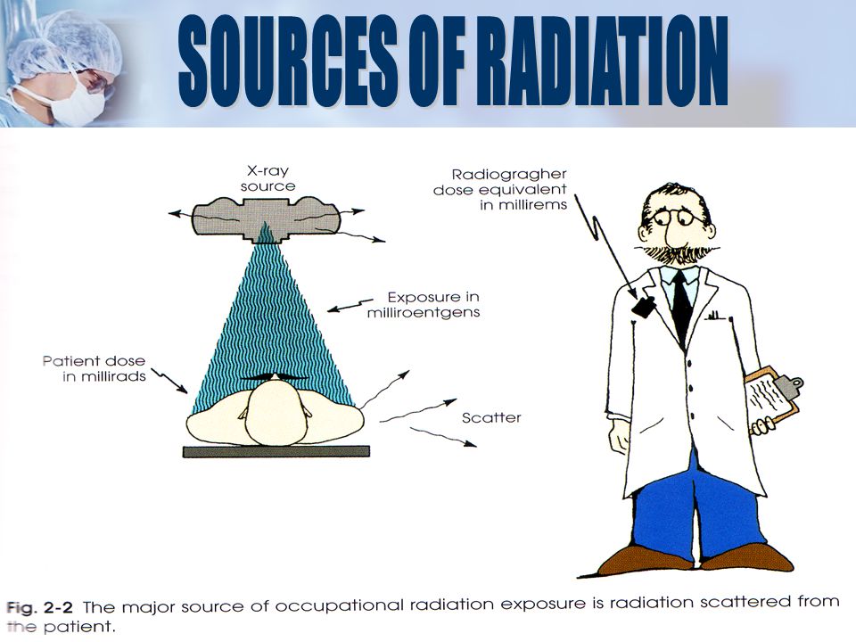
3
Be able to discuss dose limits and typical doses during different radiological procedures Be able to explain the relative risks of radiation Have a knowledge of how to reduce radiation doses, especially to yourselves

15
Over one year

18
-measured in C/kg or Roentgen(R) -amount of charge (electrons) liberated per kilogram of Air (Ionization) 1R = 2.58x10 -4 C/Kg
 -amount of charge (electrons) liberated per kilogram of Air (Ionization) 1R = 2.58x10 -4 C/Kg")
19
-measured in Gray (Gy) or Rad -amount of energy deposited/ absorbed per kilogram of tissue 1Gy = 1 Joule/Kg 100 Rad = 1 Joule/Kg 1Rad = 1/100 Gy
 or Rad -amount of energy deposited/ absorbed per kilogram of tissue 1Gy = 1 Joule/Kg 100 Rad = 1 Joule/Kg 1Rad = 1/100 Gy")
20
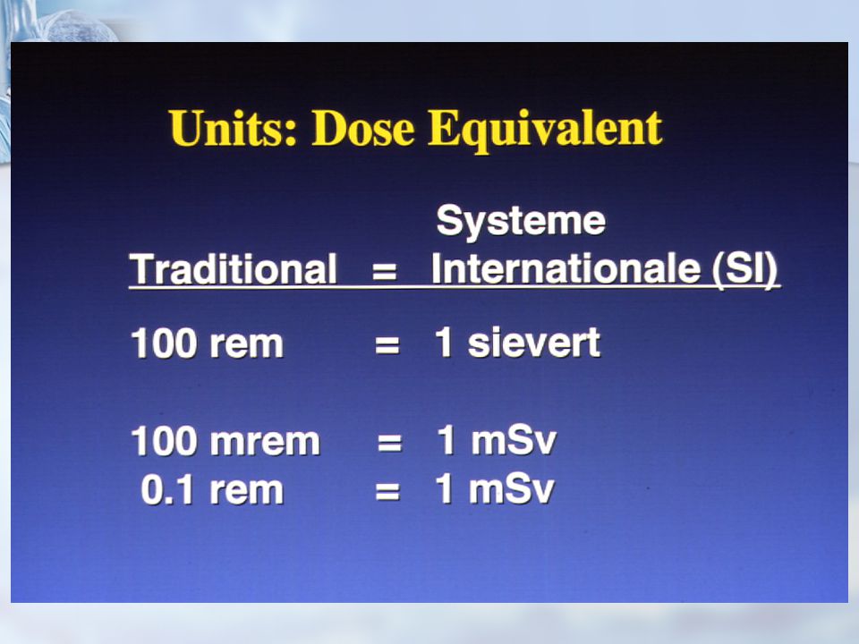
- measured in Sieverts (Sv) or Rem - amount of biological damage - gives a measure dose as if received by the whole body - used to equate dose to risk 1Sv = 1 Joule/Kg 100 Rem= 1 Joule/Kg 1Sv= 1/100 Rem
 or Rem - amount of biological damage - gives a measure dose as if received by the whole body - used to equate dose to risk 1Sv = 1 Joule/Kg 100 Rem= 1 Joule/Kg 1Sv= 1/100 Rem")
21
EXAMBONE MARROW DOSE (mRad) Gonadal Dose (mRad) MaleFemale Skull10<1 C-Spine20<1 Chest2<1 Abdomen30100200 L-Spine60175400 Pelvis20300150 Extremity2<1
 Gonadal Dose (mRad) MaleFemale Skull10<1 C-Spine20<1 Chest2<1 Abdomen L-Spine Pelvis Extremity2<1")
22
ExamTechniqueDosage Typical mammography exam 30mRem Chest120Kvp@ 100mAs 360mRem 120Kvp@ 20mAs 70mRem Abdomen120Kvp@ 160mAs 1000mRem During regular CT breast gets 700mRem Low dose mammography 160mRem Generally 1 CT exam is equivalent to 20 mammos

23
OCCUPATION EXPOSURE 1. Effective Dose Limits. a. Annual 50mSv 5 rem b. Cummulative 10mSv x age 1 rem x age 2. Equivalent Dose Annual Limits for Tissues and Organs a. Lens of the eye 150mSv 15 rem b. Localized area of the skin, hands and feet. 500mSv 50 rem

24
PUBLIC EXPOSURE (Annual) 1. Effective dose limits, continuos or frequent exposure. 1 mSv (0.1 rem) 2. Effective dose limits, infrequent exposure 5 mSv (0.5 rem) 3. Equivalent Dose Annual Limits for Tissues and Organs a. Lens of the eye 15mSv (1.5 rem) b. Localized area of the skin, hands and feet. 50mSv (5.0 rem) 4. Negligible individual dose (annual) 0.5 mSv (0.05 rem)
 2. Effective dose limits, infrequent exposure 5 mSv (0.5 rem) 3. Equivalent Dose Annual Limits for Tissues and Organs a. Lens of the eye 15mSv (1.5 rem) b. Localized area of the skin, hands and feet. 50mSv (5.0 rem) 4. Negligible individual dose (annual) 0.5 mSv (0.05 rem).")
29
More radiosensitive than adults due to sensitive cells and developing organs More radiosensitive than adults due to sensitive cells and developing organs Gonad shielding important Gonad shielding important Radiographic examinations difficult: Radiographic examinations difficult: Patient movement Patient movement Exposure technique more critical Exposure technique more critical

38
Acute Radiation SyndromeHematologic Syndrome Gastrointestinal syndrome Central nervous system Local Tissue damageSkin Gonads Extremities Hematologic depression Cytogenic damage

39
Acute Radiation SyndromeHematologic Syndrome Gastrointestinal syndrome Central nervous system Other malignant diseaseBone cancer Lung cancer Breast cancer Leukemia Genetically significant dose Lifespan shortening

40
Photograph of the patient’s back 6-8 weeks after multiple coronary angiography and angioplasty procedures. Photograph of the injury 16-21 weeks after the procedures. A small ulcerated area is present.

41
Close-up of the lesion shown in C Photograph of the patient’s back 18-21 months after the procedures. Tissue necrosis is evident

42
Photograph of the patient’s back after Grafting.

45
cancer birth defects genetic effects

46
Based upon studies of Hiroshima atomic bomb survivors, statisticians predict that an effective dose of 10 mSv (1 rem) given to a population of one million would result in 400 additional cancer deaths!
 given to a population of one million would result in 400 additional cancer deaths!")
47
Radiogenic cancers have a 20+ year latency period Radiologists during the first half of the twentieth century discovered this the hard way

48
Radiation exposure of personnel and the general public should be kept A s L ow A s R easonably A chievable.

49
correct exposure factors correct radiographic technique appropriate radiation protection appropriate development/viewing techniques appropriate radiographic positions for examination minimize repeat examinations continuing education

50
Only a physician or a registered x-ray technologist under the direct supervision of a physician may perform fluoroscopy.

51
Time Time Distance Distance Shielding Shielding 3 Basic Categories

52
Reduce of your exposure Increase from the source Make use of available TIME DISTANCE SHIELDING

53
minimize time in radiography or fluoroscopy rooms minimize time in radiography or fluoroscopy rooms minimize time spent with patients who are undergoing therapy treatment eg. nuclear medicine procedures, radioactive implants minimize time spent with patients who are undergoing therapy treatment eg. nuclear medicine procedures, radioactive implants Know Your Protocol Know Your Protocol Read the procedure through carefully Understand the steps clearly or Have the protocol displayed where you can see it Practice the technique beforehand Practice the technique beforehand

54
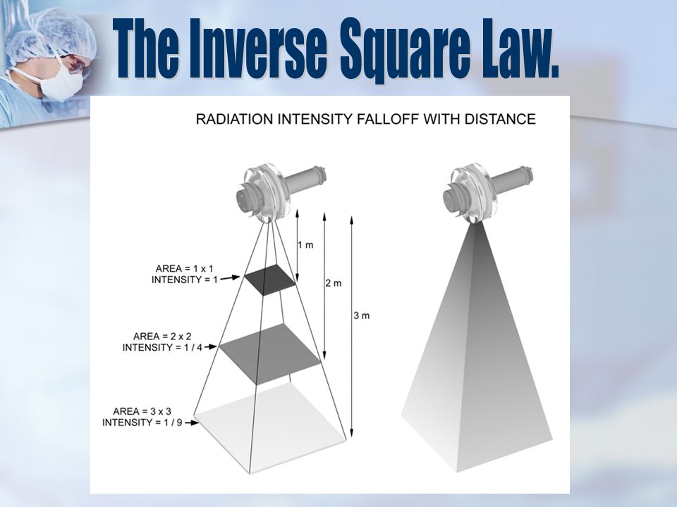
The distance between you and the isotope is of paramount importance for high energy emitters and penetrating radiations. The intensity of radiation at different distances is represented by the formula: This is the Inverse Square Law.

57
Inverse Square Law - double the distance from the source of radiation - reduce dose by a factor of 4 Inverse Square Law - double the distance from the source of radiation - reduce dose by a factor of 4 General rule - 3 meters (approximately 10 ft) from the source of radiation - dose is insignificant General rule - 3 meters (approximately 10 ft) from the source of radiation - dose is insignificant
 from the source of radiation - dose is insignificant General rule - 3 meters (approximately 10 ft) from the source of radiation - dose is insignificant")
59
Fluoro only when viewing monitor Use pulsed fluoroscopy when possible Use last image hold

60
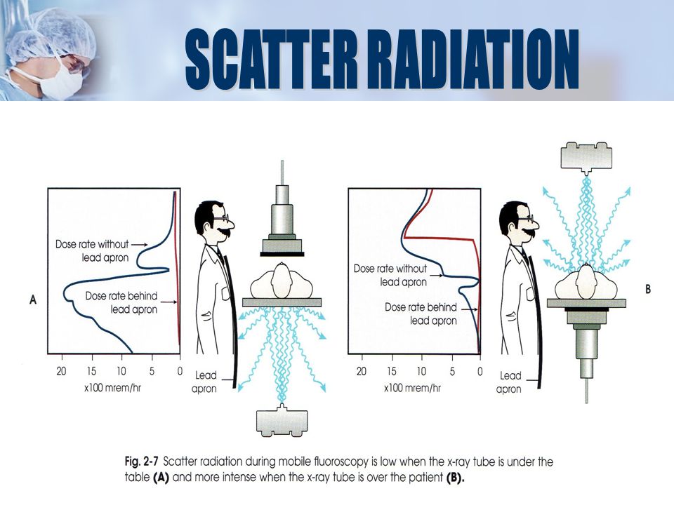
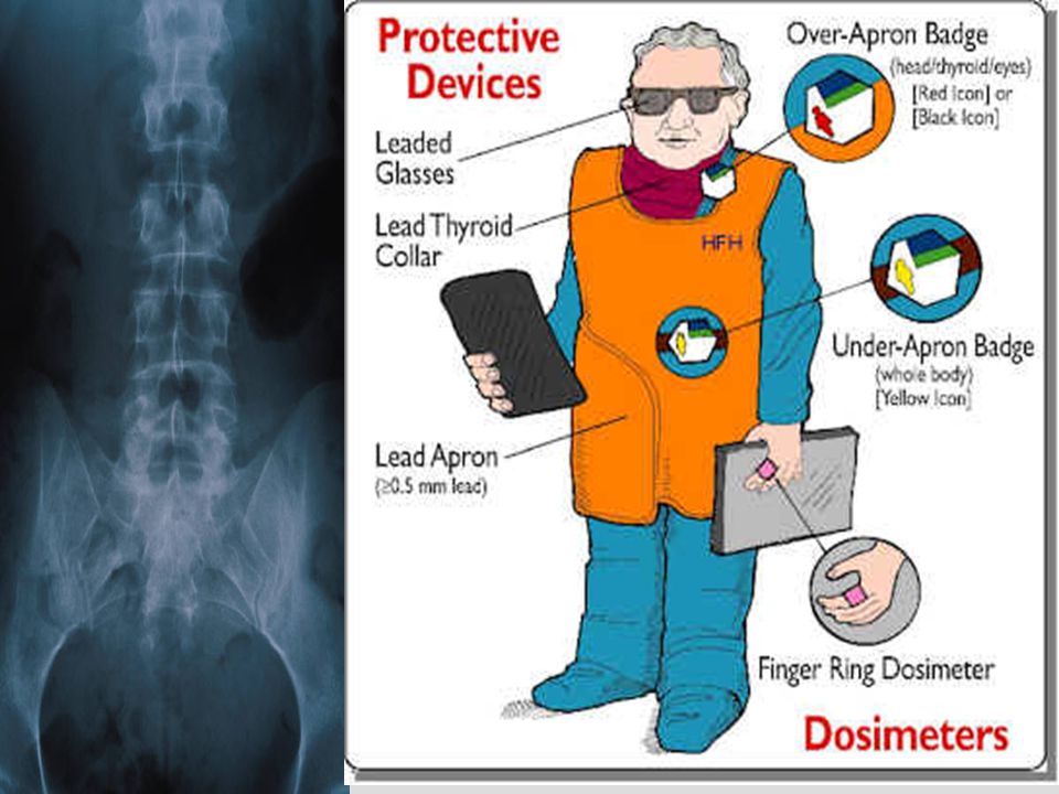
Personal shielding lead aprons - at least 3 - 5mm Pb equivalent provide up to 90% shielding. thyroid / eye shielding during fluoroscopy lead glove (5mm + Pb eq.) if hands are likely to be in the beam Lead drape on fluoro tower provides an additional 90% protection of the remaining 10% from lead aprons above.
 if hands are likely to be in the beam Lead drape on fluoro tower provides an additional 90% protection of the remaining 10% from lead aprons above..")
61
Protective barriers lead glass / acrylic for windows, lead sheets in doors or plaster board walls, brick walls lead glass / acrylic for windows, lead sheets in doors or plaster board walls, brick walls minimize directing primary beam at widows / doors minimize directing primary beam at widows / doors best position to be located during x-ray exposure best position to be located during x-ray exposure

62
State law requires that, during fluoroscopy, one badge must be worn outside the apron at the collar level. Some institutions provide additional badges, usually upon request during pregnancy

64
Badge readings are reviewed regularly by the RSO Institutional investigation levels are set below regulatory limits Personnel are notified regularly and badge readings are posted

65
IncidenceMortality ALL TUMORS Total (%) Total (%) 9.014 (100%) 5859 (100%) SOLID TUMOR TOTAL 95.586125529 Digestive system 53.247963534 Respitory system 11.41027828 Female Breast 5.9529141 Female Genitals 9.9891402 Male Genital 1.816068 Other solid cancers 13.31209556 HEMATO-LYMPHOPOIETIC4.5402330
 Total (%) (100%) 5859 (100%) SOLID TUMOR TOTAL Digestive system Respitory system Female Breast Female Genitals Male Genital Other solid cancers HEMATO-LYMPHOPOIETIC")
66
Sources: American College of Radiology, David J. Brenner/Columbia University Medical Center, U.S. Food and Drug Administration, David C. Levin/Thomas Jefferson University Hospital, National Institutes of Health, Nuclear Energy Institute, Yale University School of Medicine 1,300 Radiation dose, in millirems (mrem), from a single full-body computed tomography (CT) scan 1.5 Miles Distance you’d need to have been from the Hiroshima atomic explosion to receive an equivalent dose 29 Radiation dose, in mrem, from smoking a pack of cigarettes 300 Average annual radiation dose from natural sources, in mrem, per person in the U.S. 1 Average annual radiation dose, in mrem, from eating one or two bananas a week 0.08% Increase in risk of death from cancer after a full-body CT scan 3.75% Increase in risk of death from cancer if you receive a full-body CT scan annually starting at age 25 57 MILLION Number of full-body CT scans performed in 2003 $16 BILLION Estimated annual cost of unnecessary diagnostic imaging 7 Percentage of patients informed of the risks of their CT scans
, from a single full-body computed tomography (CT) scan 1.5 Miles Distance you’d need to have been from the Hiroshima atomic explosion to receive an equivalent dose 29 Radiation dose, in mrem, from smoking a pack of cigarettes 300 Average annual radiation dose from natural sources, in mrem, per person in the U.S. 1 Average annual radiation dose, in mrem, from eating one or two bananas a week 0.08% Increase in risk of death from cancer after a full-body CT scan 3.75% Increase in risk of death from cancer if you receive a full-body CT scan annually starting at age 25 57 MILLION Number of full-body CT scans performed in 2003 $16 BILLION Estimated annual cost of unnecessary diagnostic imaging 7 Percentage of patients informed of the risks of their CT scans.")
69
1990 Recommendations of the International Commission on Radiological Protection, ICRP Publication 60, Permagon Press, Oxford 1990 Recommendations of the International Commission on Radiological Protection, ICRP Publication 60, Permagon Press, Oxford Brennen S.E. and Putney R.G., eds (1983) Dose reduction in diagnostic radiology, The Hospital Physicist’s Associtaion Plaut S., (1993) Radiation protection in the x-ray department, Butterworth- Heinemann, Oxford Brennen S.E. and Putney R.G., eds (1983) Dose reduction in diagnostic radiology, The Hospital Physicist’s Associtaion Plaut S., (1993) Radiation protection in the x-ray department, Butterworth- Heinemann, Oxford Bushong S.C., (2004) Radiological science for technologists: Physics, Biology and Protection, 8th Ed. Mosby, St Louis Bushong S.C., (2004) Radiological science for technologists: Physics, Biology and Protection, 8th Ed. Mosby, St Louis Faulkner K. and Wall B.F., eds (1988) Are x-rays safe enough? Patient dose and risks in diagnostic radiology, The Institute of Physical Science in Medicine, Report No. 55 Faulkner K. and Wall B.F., eds (1988) Are x-rays safe enough? Patient dose and risks in diagnostic radiology, The Institute of Physical Science in Medicine, Report No. 55 Norris, Teresa G., Radiation Safety in Fluoroscopy. Radiologic Technology. Vol 73 No 6, August 2002, 511 Norris, Teresa G., Radiation Safety in Fluoroscopy. Radiologic Technology. Vol 73 No 6, August 2002, 511 Seeram E., (1997) Radiation protection, Lippincott, Philadelphia Seeram E., (1997) Radiation protection, Lippincott, Philadelphia Sherer, Mary Alice., Visconti, Paul J., Ritenour, Russell.,(2002) Radiation Protection in Medical Radiography, 8th Ed. Mosby, St Louis. Sherer, Mary Alice., Visconti, Paul J., Ritenour, Russell.,(2002) Radiation Protection in Medical Radiography, 8th Ed. Mosby, St Louis. Web D.V., Solomon S.B. and Thomson J.E.M., (1999) Background radiation levels and medical exposure in Australia, Radiation Protection in Australia, Vol 16 No 2 pp.25-32 Web D.V., Solomon S.B. and Thomson J.E.M., (1999) Background radiation levels and medical exposure in Australia, Radiation Protection in Australia, Vol 16 No 2 pp.25-32
 Dose reduction in diagnostic radiology, The Hospital Physicist’s Associtaion Plaut S., (1993) Radiation protection in the x-ray department, Butterworth- Heinemann, Oxford Brennen S.E. and Putney R.G., eds (1983) Dose reduction in diagnostic radiology, The Hospital Physicist’s Associtaion Plaut S., (1993) Radiation protection in the x-ray department, Butterworth- Heinemann, Oxford Bushong S.C., (2004) Radiological science for technologists: Physics, Biology and Protection, 8th Ed. Mosby, St Louis Bushong S.C., (2004) Radiological science for technologists: Physics, Biology and Protection, 8th Ed. Mosby, St Louis Faulkner K. and Wall B.F., eds (1988) Are x-rays safe enough. Patient dose and risks in diagnostic radiology, The Institute of Physical Science in Medicine, Report No. 55 Faulkner K. and Wall B.F., eds (1988) Are x-rays safe enough. Patient dose and risks in diagnostic radiology, The Institute of Physical Science in Medicine, Report No. 55 Norris, Teresa G., Radiation Safety in Fluoroscopy. Radiologic Technology. Vol 73 No 6, August 2002, 511 Norris, Teresa G., Radiation Safety in Fluoroscopy. Radiologic Technology. Vol 73 No 6, August 2002, 511 Seeram E., (1997) Radiation protection, Lippincott, Philadelphia Seeram E., (1997) Radiation protection, Lippincott, Philadelphia Sherer, Mary Alice., Visconti, Paul J., Ritenour, Russell.,(2002) Radiation Protection in Medical Radiography, 8th Ed. Mosby, St Louis. Sherer, Mary Alice., Visconti, Paul J., Ritenour, Russell.,(2002) Radiation Protection in Medical Radiography, 8th Ed. Mosby, St Louis. Web D.V., Solomon S.B. and Thomson J.E.M., (1999) Background radiation levels and medical exposure in Australia, Radiation Protection in Australia, Vol 16 No 2 pp Web D.V., Solomon S.B. and Thomson J.E.M., (1999) Background radiation levels and medical exposure in Australia, Radiation Protection in Australia, Vol 16 No 2 pp")
Similar presentations


>")


>")


