Download presentation
Presentation is loading. Please wait.

1
Nursing Management of Patients with Respiratory Disorders
Winter Quarter 2012 Teresa M. Champion, RN MSN

2
ASSESSMENT OF PATIENTS WITH RESPIRATORY DISORDERS

3
Anatomy Physiology of Pulmonary System
Ventilation – movement of air in and out of lungs Respiration – consists of diffusion of oxygen across alveolar-capillary membrane into pulmonary circulation and release of carbon dioxide molecules across the alveolar- capillary membrane through the airways out into the environment

4
Exchange of Gases during Respiration
Respiration Perfusion – the exchange of O2 and CO2 across the alveolar membrane Alveoli – place in lungs where exchange occurs and must be adequately expanded by air to have adequate contact with hemoglobin If alveoli are expanded adequately but unable to exchange due to edema or secretions – a ventilation(V)/perfusion(Q) mismatch occurs If alveoli are not expanded adequately despite blood flow – Ventilation (V) and Perfusion (Q) mismatch will also occur Anemic patients have fewer binding sites for O2. Other compromising conditions – shock, heart failure can also affect gas exchange because blood flow Is diverted from the alveoli and decreases gas exchange
/perfusion(Q) mismatch occurs. If alveoli are not expanded adequately despite blood flow – Ventilation (V) and Perfusion (Q) mismatch will also occur. Anemic patients have fewer binding sites for O2. Other compromising conditions – shock, heart failure can also affect gas exchange because blood flow Is diverted from the alveoli and decreases gas exchange.")
5
Mechanisms of Respiration
Ventilation – dependent on neuromuscular and musculoskeletal integrity CNS – medulla and Pons respond to changes to carbon dioxide and oxygen levels in the blood by increasing or decreasing rate and depth of respiration Musculoskeletal – assist and influence respiration - intercostal muscles, diaphragm, abdominal muscles, thoracic muscles (scalene, sternomastoid and trapezius)
")
6
Inspiration and Expiration
Influenced by intrapleural pressures When the pressure of air in the lungs reaches capacity during inspiration, expiration begins Inspiration is active Expiration is passive Movement of air in one breath is the tidal volume Movement of air over one minute is minute ventilation Normal tidal volume at rest is ~500ml, but can increase if more is demanded by the body (i.e. exercise and stress)
")
7
Collection of Patient Data
History Biographic and demographic data Chief complaint - dyspnea Past medical history, allergies Family history Risk factors Social history, Cultures Medications Nutrition Travel and Areas of Residence Biographic and Demographic – age, race, gender and cultures that affect perceptions of health conditions like pulmonary disorders. Chief complaint – s/s of respiratory malfunctions (dyspnea, fatigue) Systems that affect the patients ability to carry out or perform normal daily activities Past medical history – cancers like breast and testicular – mets to lungs OR other prior illnesses. Cardiovascular complications and medication related pulmonary fibrosis that occurs with amiodarone. Thyroid disorders (myxedema or thyroitoxosis) Childhood diseases – cystic fibrosis, asthma, lack of immunizations, premature infants that were intubated and mechanically ventilated at birth. Childhood or past trauma. Past pulmonary dysfunctions, history of DVT. Allergies – allergies can trigger respiratory problems – Pts that are allergic to soy or nuts should not use atrovent and combivent Family history – genetic related conditions or family members that smoke or family history of lung disease, cardiovascular disease and lung cancers. Risk factors – smoking, substance abuse, lack of physical activity and obesity Social – lifestyles and habits – occupational exposures via work (asbestos, pesticides) – exposures in the home (mold, pet dander, cock roach infestations, meth labs) Cultures – affect how and when people seek and accept diagnostics and treatments. Cultural Practices like use of amulets, cupping, coining, and herbal remedies Medication like ACE inhibitors cause cough, amiodarone cause pulmonary fibrosis, address OTC like cough suppressants, expectorants, antihistamines Herbals Proper use of inhaled treatment – bronchodilators first then steroids, Advair a combo of the two should not be given with a spacer. Traveling to other countries like the (SARS) outbreak was traced to a specific location in the world. High altitudes (HAPE), living near livestock or factories that emit possible environmental exposures
 Systems that affect the patients ability to carry out or perform normal daily activities. Past medical history – cancers like breast and testicular – mets to lungs OR other prior illnesses. Cardiovascular complications and medication related pulmonary fibrosis that occurs with amiodarone. Thyroid disorders (myxedema or thyroitoxosis) Childhood diseases – cystic fibrosis, asthma, lack of immunizations, premature infants that were intubated and mechanically ventilated at birth. Childhood or past trauma. Past pulmonary dysfunctions, history of DVT. Allergies – allergies can trigger respiratory problems – Pts that are allergic to soy or nuts should not use atrovent and combivent. Family history – genetic related conditions or family members that smoke or family history of lung disease, cardiovascular disease and lung cancers. Risk factors – smoking, substance abuse, lack of physical activity and obesity. Social – lifestyles and habits – occupational exposures via work (asbestos, pesticides) – exposures in the home (mold, pet dander, cock roach infestations, meth labs) Cultures – affect how and when people seek and accept diagnostics and treatments. Cultural Practices like use of amulets, cupping, coining, and herbal remedies. Medication like ACE inhibitors cause cough, amiodarone cause pulmonary fibrosis, address OTC like cough suppressants, expectorants, antihistamines Herbals. Proper use of inhaled treatment – bronchodilators first then steroids, Advair a combo of the two should not be given with a spacer. Traveling to other countries like the (SARS) outbreak was traced to a specific location in the world. High altitudes (HAPE), living near livestock or factories that emit possible environmental exposures.")
8
PHYSICAL EXAMINATION Inspection General Appearance Mentation
Rate, Depth and Rhythm of Respirations Tachypenea, Bradypnea, Orthopnea, Apnea, Hyperpnea Thoracic Size and Shape Thoracic Expansion and Symmetry Use of Accessory Muscles Color and Appearance of Skin and Extremities Pallor Cyanosis Neck Inspection – Tracheal Deviation General Appearance – does patient appear capable to take care of themselves, comfortable. Facial appearance non-distressed. No evidence of tripod posture – leans forward resting on both arms to allow chest expansion. Nasal flaring, pursed lip breathing? Mentation – the brain needs oxygen…any confusion or agitation, lethargy? Decreased level of consciousness Regular rate of breathing is between 12 and 20 per minute. FOR EACH INCREASE IN ONE DEGREE OF TEMPERATURE – HEART RATE WILL INCREASE 4 BEATS PER MIN AND RESPIRATORY RATE INCREASES ONE BREATH PER MINUTE. Orthopnea – difficulty breathing while lying down Hyperpnea – increased rate and depth of respirations Thoracic size and shape – clues for pulmonary disease are structural abnormalities like sternal depression, pigeon chest, barrel chest and scoliosis or kyphoscoliosis. These abnormalities will limit chest expansion needed for breathing Chest expansion and symmetry – inspiration diaphragm flattens and rises with expiration Accessory muscles – trapezius, sternomastoid, scalenous, abdominal muscles Color and appearance – clubbing of fingers, pallor – pale, cyanosis - blue

10
ABNORMAL BREATHING PATTERNS
Cheyenne Stokes Breaths are deep than become shallow followed by periods of apnea Causes: severe brain pathology - brain stem herniation, Increased ICP, compression on Brain Stem Kussmal’s Breaths are deep, rapid and labored Rates are >20 bpm Causes: metabolic acidosis, renal failure, diabetic ketoacidosis

11
PHYSICAL EXAMINATION Palpatation of Skin and Extremities
Edema – Caused by Pulmonary HTN 8mm – 4+, 6mm – 3+, 4mm – 2+, 2 mm – 1+ Skin Temperature & Moisture warm moist skin r/t increased effort of breathing, possible fever from pulmonary infection Dry skin-moisture lost from increased respirations Clinical Reference Points – landmarks Trachea, nipple line, sternum, intercostals, axillary line, midaxillary line, midclavicular line Chest Excursion – should be equal and up to 5-10 cm Tactile Fremitus – palpations of vibration in thorax Tenderness Crepitus – also called “subcutaneous emphysema – air trapped under the subcutaneous tissue EDEMA – press finger down over 5 seconds – depth related to the rating of pitting – caused by Right sided Heart Failure or Right and Left Heart Failure Right sided heart failure is caused by Pulmonary Hypertension and the increased resistance of forward flow causes the right ventricle to hypertrophy in an attempt to increase the flow through the pulmonary tree. SKIN TEMP and MOISTURE – COPD and Asthma Exacerbations may have dry skin due to moisture lost through the respiratory tract from increased breathing rates. Causes dehydration as they may not be able to replace fluids. Clinical Reference points during the pulmonary assessments and lesions, adventitious lung sounds heard along these reference points/lines. Tactile Fremitis – palpated over the thorax by having the patient vocalize “99” . Palpate with ulnar side of hand from scapula (back) Clavicles on front of chest – move down bilaterally. Vibrations should be the strongest over large airways on upper thorax, and decrease as you move down. Unequal fremitis could indicate unilateral airway obstruction, pneumothorax, or pleural effusion. Decreased fremitis could also be palpated with pneumonia or consolidations Tenderness – from coughing, intercostals are strained and thoracic muscles. Pneumonia and consolidation can also cause tenderness, people may not breath as deep Crepitus – caused by ruptured pulmonary bullae, pneumothorax or around chest tube sites.
 Clavicles on front of chest – move down bilaterally. Vibrations should be the strongest over large airways on upper thorax, and decrease as you move down. Unequal fremitis could indicate unilateral airway obstruction, pneumothorax, or pleural effusion. Decreased fremitis could also be palpated with pneumonia or consolidations. Tenderness – from coughing, intercostals are strained and thoracic muscles. Pneumonia and consolidation can also cause tenderness, people may not breath as deep. Crepitus – caused by ruptured pulmonary bullae, pneumothorax or around chest tube sites.")
13
PHYSICAL EXAMINATION Auscultation of the lungs – 4 types of breath sounds Tracheal Breath Sounds – loud and high pitched – over the largest airway and are the loudest – length of time heard is equal during expiration and inspiration Bronchial Breath Sounds – loud and high pitch, harsh and less turbulent and lower in frequency than tracheal – Expiration is heard longer than inspiration Bronchovesicular – Midway in pitch between Bronchial and Vesicular and are heard during inspiration and expiration Vesicular Soft and low pitched – heard longer during expiration, heard over most of thorax Tracheal – best heard over the trachea and neck – Largest airway Bronchial - from 2nd to 4th intercostals on anterior chest and between 3rd and 6th on posterior chest - Bronchovesicular best heard over 1st and 2nd intercostals on anterior chest and between scapulae of the posterior chest – air movement in the moderate airways between the bronchi and the smaller airways. Vesicular – air moving through the smaller airways Expiration last longer than inspiration so most breath sounds are heard longer during expiration than inspiration.

14
Bronchial – 2nd to 4th intercostals
Bronchovesicular – 1st and 2nd intercostals Vesicular – heard all over thorax

15
Bronchial – 3rd and 6th intercostals
Bronchovesicular – between scapulas

16
ADVENTITIOUS BREATH SOUNDS
Crackles (Rales) Indicate fluid, inflammation in airways – snapping sound when airways open – can be heard when airways close too but softer sounding than on inspiration Intermittent or discontinuous. Fine or Course Wheezes High-pitched musical sounds caused by inflammation in narrowing airways or bronchospasms Rhonchi – indicate mucus secretions in the airways Caused by air passing through mucus strands Can be heard on inspiration and expiration Continuous/ discontinuous (intermittent), Mild/moderate/severe Crackles are described as snapping, popping sounds Rales and Rhonchi terms not used according to this book. Rhonchi is classified as Wheezes, according to this book. Low pitched wheezes – sounds like rattling, snoring, gurgling and usually clear with coughing and or suctioning.
 Indicate fluid, inflammation in airways – snapping sound when airways open – can be heard when airways close too but softer sounding than on inspiration. Intermittent or discontinuous. Fine or Course. Wheezes. High-pitched musical sounds caused by inflammation in narrowing airways or bronchospasms. Rhonchi – indicate mucus secretions in the airways. Caused by air passing through mucus strands. Can be heard on inspiration and expiration. Continuous/ discontinuous (intermittent), Mild/moderate/severe. Crackles are described as snapping, popping sounds. Rales and Rhonchi terms not used according to this book. Rhonchi is classified as Wheezes, according to this book. Low pitched wheezes – sounds like rattling, snoring, gurgling and usually clear with coughing and or suctioning.")
17
ADVENTITIOUS BREATH SOUNDS
Stridor Heard only during inspiration as air attempts to flow though an obstruction, high pitched crowing sound – needs immediate intervention Pleural Friction Rub Indicate inflamed pleural surfaces – easily heard on inspiration – hold breath to determine it is not pericardial

18
Page 878 in book

19
USING THE STEHASCOPE Diaphragm - best for higher pitched sounds, like breath sounds and normal heart sounds. Bell - is best for detecting lower pitch sounds, like some heart murmurs, and some bowel sounds. It is used for the detection of bruits, and for heart sounds (for a cardiac exam, listen with the diaphragm, and repeat with the bell).
.")
20
PHYSICAL EXAMINATION Percussion Dull - Abnormal Finding
Heard over solid tissue, occurs when air is absent, can be heard over consolidation areas with pneumonia, pleural effusion, hemothorax, solid tumors Dull thumping sound without vibration Resonant – Normal Finding heard over lung fields during inspiration while lungs are full of air low pitched clear sounds – Normal Finding Hyperresonant – Abnormal Finding Very loud, lower pitched longer sound than resonance Drum like sound with vibration indicates hyperinflamation – emphysema, pneumothorax Tympanic – like hyperresonance like tapping on air filled cheeks

21
PAIN Pain in association with breathing may be related to Pulmonary Embolism, Pneumothorax, Pleural Disease, Pericarditis, Musculoskeletal Disease or Pneumonia Sudden onset shortness of breath may be related to Pulmonary Embolism or pneumothorax Pain during respiration may decrease tidal volumes Pain management enables participation in rehabilitative activities and promotes deep breathing to prevent pneumonia and atelectasis Use cough suppressants with caution

22
GERONTOLOGICAL CONSIDERATIONS
Aging decreases respiratory function Osteoporosis – stooped posture, decreased rib expansion Anterior to posterior diameter increases Alveolar surface decreases Decreased elasticity Increased atelectisis Lower arterial oxygen values – decreased exchange of O2/CO2 Increase risk of pneumonia – Decreased tidal volumes, ineffective cough Risk of aspiration may increase with aging Aging may affect patient comfort needs during the examination

23
HEALTH PROMOTION Smoking cessation
Decrease exposure to second-hand smoke Hand Hygiene Flu and Pneumonia Vaccines Instruction and use of Personal Protective Equipment (PPE) especially in the workplace for workers exposed to allergens, mold, bird, bat and rat feces and other toxins like asbestos
 especially in the workplace for workers exposed to allergens, mold, bird, bat and rat feces and other toxins like asbestos.")
24
STANDARD OF CARE For patients with cardiac and respiratory illness, standard is: Continuous or intermittent observation of the patient’s oxygen saturation (most cost effective) End-tidal carbon dioxide levels (being used more, but is more costly but most accurate). Monitoring Peak Flow results is utilized to trend treatment effectiveness in patients with asthma
 End-tidal carbon dioxide levels (being used more, but is more costly but most accurate). Monitoring Peak Flow results is utilized to trend treatment effectiveness in patients with asthma.")
25
RESPIRATORY MONITORING
Pulse Oximetry Measures saturation of hemoglobin May NOT be accurate with patients with low Hgb, hypovolemia and shock states Nail polish, ambient light may interfere with reading Wave forms should match pulse rate and should not be dampened Pulse Ox Probes should not be placed on extremities with blood pressure cuffs, fistulas or arterial lines due to lack of blood flow Pulse Ox Saturations do not necessarily match calculated Oxygen Saturation in ABG – SPO2%

26
RESPIRATORY MONITORING
Peak Flow Meters Evaluate air movement to determine severity of asthma exacerbation Measure Peak Expiratory Flow Rate Measurements are based on age and body size Red Zone (Dangerous) – less than 50% of the normal value Yellow Zone (caution) – Between 50% to 80% below normal value Green Zone (Good) – meets 80% to 100% of normal value
 – less than 50% of the normal value. Yellow Zone (caution) – Between 50% to 80% below normal value. Green Zone (Good) – meets 80% to 100% of normal value.")
27
RESPIRATORY MONITORING
Arterial Blood Gases Determine Respiratory Acidosis and Alkalosis PaO2 levels below 80mmHg and/or SaO2 <95% indicate hypoxemia Cost $800 to $ per draw Invasive procedure

28
RESPIRATORY MONITORING
Capnography Measures exhaled carbon dioxide or End Tidal CO2 (ETCO2) Small disposable capnographers are used to check ET Tube placement after intubation and/or continuous monitoring of tube placement Capnography monitoring has been added to the – 2015 ACLS Guidelines for Compression Effectiveness. Normal ETCO2 values are 35mmHg to 45mmHg. ETCO2 values between 10-20mmHg indicate high quality compressions ETCO2 less than 10mmHg – quality of chest compressions need improvement
 Small disposable capnographers are used to check ET Tube placement after intubation and/or continuous monitoring of tube placement. Capnography monitoring has been added to the 2010 – 2015 ACLS Guidelines for Compression Effectiveness. Normal ETCO2 values are 35mmHg to 45mmHg. ETCO2 values between 10-20mmHg indicate high quality compressions. ETCO2 less than 10mmHg – quality of chest compressions need improvement.")
29
MONITOR SHOWING ETCO2 WAVEFORM

30
RESPIRATORY PATHWAY DISORDERS
SLEEP APNEA AIRWAY OBSTRUCTION TRACHEOSTOMY

31
ANATOMY OF RESPIRATORY TRACT
Nose to pharynx-behind the mouth to esophagus (approx. 5 inches) Epiglottis – above Larynx covers when swallowing Larynx - voice box: air passes between pharynx and trachea Trachea - windpipe Bronchi - main branch that air passes through divides into left and right branch Bronchioles - subdivides and connects with alveoli for gas exchange
 Epiglottis – above Larynx covers when swallowing. Larynx - voice box: air passes between pharynx and trachea. Trachea - windpipe. Bronchi - main branch that air passes through divides into left and right branch. Bronchioles - subdivides and connects with alveoli for gas exchange.")
32
SLEEP APNEA Defined – a person stops breathing for more than 10 seconds, more that times in an hour 3 types – Central Brain fails to send signal to the breathing muscles to initiate respirations (less common) Obstructive Physical obstruction from tissues in upper airway Combination of both or Mixed Sleep Apnea
 Obstructive. Physical obstruction from tissues in upper airway. Combination of both or Mixed Sleep Apnea.")
33
RISK FOR SLEEP APNEA Overweight/Obesity High Blood Pressure
Decreased Airway Size – congestion, inflammation (allergies), anatomical abnormalities Family History
, anatomical abnormalities. Family History.")
34
MEDICAL MANAGEMENT FOR SLEEP APNEA
Weight loss Avoid alcohol, tobacco and sleeping pills (sedatives) Use side-lying positions when sleeping Dental devises that move tongue or mandible forward Continuous Positive Airway Pressure (CPAP) Machines Surgical Interventions – UVPPP – resection of the uvula and soft palate, Tracheotomy Focus is on airway patency
 Use side-lying positions when sleeping. Dental devises that move tongue or mandible forward. Continuous Positive Airway Pressure (CPAP) Machines. Surgical Interventions – UVPPP – resection of the uvula and soft palate, Tracheotomy. Focus is on airway patency.")
35
AIRWAY OBSTRUCTION Potentially life threatening – requires immediate intervention Types: foreign object, allergy, lesions, stenosis, swelling Causes: Viral and Bacterial Infections, fire or inhalation burns, allergic reactions (foods/medications/bee stings) Infections after dental extractions Laryngeal trauma-MVA, Strangulation or surgical procedures Large tumors Aspiration of foreign objects
 Infections after dental extractions. Laryngeal trauma-MVA, Strangulation or surgical procedures. Large tumors. Aspiration of foreign objects.")
36
Clinical Manifestations of Airway Obstruction
STRIDOR Inability to speak (partial or complete) Labored breathing and use of accessory muscles Air Hunger (mild) Cyanosis (severe)
 Labored breathing and use of accessory muscles. Air Hunger (mild) Cyanosis (severe)")
37
Medical Management of Airway Obstruction
Diagnosis and treat the cause Provide Oxygenation Support!! Sit in upright position Keep patient’s airway patent (if partial or mild obstructions get worse – need immediate intervention) Secure and protect airway – Endotracheal Intubation, Cricothyroidotomy or tracheotomy – bag/mask ventilation will not work with obstruction! Treat cause – if infection provide anti-infectives, .steroids, bronchodilators, epinephrine if anaphylaxis allergy with airway swelling
 Secure and protect airway – Endotracheal Intubation, Cricothyroidotomy or tracheotomy – bag/mask ventilation will not work with obstruction! Treat cause – if infection provide anti-infectives, .steroids, bronchodilators, epinephrine if anaphylaxis allergy with airway swelling.")
38
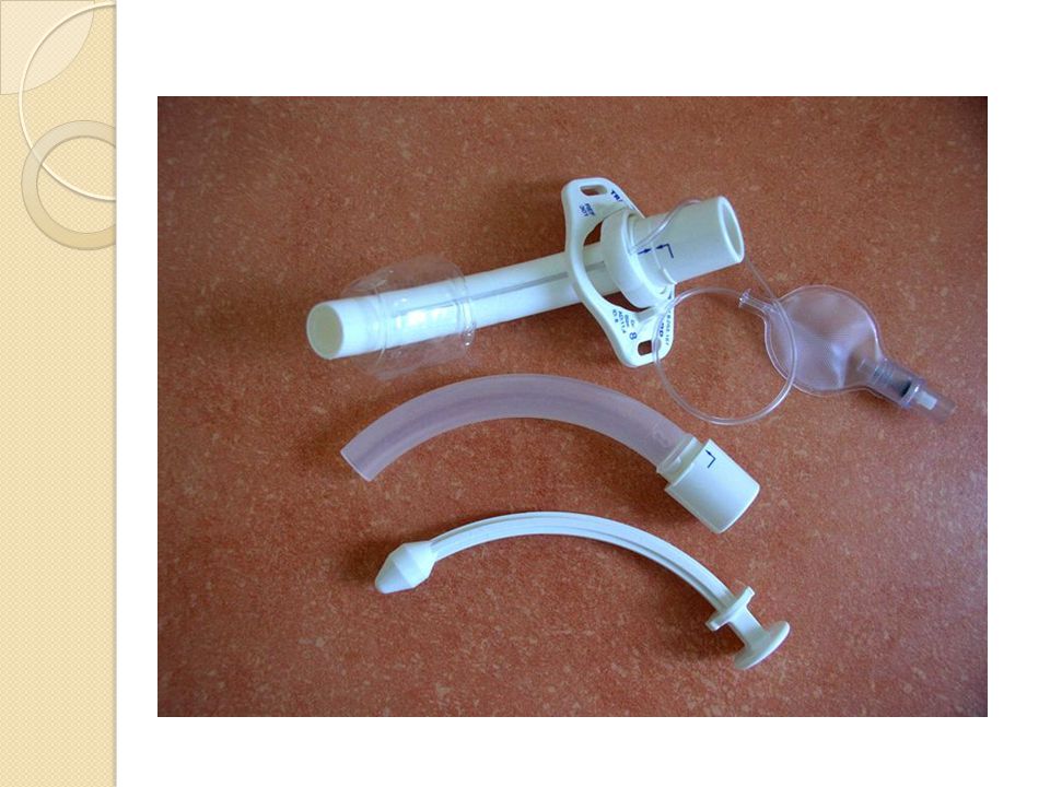
Tracheotomy Insertion of artificial airway in the trachea
Recommended for oral/nasal endotracheal intubations lasting longer than 1 – 2 weeks. (book says 7 – 10 days) Usually is temporary to protect airway until underlying cause can be fixed or corrected Decreases risk of permanent injury to the airway, and vocal cords
 Usually is temporary to protect airway until underlying cause can be fixed or corrected. Decreases risk of permanent injury to the airway, and vocal cords.")
39
Tracheotomy Incision is below the prominent thyroid cartilage (Adam’s Apple) and below the cricoid cartilage – between 2 – 3 OR 3 – 4 tracheal cartilages A tube is placed and secured (Sutured) to keep the tracheal stoma open The tube is also secured with ties The tube remains in place until the tracheal stoma is well established and won’t close back up A post tracheotomy kit is kept at bedside or on the nursing unit if accidental decannulation occurs An Obturator is kept at bedside to prevent stoma closure if decannulation occurs until new tracheotomy tube can be put in immediately
 and below the cricoid cartilage – between 2 – 3 OR 3 – 4 tracheal cartilages. A tube is placed and secured (Sutured) to keep the tracheal stoma open. The tube is also secured with ties. The tube remains in place until the tracheal stoma is well established and won’t close back up. A post tracheotomy kit is kept at bedside or on the nursing unit if accidental decannulation occurs. An Obturator is kept at bedside to prevent stoma closure if decannulation occurs until new tracheotomy tube can be put in immediately.")
40
Tracheostomy tubes can be fitted with an inflatable cuff that blocks off the top of the windpipe, preventing aspiration. Most people do better with uncuffed tubes, since they can push secretions up and out, and prevent accumulation of unswallowed food above the cuff. Also, cuff inflation interferes with passage of air up to vocal cords needed for speech.

41
TYPES OF TRACHEAL TUBES
Made of silicone, plastic, stainless steel or silver With or without an inner cannula With or with out a cuff Mechanical ventilation requires cuffed tubes to seal the airway to maintain pressures for ventilation Cuffed tracheal tubes decrease aspiration risk Inner cannulas prevent tube obstructions from thick crusted secretions Average adult size are 7 to 8 Shiley or Bivona are most common

44
Assessment of New Tracheal Artificial Airway
Auscultation of the lungs Monitoring Oxygenation saturations Assessment of increased amount of blood in the sputum and around site Subcutaneous emphysema (crepitis) around the neck Respiratory distress Patency of tracheal tube Postoperative pulmonary edema (POPE) Post operative edema (POPE) is a potentially life-threatening complication of acute airway obstruction. The predominant mechanism is forced inspiration against a closed or occluded airway. Forceful attempts to inhale against an obstruction create highly negative intrathoracic pressure, which causes increased venous return, decreased cardiac output and fluid transudation into the alveolar space.
 around the neck. Respiratory distress. Patency of tracheal tube. Postoperative pulmonary edema (POPE) Post operative edema (POPE) is a potentially life-threatening complication of acute airway obstruction. The predominant mechanism is forced inspiration against a closed or occluded airway. Forceful attempts to inhale against an obstruction create highly negative intrathoracic pressure, which causes increased venous return, decreased cardiac output and fluid transudation into the alveolar space.")
45
Interventions following Tracheostomy
Encourage cough and deep breathing Suction as necessary, but keep to a minimum Provide supplemental oxygen Pre-oxygenate with 100% Oxygen when suctioning Hyperventilate (bag – suction) when necessary Suction limited to 5-10 seconds with each pass Insert catheter till patient starts to cough or meet slight resistance – do not use force
 when necessary. Suction limited to 5-10 seconds with each pass. Insert catheter till patient starts to cough or meet slight resistance – do not use force.")
46
Tracheostomy Cares Tracheotomy cares should be done every 8 to 12 hours with cleaning the inner cannula or changing the disposable cannula Most medical institutions no longer use Hydrogen peroxide to clean around tracheostomy site (book says use half- strength) Aseptic technique is used when cleaning the inner cannula (sterile gloves)
 Aseptic technique is used when cleaning the inner cannula (sterile gloves)")
47
Laryngectomy Trachea is sutured to the stoma
Oral airway is permanently bypassed Patients lose the ability to speak because the vocal cords are bypassed permanently Patient only breath out of their stoma The esophagus still follows the normal pathway and patients can still eat normally

48
LOWER AIRWAY DISORDERS

49
Restrictive lung diseases (interstitial lung diseases)
Result in reduced lung volumes Alteration in lung parenchyma (alveolar tissue w/ terminal bronchioles, respiratory bronchioles, alveolar ducts) Disease of pleura, chest wall or neuromuscular apparatus Characterized by reduced total lung capacity, vital capacity, or resting lung volume
 Disease of pleura, chest wall or neuromuscular apparatus. Characterized by reduced total lung capacity, vital capacity, or resting lung volume.")
50
Obstructive lung diseases
Common characteristic – chronic and recurring blockage of airways Limit airflow through the airways and out of the lungs

51
LOWER AIRWAY DISORDERS
Infections and Inflammatory Disorders of the Lungs Acute or Chronic Bronchitis Influenza – complication - pneumonia Pneumonia – 6th leading cause of death Tuberculosis – Mycrobacterium tuberculosis Lung Abcesses/Empyema Globally, pneumonia causes more deaths than any other infectious disease. Empyema is a collection of pus in the space between the lung and the inner surface of the chest wall (pleural space).
.")
52
Pneumonia Inflammatory process that results in edema of the parenchymal lung tissue (aveoli and bronchioles) caused by inhalation of bacteria, viruses, fungi, protozoa or parasites or enter lungs via the blood stream Aspiration of water, food, vomitus, toxic gases, chemicals and smoke Radiation therapy Effects terminal gas exchange
 caused by inhalation of bacteria, viruses, fungi, protozoa or parasites or enter lungs via the blood stream. Aspiration of water, food, vomitus, toxic gases, chemicals and smoke. Radiation therapy. Effects terminal gas exchange.")
53
Classifications of pneumonia
Type of agent causing infection Distribution of the infection (lobar - consolidation of a lobe, or bronchial – patchy consolidation) Setting (community or healthcare (hospital) aquired
 Setting (community or healthcare (hospital) aquired.")
54
Community Acquired Pneumonia (CAP)
CAP: begins outside hospital or is diagnosed w/in 48 hours after admission Patient did not reside in a long-term facility prior to admission Incidence of CAP is highest in winter months Smoking an important risk factor

55
Healthcare (Hospital) Acquired Pneumonia (HAP)
HAP: occurs > 48 hours after hospital admission HAP has a mortality rate of 20% to 50% 90% of HAP infections are bacterial Compromised immune systems, chronic lung disease, intubation and mechanical ventilation increase risk

56
Severe Acute Respiratory Syndrome
Severe acute respiratory syndrome (SARS) is a serious form of pneumonia. It is caused by a virus that was first identified in Infection with the SARS virus causes acute respiratory distress (severe breathing difficulty) and sometimes death. SARS is a dramatic example of how quickly world travel can spread a disease. It is also an example of how quickly a connected health system can respond to a new health threat World Health Organization (WHO) physician Dr. Carlo Urbani identified SARS as a new disease in
 is a serious form of pneumonia. It is caused by a virus that was first identified in Infection with the SARS virus causes acute respiratory distress (severe breathing difficulty) and sometimes death. SARS is a dramatic example of how quickly world travel can spread a disease. It is also an example of how quickly a connected health system can respond to a new health threat. World Health Organization (WHO) physician Dr. Carlo Urbani identified SARS as a new disease in")
57
Clinical Manifestations of Pneumonia
Fever, chills Increased respiratory rates Rusty bloody sputum Crackles X-ray abnormalities Chest discomfort Cough Fatigue, muscle aches, headache, nausea

58
Nursing Management of Pneumonia
Administer antibiotics (prime treatment) Antibiotic Type depend on organism Primary nursing intervention: Maintain airway and O2 saturation above 93% Promote nutrition and hydration Provide small, frequent, high-carb, high- protein meals
 Antibiotic Type depend on organism. Primary nursing intervention: Maintain airway and O2 saturation above 93% Promote nutrition and hydration. Provide small, frequent, high-carb, high- protein meals.")
59
Discharge Priorities/Prevention
Teach patient about Continue deep breathing and coughing exercises 4x/day, 6-8 weeks Signs and symptoms to report to health care provider Continue and complete antibiotic therapy as directed Rest, fluids and nutrition important

60
Pulmonary Tuberculosis
Mycobacterium tuberculosis (bacteria) Transmitted via aerosolization (i.e., an airborne route) Affects people with repeated close contact with an infected but undiagnosed person TB an opportunistic infections common with HIV/AIDS The newest form of TB is multidrug-resistant tuberculosis (MDRTB) Resistant TB is difficult and costly to treat and can be fatal
 Transmitted via aerosolization (i.e., an airborne route) Affects people with repeated close contact with an infected but undiagnosed person. TB an opportunistic infections common with HIV/AIDS. The newest form of TB is multidrug-resistant tuberculosis (MDRTB) Resistant TB is difficult and costly to treat and can be fatal.")
61
Clinical Manifestations
Dyspnea Weight loss Cough Sputum production, streaked with blood Sleep disturbances Lethargy, exhaustive fatigue, activity intolerance, nausea, irregular menses Low-grade fever may have occurred for weeks or months Fever also may be accompanied by night sweats

62
Laboratory and Diagnostic Procedures
Tuberculin skin test Chest x-ray Acid-fast bacillus smear Sputum culture

63
Nursing Management Administer drug therapy as ordered by health care provider Report the diagnosis to the local health department Keep patient in negative pressure room with respiratory airborne isolation Maintain isolation until three consecutive sputum cultures have tested negative Focus on preventing the spread of the infection Discuss pain management, handling fatigue, importance of good nutrition

64
Health Promotion and Prevention of TB
The main focus of TB management is preventing spread of the infection Patient typically must take drugs for 9 months Test and treat all persons in close contact with the infected individual

65
Lung Abcess Most often abscess is secondary to anaerobic and aerobic organisms that colonize the upper respiratory tract. Formation of multiple abscesses and cavities occurs commonly in patients with TB or fungal infections of the lung. Pulmonary Empyema - is a collection of pus in the space between the lung and the inner surface of the chest wall (pleural space).
.")
66
Clinical Manifestations of Lung Abcess
Spiking temperature with rigors and night sweats Cough with foul sputum Pleural chest pain Tachycardia Dullness on percussion over the abcessed area. Oxygen saturation may decrease with larger abcesses

67
Laboratory and Diagnostic Procedures for Lung Abcess
CT scan Pleural fluid and blood cultures may be obtained (thoracentesis) Bronchoscopy Transtracheal aspiration via suction sputum collection for cultures
 Bronchoscopy. Transtracheal aspiration via suction sputum collection for cultures.")
68
Nursing Management of Lung Abcess
Penicillin G or clindamycin is the pharmacologic therapy of choice Administer antipyretic, antibiotic, and pain medications Assess for recent history of influenza, pneumonia, febrile illness, cough, and sputum production Space physical care to allow for periods of rest between activities

69
Chronic Obstructive Pulmonary Disease (COPD)
Characterized by chronic, recurrent obstruction in pulmonary airways Encompasses chronic bronchitis and emphysema Obstruction is generally permanent and progressive Unifying symptoms Dyspnea Wheezing Use of accessory muscles Ventilation/perfusion (V/Q) mismatching Decreased forced expiratory volume
 mismatching. Decreased forced expiratory volume.")
70
Chronic Obstructive Pulomary Disease (COPD)
Emphysema: abnormal, permanent enlargement of the aveoli (air sacs) accompanied by destruction of their walls Chronic bronchitis: characterized by hypersecretion of mucus and chronic productive cough that continues at least 3 months of the year for at least two consecutive years
 accompanied by destruction of their walls. Chronic bronchitis: characterized by hypersecretion of mucus and chronic productive cough that continues at least 3 months of the year for at least two consecutive years.")
71
Etiology of COPD The primary cause of COPD is exposure to tobacco smoke. Clinically significant COPD develops in 15% of cigarette smokers. Age of initiation, total pack-years, and current smoking status predict COPD mortality

72
Pathophysiology of COPD
Obstructed airways close on expiration and traps air in the distal portions of the lung, causing: Hypoventilation (increased PaCO2) Ventilation/perfusion mismatching Hypoxemia Edema and accumulation of inflammatory cells lead to bronchial wall inflammation and thickening Airway enlargement, loss of elastic recoil in the alveoli trap air, limit outflow Enzymes called proteases break down elastin, cause alveolar destruction
 Ventilation/perfusion mismatching. Hypoxemia. Edema and accumulation of inflammatory cells lead to bronchial wall inflammation and thickening. Airway enlargement, loss of elastic recoil in the alveoli trap air, limit outflow. Enzymes called proteases break down elastin, cause alveolar destruction.")
73
Nursing Management Assess for dyspnea, muscle fatigue,↑ work of breathing, worsening symptoms Monitor ABG results Manage the anxiety A major role of the nurse is patient and family education Breathing retraining Use of postural drainage techniques Energy conservation Single most important factor in preventing COPD – smoking cessation

74
Cystic Fibrosis (CF) A person is born with CF, and it affects boys more than girls Affects Caucasians 5 times more often than African American people Typical features: mucous plugging, chronic inflammation, infection Peripheral bullae or blebs may develop due to obstruction, airway wall weakening Affects mucous glands of the lungs, liver, pancreas, and intestines Causes progressive disability due to multiple- system failure Bullae or blebs – cyst like air pockets

75
Clinical Manifestations of CF
Acute exacerbation characterized by: Increasing breathlessness Change in sputum volume, color, and viscosity Tiredness Loss of appetite Weight loss Include barrel chest and digital clubbing GI: malabsorptive symptoms e.g. frequent loose and oily stools, cramping, rectal prolapse Signs and symptoms of diabetes including abnormal glucose tolerance, polydipsia, polyuria, and polyphagia Subtle manifestations: chronic sinusitis, nasal polyps

76
Nursing Management of CF
Assist patient to maintain adequate airway clearance, reduce risk factors, perform ADLs Involve patient/family in planning and implementing the therapeutic regimen Encourage use of corticosteroids, bronchodilators, and antibiotics Postural drainage techniques – percussion and vibration

77
Diagnostic Tests for CF
Possibly abnormal ABGs and PFTs Abnormal sweat chloride test >60 mEq/L Chest x-ray – densities w/o consolidation Fecal fat analysis – fat concentration is elevated Pacreatic Enzymes decreased Serum Glucose Increased Sperm count low Genetic Analysis – positive for CF Liver enzymes - elevated

78
Pulmonary Embolism (PE)
Thrombus breaks loose and blocks a pulmonary artery Produces widespread pulmonary vasoconstriction and impairs ventilation and perfusion (V/Q) resulting in life- threatening hypoxemia, pulmonary ischemia and pulmonary infarction
 resulting in life- threatening hypoxemia, pulmonary ischemia and pulmonary infarction.")
79
Epidemiology of PE Occurs mostly in older individuals, males more than females Highest incidence in hospitalized patients In patients younger than 55 yrs of age occurs more in females Patients who have survived PE have higher incidence of recurring PE and the development of pulmonary hypertension and cor pulmonale

80
Etiology of PE Complication of a Deep Vein Thrombosis (DVT) common after surgery, trauma, childbirth, stroke, heart failure, Myocardial Infarction (MI), Atrial Fibrillation, Cancer and prolonged immobilization Arise from thrombi in proximal deep veins Can also arise from pelvis from childbirth or pelvic fractures
 common after surgery, trauma, childbirth, stroke, heart failure, Myocardial Infarction (MI), Atrial Fibrillation, Cancer and prolonged immobilization. Arise from thrombi in proximal deep veins. Can also arise from pelvis from childbirth or pelvic fractures.")
81
Risk Factors of PE Most common – prior history of DVT or PE
Venous stasis with hypercoagulation states or a clotting tendency of the blood Long trips in airplanes, trains and cars Oral contraception Pelvic, Hip or femur fractures Central venous catheters Genetic conditions causing increased clotting disorders Genetic factors – Mutations in Factor V, antithrombin II deficiency, protein C or protein S deficiency, fibrinolysis defects

82
Pathophysiology of PE Pulmonary occlusion occurs when a bloodborne substance occludes a branch of the pulmonary artery and obstructs blood flow Embolism – thrombus, air (accidental air injection), fat from bone marrow after a fracture, amniotic fluid that enters the mother’s blood stream after rupture of membranes at birth, piece of an IV catheter that sheared off Atelectasis (lung collaspe) may occur from loss of surfactant, pulmonary infiltrates can occur Systemic hypotension, decreased cardiac output, pulmonary hypertension and right ventricular failure and death.
, fat from bone marrow after a fracture, amniotic fluid that enters the mother’s blood stream after rupture of membranes at birth, piece of an IV catheter that sheared off. Atelectasis (lung collaspe) may occur from loss of surfactant, pulmonary infiltrates can occur. Systemic hypotension, decreased cardiac output, pulmonary hypertension and right ventricular failure and death.")
83
Four types of PE Occurances
Massive occlusion of pulmonary circulation Infarction of a portion of the lung Embolus without infarction Multiple pulmonary emboli that may be chronic or recurrent

84
Clinical Manefestations
Triad of symptoms – hemoptysis, dyspnea (sudden onset), and chest pain Many signs and symptoms are atypical – pleural chest pain, chest wall tenderness, friction rub Small repeating emboli – decreases pulmonary bed Massive PE – tachypnea, S3 and S4 gallop, tachycardia, sudden crushing chest pain and are usually fatal
, and chest pain. Many signs and symptoms are atypical – pleural chest pain, chest wall tenderness, friction rub. Small repeating emboli – decreases pulmonary bed. Massive PE – tachypnea, S3 and S4 gallop, tachycardia, sudden crushing chest pain and are usually fatal.")
85
Diagnostic Testing for PE
AGB’s – abnormal in some cases Pulse Oximetry – sometimes abnormal WBC – may be elevated ECG – tachycardia, peaked T-waves in lead II, right axis deviation, right bundle branch block D-dimer – positive >500 mg/L V/Q scan – somes shows abnormal perfusion patern Pulmonary Angiogram – positive Lower extremity dopplers (U/S) – positive for DVT Echocardiography – right sided heart failure Spiral CT - positive If d-dimer is positive and is combined with tachycardia, crackes, dyspnea, pleuritic chest pain and cough PE is diagnosed
 – positive for DVT. Echocardiography – right sided heart failure. Spiral CT - positive. If d-dimer is positive and is combined with tachycardia, crackes, dyspnea, pleuritic chest pain and cough PE is diagnosed.")
86
Nursing Management of PE
Evaluation of risk factors on admission and during hospital stay Encourage maximal mobility, range of motion and ambulation when appropriate or leg compression devices if on bed rest Administer anticoagulant medication - heparin continuous IV drip until coumadin started and PT/INR is theraputic Monitor liver function when patients receive anticoagulants Monitor Lab for anticoagulant effectiveness (Heparin - PTT q 6 hrs till in range then q day) Assess for symptoms of bleeding and heparin-induced thrombocytopenia (HIT) IVC Filter – vena cava filter
 Assess for symptoms of bleeding and heparin-induced thrombocytopenia (HIT) IVC Filter – vena cava filter.")
87
Discharge Teaching Discharge priorities include educating the patient and family about risk factors and following treatment regimes (anticoagulant therapy) Coumadin – routine INR Diet – low in vitamin K
 Coumadin – routine INR. Diet – low in vitamin K.")
89
Saddle Pulmonary Embolism

90
Cor Pulmonale Alteration in the structure and function of the right ventricle caused by a primary disorder of the respiratory system Chronic lung disease Pulmonary embolism Interstitial lung disease Primary pulmonary hypertension Right sided heart failure caused by the left side of the heart or congenital heart disease is NOT considered cor pulmonale

91
Pathophysiological causes of Cor Pulmonale
Pathophysiological respiratory mechanisms lead to Primary Pulmonary Hypertension (PPH) then to cor pulmonale these include: Pulmonary vasoconstriction due to alveolar hypoxia Anatomic compromise of the pulmonary vascular bed Increased blood viscosity secondary to blood disorders Idiopathic primary pulmonary hypertension
 then to cor pulmonale these include: Pulmonary vasoconstriction due to alveolar hypoxia. Anatomic compromise of the pulmonary vascular bed. Increased blood viscosity secondary to blood disorders. Idiopathic primary pulmonary hypertension.")
92
Two types of Cor Pulmonale
Acute: usually results from massive PE or injury d/t mechanical ventilation for ARDS Chronic cor pulmonale usually caused by COPD

93
Diagnostic Testing for Cor Pulmonale
Echocardiography gives information about the size of the heart Chest x-rays and CAT scan PFT evaluate ventilation/perfusion mismatch ABG tests identify gas exchange, presence of acidosis and alkalosis

94
Clinical Manifestations of Cor Pulmonale
Asymptomatic initially Later, as right ventricular (RV) pressures increase, physical signs commonly include: Left parasternal systolic lift (visible pulsations to left midsternal) Loud pulmonic component of the second heart sound (S2) Murmurs of functional tricuspid and pulmonic insufficiency Then even later, an RV gallop rhythm (third [S3] and fourth [S4] heart sounds) Distended jugular veins, hepatomegaly Lower extremity edema, fatigue, dyspnea, chest pain on exertion, cough In advanced stages, hepatic congestion leads to anorexia, RUQ abdominal discomfort
 pressures increase, physical signs commonly include: Left parasternal systolic lift (visible pulsations to left midsternal) Loud pulmonic component of the second heart sound (S2) Murmurs of functional tricuspid and pulmonic insufficiency. Then even later, an RV gallop rhythm (third [S3] and fourth [S4] heart sounds) Distended jugular veins, hepatomegaly. Lower extremity edema, fatigue, dyspnea, chest pain on exertion, cough. In advanced stages, hepatic congestion leads to anorexia, RUQ abdominal discomfort.")
95
Physical Assessment Findings and Nursing Management
Increased chest diameter Labored respirations with retractions of the chest wall and use of accessory muscles Hyperresonance to percussion Diminished breath sounds Cyanosis Manage dyspnea by administration of oxygen Administer medications to treat right ventricular hypertrophy and pulmonary hypertension Provide patient education re: managing equipment and medications Refer to home health and pulmonary rehabilitation

96
Nursing Management of Cor Pulmonale
Manage dyspnea by administration of oxygen Administer medications to treat right ventricular hypertrophy and pulmonary hypertension (vasodilators, calcium channel blockers) Provide patient education re: managing equipment and medications Refer to home health and pulmonary rehabilitation Regularly assess oxygen needs and medications Single most preventive measure – encourage smoking cessation Avoid exposure to secondhand smoke and respiratory pollutants
 Provide patient education re: managing equipment and medications. Refer to home health and pulmonary rehabilitation. Regularly assess oxygen needs and medications. Single most preventive measure – encourage smoking cessation. Avoid exposure to secondhand smoke and respiratory pollutants.")
97
Complex Respiratory Disorders

98
The Alveolar-Capillary (A-C) Membrane
 Membrane")
99
Ventilation/Perfusion
Ventilation (V) – movement of air Perfusion (Q) – the movement of blood carrying oxygen Near equal relationship of ventilation is 4L/min and perfusion 5L/min Acute Respiratory Failure commonly caused by mismatch of ventilation and perfusion
 – movement of air. Perfusion (Q) – the movement of blood carrying oxygen. Near equal relationship of ventilation is 4L/min and perfusion 5L/min. Acute Respiratory Failure commonly caused by mismatch of ventilation and perfusion.")
100
Ventilation is 4L/Min and Perfusion is 5L/min
Normal ventilation to perfusion is 4/5 or 0.8 V/Q is equal to 0.8 – no miss match V/Q is >0.8 – there is ventilation but no perfusion V/Q is <0.8 – there is perfusion but little or no ventilation V/Q no perfusion and no ventilation

101
Acute Respiratory Failure
Respiratory system unable to provide O2 and remove CO2 Lungs can not meet the physiological needs of the body due to failure of heart, lungs or both. Hallmark of Acute Respiratory Failure is respiratory difficulty with abnormal ABG’s

102
Three main categories of Acute Respiratory Failure
Hypoxemia (deprived of oxygen) Caused by failure of oxygenation Hypercapnea (high CO2 in blood) Caused by failure of respiratory system to ventilate Failure of respiratory centers in the brain
 Caused by failure of oxygenation. Hypercapnea (high CO2 in blood) Caused by failure of respiratory system to ventilate. Failure of respiratory centers in the brain.")
103
ABG Findings for ARF Hypoxemia: Hypercapnea:
PaO2 below normal (<60 mmHg) SaO2 <90% on room air Hypercapnea: PaCO2 above normal (>50 mmHg) pH <7.3
 SaO2 <90% on room air. Hypercapnea: PaCO2 above normal (>50 mmHg) pH <7.3.")
104
Pathophysiology of Acute Respiratory Failure
Alteration in oxygenation is most common form of Acute Respiratory Failure Perfusion (Q)exceeds ventilation (V) A low V/Q ratio causes decreased oxygenation of venous blood & a mixing of less oxygenated blood with arterial blood Reduced arterial oxygen value (hypoxemia) Hypoventilation
exceeds ventilation (V) A low V/Q ratio causes decreased oxygenation of venous blood & a mixing of less oxygenated blood with arterial blood. Reduced arterial oxygen value (hypoxemia) Hypoventilation.")
105
Pulmonary Edema Types Acute Pulmonary Edema
Cardiogenic Pulmonary Edema (CPE) Noncardiogenic Pulmonary Edema (NCPE) Neruogenic Pulonary Edema Negative Pressure Pulmonary Edema Pulmonary Edema in Specific Populations
 Noncardiogenic Pulmonary Edema (NCPE) Neruogenic Pulonary Edema. Negative Pressure Pulmonary Edema. Pulmonary Edema in Specific Populations.")
106
Acute Pulmonary Edema Abnormal accumulation of fluid in the lungs
Occurs rapidly – over minutes or hours Etiologies – all relate to failure of heart and/or lungs

107
Cardiogenic Pulmonary Edema (CPE)
Initial insult is caused by heart failure ↑ Pulmonary venous pressure leads to ↑ Hydrostatic pressure in pulmonary capillaries Result: pulmonary edema Cardiac dysfunction is most common factor Fluid overload, and chronic hypoxemia may also be present

108
Noncardiogenic Pulmonary Edema (NCPE)
Insult to the A-C membrane Changes the permeability of the A-C membrane Major causes: sepsis, inflammation, inhaled toxins, drugs

109
Clinical Manifestations of Cardiogenic PE/Non-Cardiogenic PE
Respiratory clues are identical Agitation, confusion common to both CPE and NCPE Distinguishing factors are subtle Most evident in cardiac assessment, skin appearance

110
Differentiating CPE/Non-CPE
Mostly evident in cardiac assessment, skin appearance Example 1: tachycardia with hypotension and cool diaphoretic skin suggests CPE Example 2: tachycardia with hypertension, bounding pulses and dry skin suggests NCPE

111
Differentiating CPE/Non-CPE
Other Distinguishing Factors Jugular Vein Distension more common in CPE If coronary artery catheter is used, Pulmonary Artery Occlusion Pressures (PAOP) or Pulmonary Capillary Wedge Pressure (PCWP) above 18mmHg confirms CPE
 or Pulmonary Capillary Wedge Pressure (PCWP) above 18mmHg confirms CPE.")
112
Neurogenic Pulmonary Edema
Direct insult to central nervous system Examples: seizures, cerebral hemorrhage, head injury Dyspnea (shortness of breath) is primary presenting symptom Other symptoms may be present Crackles, pink frothy sputum
 is primary presenting symptom. Other symptoms may be present. Crackles, pink frothy sputum.")
113
Negative Pressure Pulmonary Edema
Caused by ventilation with airway obstruction High pressures required When obstruction is relieved Hydrostatic pressure pushes fluid into lungs

114
Pulmonary Edema and Specific Populations
Mountain climbers – HAPE (high altitude pulmonary edema) – causes vasoconstriction and hypoxemia and confusion – vasoconstriction causes increased pulonary pressures and forses fluid in the aveoli and interstical spaces Heroin users Scuba divers/hyperbaric chamber users Excessive intravenous fluid administration
 – causes vasoconstriction and hypoxemia and confusion – vasoconstriction causes increased pulonary pressures and forses fluid in the aveoli and interstical spaces. Heroin users. Scuba divers/hyperbaric chamber users. Excessive intravenous fluid administration.")
115
Acute Respiratory Distress Syndrome (ARDS)
Most severe type of respiratory failure Caused by injury to A-C membrane Mortality rate = 40% Acute lung injury (ALI) less severe than ARDS
 less severe than ARDS.")
116
Acute Respiratory Distress Syndrome (ARDS)
ARDS leads to a buildup of fluid in the air sacs. This fluid prevents enough oxygen from passing into the bloodstream. The fluid buildup also makes the lungs heavy and stiff, and decreases the lungs' ability to expand. The level of oxygen in the blood can stay dangerously low, even if the person receives oxygen from a breathing machine (mechanical ventilator) through a breathing tube (endotracheal tube). ARDS often occurs along with the failure of other organ systems, such as the liver or kidneys. Cigarette smoking and heavy alcohol use may be risk factors.
 through a breathing tube (endotracheal tube). ARDS often occurs along with the failure of other organ systems, such as the liver or kidneys. Cigarette smoking and heavy alcohol use may be risk factors.")
117
Treatment for ARDS Typically people with ARDS need to be in an intensive care unit (ICU). The goal of treatment is to provide breathing support and treat the cause of ARDS. This may involve medications to treat infections, reduce inflammation, and remove fluid from the lungs. A breathing machine is used to deliver high doses of oxygen and continued pressure called PEEP (positive end- expiratory pressure) to the damaged lungs. Patients often need to be deeply sedated with medications when using this equipment. Some research suggests that giving medications to temporarily paralyze a person with ARDS will increase the chance of recovery. Treatment continues until you are well enough to breathe on your own.
 to the damaged lungs. Patients often need to be deeply sedated with medications when using this equipment. Some research suggests that giving medications to temporarily paralyze a person with ARDS will increase the chance of recovery. Treatment continues until you are well enough to breathe on your own.")
118
Causes of ARDS ARDS can be caused by any major injury to the lung. Some common causes include: Breathing vomit into the lungs (aspiration) Inhaling chemicals Lung transplant Pneumonia Septic shock (infection throughout the body) Trauma
 Trauma.")
119
How the Ventilator Works
Monitors respiratory rate, pressure, volume Delivers specified volume, pressure, or both Controls concentration of oxygen Mixes compressed air with oxygen to reach desired FiO2

120
Nursing Issues Complexity of equipment is increasing
Variety of equipment is increasing No standard terminology among manufacturers

121
Terminology Spontaneous breaths Mandatory breaths Assisted breaths
Types of ventilation Modes

122
Types of Breath Spontaneous breaths Assisted breaths
Patient initiates breath Patient controls switch from inspiration to expiration Assisted breaths Ventilator controls switch to expiration Ventilator controls volume and pressure

123
Terminology Mandatory breaths – controlled entirely by ventilator
Inspiration Expiration Volume/pressure of gas delivery

124
Types of Ventilation Volume – clinician controls tidal volume; pressure can vary – can set rate, set volume Pressure – clinician controls pressure; tidal volume can vary - set rate, set pressure, need to monitor minute volumes – pressure is determined by lung compliance – used for ARDS No clinical consensus on preferred type

125
Common Ventilator Modes
Mode: describes the pattern of breath delivery Common modes Assist control mode (ACM) (A/C) Synchronized mandatory intermittent ventilation (SIMV) Pressure support (PS or PSV) – assists spontaneous breathing Pressure controlled ventilation (PCV)
 (A/C) Synchronized mandatory intermittent ventilation (SIMV) Pressure support (PS or PSV) – assists spontaneous breathing. Pressure controlled ventilation (PCV)")
126
Assist Control Mode ACM delivers a preset volume or a preset pressure for each breath Patient can trigger a breath or the breath can be time triggered (CMV, A/C) Commonly used in care of in the postoperative patient
 Commonly used in care of in the postoperative patient.")
127
Assist Control Mode Nursing Implications of ACM
As patient awakens, she or he may begin initiating breaths Machine may not have time to deliver set volume Patient can become hypoxic by attempt to breathe faster, stacking breaths Pressure builds; lungs may be injured

128
Assist Control Mode Nursing Implications of ACM
Nurse must monitor to assure that patient and machine are working together At high respiratory rates – air trapping may occur and cause high pressures and the high pressure to alarm on the ventilator

129
Synchronized Intermittent Mandatory Ventilation
Very common mode in US SIMV sets the mandatory respiratory rate (VE ) Ventilator will deliver a set volume or pressure Patient can also initiate a breath Ventilator waits for the patient, to breathe Synchronizes delivery of breath in concert with the patient
 Ventilator will deliver a set volume or pressure. Patient can also initiate a breath. Ventilator waits for the patient, to breathe. Synchronizes delivery of breath in concert with the patient.")
130
Synchronized Intermittent Mandatory Ventilation
Nursing Implications of SIMV Desirable for patient to “overbreathe” the machine; i.e. breathe faster than the VE In SIMV, patient may initiate breaths, some are assisted and some are not Team should evaluate VE, level of sedation or analgesia

131
Pressure Support PS is a form of assisted ventilation
Requires stable respiratory effort from patient IF ventilator senses negative pressure on inspiration THEN ventilator supports the patient- initiated breath

132
Pressure Support Does not control the rate or tidal volume
Therefore, usually used with SIMV, CPAP mode PS not triggered unless patient breathes above the VE (mandatory rate)
")
133
Pressure Support Nursing Implications of PS with SIMV
If patient does not “overbreathe” the machine, no benefit from PS The nurse should assess the patient and talk to the team to determine a course of action

134
Pressure Control Ventilation
Clinician sets rate and pressure Tidal volume is allowed to vary Usually reserved for patients with noncompliant lungs, difficult to ventilate and oxygenate Gas delivery distinguishes PCV from PS Breath triggers rapid delivery of gas to reach set pressure, then the flow is decelerated

135
Pressure Control Ventilation
Nursing Implications of PCV The nurse should trend the VE and the expiratory volume over time Volume decrease may indicate lungs are becoming less compliant Adjust Pressure to Achieve the Same Volume

136
Positive End-Expiratory Pressure
PEEP is a ventilator setting, not a mode Provides resistance at end of exhalation Prevents alveoli from collapsing CPAP – continuous positive airway pressure – related to PEEP

137
Three Types of PEEP Physiological PEEP – 5 cm of H2O
Treatment PEEP – >5 cm of H2O Auto-PEEP - gas trapped in alveoli at end expiration, due to inadequate time for expiration, bronchoconstriction or mucus plugging. It increased the work of breathing. For most ventilated patients, PEEP of at least 5 cm of H2O required to prevent alveolar collapse

138
Nursing Implications of PEEP
PEEP of greater than 5 cm of H2O can cause decreased cardiac output – and CAUSE HYPOTENSION Pneumothorax at higher levels of PEEP The nurse should be aware of the level of PEEP, especially if patient is hypotensive

139
Auto-PEEP Potential problems Result: pressure builds in the lungs
Ventilator set rate is too high Overaggressive use of an Ambu bag Result: pressure builds in the lungs Disconnect the ventilator or Ambu briefly Allows the excess pressure to dissipate

140
CPAP Commonly used prior to extubation
Patient is breathing spontaneously Ventilator support at end of expiration only

141
Nursing Assessment in ARF
Priorities are airway and oxygenation status Frequent, ongoing assessment is vital

142
Assessment Data Ask if the patient feels s/he is getting enough air
Evaluate for anxiety Respiratory rate, work of breathing, SO2, vital signs Assess skin and nail beds for cyanosis and pallor

143
Nursing Diagnosis Impaired gas exchange
Ineffective tissue perfusion: cardiopulmonary and peripheral Deficient knowledge related to the disease process Self-care deficit Ineffective airway clearance Ineffective breathing pattern

144
Nursing Interventions in ARF
Encourage deep breathing and coughing Encourage incentive spirometer use, if ordered Frequent turning and repositioning

145
Indications for Endotracheal Intubation
Inability to maintain oxygenation/ ventilation Airway protection Elective surgery

146
Nurse’s Role Know the proper equipment and its use
Anticipate the health provider’s needs Position the patient Preoxygenate the patient Provide suction as necessary Monitor the patient Provide information and reassurance

147
Figure 36.6 Endotracheal tube.
How Intubation Works Figure Endotracheal tube.

148
Documentation Size of ET tube Location of ET tube in airway
Medications administered Patient’s tolerance of procedure

149
Suctioning Performed based on assessment only Never routinely ordered
DO: Hyperoxegenate before/after suctioning DON’T: Routinely instill normal saline before suctioning 10 seconds for each pass, up to three times only

150
Complications Hypoxemia Bronchospasm Cardiac arrhythmias Tissue injury
Increased risk of infection

151
Closed Suctioning System
Patient with high PEEP, high FiO2 Closed system keeps pressure up Patient cannot tolerate use of open system Patient with airborne infectious disease Avoids exposing others to aerosolized infectious secretions

Similar presentations


>")


 Dr. Walaa Nasr Lecturer of Adult Nursing Second year.>")


