Download presentation
Presentation is loading. Please wait.

1
昆蟲學系網頁 – 實驗室網頁 昆蟲生理生化研究室 – 中文 ~ 開設課程 ~ 生物化學 –Name: biochem; Password: 159753

2
Principles of Biochemistry Fourth Edition Chapter 19 Nucleic Acids Copyright © 2006 Pearson Prentice Hall, Inc. Horton Moran Scrimgeour Perry Rawn

3
Chapter 19 Nucleic Acids Nucleic acids represent the fourth major class of biomolecules (other major classes of biomolecules are proteins, carbohydrates, fats) Deoxyribonucleic acid (DNA) Ribonucleic acid (RNA) Genome - the genetic information of an organism One complete set of DNA molecules found in the nucleus (in eukaryotes)
 Deoxyribonucleic acid (DNA) Ribonucleic acid (RNA) Genome - the genetic information of an organism One complete set of DNA molecules found in the nucleus (in eukaryotes)")
4
Information Specifying Protein Structure Information flow: DNA RNAPROTEIN Transcription - copying of the DNA sequence information into RNA Translation - information in RNA molecules is translated during polypeptide chain synthesis

5
19.1 Nucleotides Are the Building Blocks of Nucleic Acids Nucleic acids are polynucleotides Nucleotides have three components: 1)A five-carbon sugar - pentose 2)A weakly basic nitrogen base 3)1~3 Phosphate Nucleotides are phosphate esters of nucleosidesesters
A five-carbon sugar - pentose 2)A weakly basic nitrogen base 3)1~3 Phosphate Nucleotides are phosphate esters of nucleosidesesters")
6
Fig 19.1 Chemical structure of a nucleotide

7
A. Ribose and Deoxyribose Figure 19.2

8
B. Purines and Pyrimidines Figure 19.3

9
Fig 19.4 Major pyrimidines and purines

10
Fig 19.5 Tautomers of adenine, cytosine, guanine, thymine and uracil Tautomers ( 互變異構物 ) – organic compounds that are interconvertible by a chemical reaction called tautomerization Tautomerization – the reaction results in the formal migration of a hydrogen atom or proton, accompanied by a switch of a single bond and adjacent double bond.
 – organic compounds that are interconvertible by a chemical reaction called tautomerization Tautomerization – the reaction results in the formal migration of a hydrogen atom or proton, accompanied by a switch of a single bond and adjacent double bond.")
11
Fig 19.5 Tautomers of adenine, cytosine, guanine, thymine and uracil

12
Fig 19.5 (cont)
")
13
Fig 19.6 Hydrogen bond sites of bases in nucleic acids

14
C. Nucleosides Figure 19.7 (a) Nucleoside structures
 Nucleoside structures")
15
Figure 19.7 (b)
")
16
D. Nucleotides Nucleotides are phosphorylated derivatives of nucleosides Ribonucleosides contain three potential hydroxyl groups (2’, 3’ and 5’) Deoxyribonucleosides can be phosphorylated at the 3’ and 5’ positions A nucleotide is assumed to be 5’-phosphate unless specified otherwise
 Deoxyribonucleosides can be phosphorylated at the 3’ and 5’ positions A nucleotide is assumed to be 5’-phosphate unless specified otherwise.")
17
Fig 19.8 Syn and anti conformations of adenosine

19
Fig 19.9 Structures of the deoxyribonucleoside-5’-monophosphates

20
Fig 19.9 (continued)
")
21
Fig 19.10 Deoxyguanosine-5’-monophosphate (dGMP) Carbon (gray), nitrogen (blue), oxygen (red), phosphorous (purple) (Hydrogens omitted)
 Carbon (gray), nitrogen (blue), oxygen (red), phosphorous (purple) (Hydrogens omitted)")
22
19.2 DNA Is Double-Stranded Table 19.2

23
Fig 19.11 Structure of the tetranucleotide pdApdGpdTpdC A. Nucleotides are Joined by 3’-5’ Phosphodiester Linkages

25
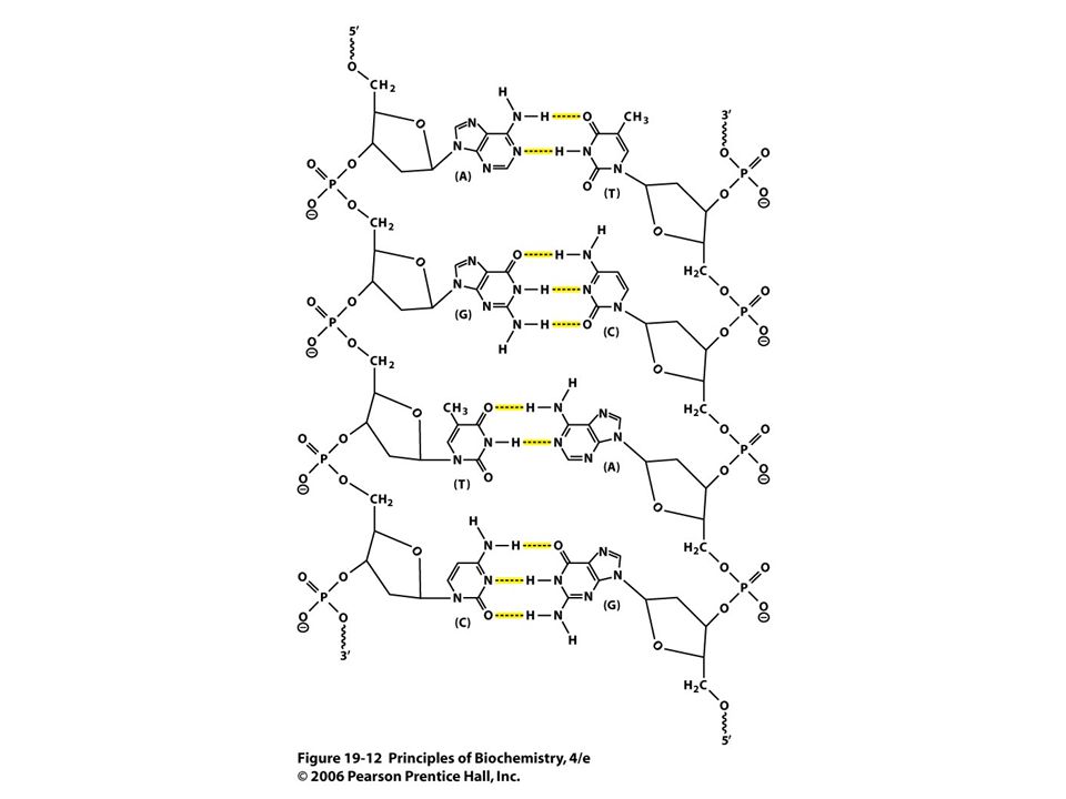
B. Two Antiparallel Strands Form a Double Helix Figure 19.12 (next slide) Two strands run in opposite directions Bases in opposite strands pair by complementary hydrogen bonding Adenine (A) - Thymine (T) Guanine (G) - Cytosine (C)
 Two strands run in opposite directions Bases in opposite strands pair by complementary hydrogen bonding Adenine (A) - Thymine (T) Guanine (G) - Cytosine (C).")
27
Fig 19.13 Complementar y base pairing and stacking in DNA

28
Three Dimensional Structure of DNA A double helix has two grooves of unequal width: major groove and minor groove Within each groove base pairs are exposed and are accessible to interactions with other molecules DNA-binding proteins can use these interactions to “read” a specific sequence

29
Fig 19.14 Structure of B-DNA Sugar phosphate backbone outside Stacking creates two unequal grooves (major and minor)
")
30
Stereo view of B-DNA B-DNA is a right-handed helix, diam. = 2.37 nm Rise (distance between stacked bases) =0.33 nm Pitch (distance to complete one turn) = 3.40 nm Base pairs nearly perpendicular to sugar- phosphate backbones Figure 19.15 Stereo views of B-DNA (next slide)
 =0.33 nm Pitch (distance to complete one turn) = 3.40 nm Base pairs nearly perpendicular to sugar- phosphate backbones Figure Stereo views of B-DNA (next slide).")
31
(a) Ball-and-stick model(b) Space-filling model
 Ball-and-stick model(b) Space-filling model")
32
C. Weak Forces Stabilize the Double Helix (1) Hydrophobic effects. Burying purine and pyrimidine rings in the double helix interior (2) Stacking interactions. Stacked base pairs form van der Waals contacts (continued next slide)
 Stacking interactions. Stacked base pairs form van der Waals contacts (continued next slide).")
33
Weak Forces (continued) (3) Hydrogen bonds. Hydrogen bonding between base pairs. (4) Charge-charge interactions. Electrostatic repulsion of negatively charged phosphate groups is decreased by cations (e.g. Mg 2+ ) and cationic proteins
 Charge-charge interactions. Electrostatic repulsion of negatively charged phosphate groups is decreased by cations (e.g. Mg 2+ ) and cationic proteins.")
34
Fig 19.16 Absorption spectra of double- stranded and single-stranded DNA Double-stranded (DS)DNA (pH 7.0), absorbance max 260 nm Denatured DNA absorbs12% -40% more than DS DNA
DNA (pH 7.0), absorbance max 260 nm Denatured DNA absorbs12% -40% more than DS DNA")
35
Denaturation of DNA Double-stranded DNA is thermodynamically more stable than the separated strands (under physiological conditions) Denaturation - Complete unwinding and separation of the 2 strands of DNA Heat or chaotropic agents (e.g. urea) can denature DNA
 can denature DNA.")
36
Heat Denaturation of DNA Melting point (T m ) - temperature at which 1/2 of the DNA has become single stranded Melting curves can be followed at Abs 260nm
 - temperature at which 1/2 of the DNA has become single stranded Melting curves can be followed at Abs 260nm")
37
Fig 19.17 Melting curve for DNA

38
D. Conformations of Double-Stranded DNA Two alternative structures to B-DNA: A-DNA (forms when DNA is dehydrated) Z-DNA (when certain sequences are present) A-DNA is more tightly wound than B-DNA, and has minor grooves of similar width Z-DNA has no grooves and a left-handed helix Both A-DNA and Z-DNA exist in vivo in short regions of DNA
 Z-DNA (when certain sequences are present) A-DNA is more tightly wound than B-DNA, and has minor grooves of similar width Z-DNA has no grooves and a left-handed helix Both A-DNA and Z-DNA exist in vivo in short regions of DNA.")
39
Fig 19.18 Forms of DNA ABZ

40
19.3 DNA Can Be Supercoiled “Relaxed” circular DNA with the B conformation (10.4 base pairs/turn) would lie flat on a surface If strands are broken, and two ends of linear DNA twisted in opposite directions and rejoined, DNA supercoils to restore 10.4 bp/turn Each supercoil compensates for one turn of the double helix
 would lie flat on a surface If strands are broken, and two ends of linear DNA twisted in opposite directions and rejoined, DNA supercoils to restore 10.4 bp/turn Each supercoil compensates for one turn of the double helix")
41
Supercoiling Most bacterial chromosomes are supercoiled, and regions of eukaryotic DNA are supercoiled Topoisomerases - enzymes that can alter the topology of DNA helixes by: 1.Cleaving one or both DNA strands 2.Unwinding or overwinding the double helix by rotating the strands 3.Rejoining ends to create (or remove) supercoils
 supercoils")
42
Supercoiling (http://chemistry.umeche.maine.edu/CHY431/Supercoil3.gif)

43
Supercoiling an Elastic Band (http://www.maths.uq.edu.au/~infinity/Infinity7/images/supercoiling.gif)

44
Fig 19.19 Structure of supercoiled DNA

45
Fig 19.20 Human topoisomerase I bound to DNA Topoisomerases can add or remove supercoils in DNA Cleave one or both DNA strands, unwind or overwind by rotating cleaved ends, then rejoin ends

46
Supercoiling (http://www.mun.ca/biochem/courses/3107/Topics/supercoiling.html)

47
19.4 Cells Contain Several Kinds of RNA Ribosomal RNA (rRNA) - an integral part of ribosomes, accounts for ~80% of RNA in cells Transfer RNA (tRNA) - carry activated amino acids to ribosomes for polypeptide synthesis (small molecules 73-95 nucleotides long)
 - an integral part of ribosomes, accounts for ~80% of RNA in cells Transfer RNA (tRNA) - carry activated amino acids to ribosomes for polypeptide synthesis (small molecules nucleotides long)")
48
Types of RNA (continued) Messenger RNA (mRNA) - carry sequence information to the translation complex Small RNA - have catalytic activity or associate with proteins to enhance activity
 Messenger RNA (mRNA) - carry sequence information to the translation complex Small RNA - have catalytic activity or associate with proteins to enhance activity")
49
RNA Structure RNA’s are single-stranded molecules Often have complex secondary structures Can fold to form stable regions of base-paired, double-stranded RNA Example is stem-loop (hairpin) structure
 structure")
50
Fig 19.21 Stem-loop structures in RNA Stem-loops or hairpins can form from short regions of complementary base pairs Stem: base-paired nucleotides Loop: noncomplementary nucleotides

51
19.5 DNA Is Packaged in Chromatin in Eukaryotic Cells Chromatin - DNA plus various proteins that package the DNA in a more compact form The packing ratio: difference between the length of the metaphase DNA chromosome and the extended B form of DNA is 8000-fold

52
A. Nucleosomes Histones - the major proteins of chromatin Eukaryotes contain five small, basic histone proteins containing many lysines and arginines: H1, H2A, H2B, H3, and H4 Positively charged histones bind to negatively- charged sugar-phosphates of DNA

53
Table 19.3

54
Nucleosomes Nucleosome “beads” are DNA-histone complexes on a “string” of double-stranded DNA Each nucleosome is composed of: Histone H1(1 molecule) Histones H2A, H2B, H3, H4 (2 molecules each) ~200 bp of DNA
 Histones H2A, H2B, H3, H4 (2 molecules each) ~200 bp of DNA")
55
Fig 19.22 Electron micrograph of chromatin Chromatin “beads-on-a-string” organization

56
Fig 19.23 Diagram of nucleosome structure

57
Fig. 19.23 (b)
")
58
Fig 19.24 Structure of chicken nucleosome core particle Histone octame Octamer bound to DNA

59
B. Higher Levels of Chromatin Structure Packaging of DNA in nucleosomes reduces DNA length ~tenfold DNA is packaged further by coiling of the “beads-on-a-string” into a solenoid structure Achieves another fourfold reduction in chromosome length

60
Solenoid Model of 30 nm Chromatin Structure Figure 19.25 (next slide) Model of the 30 nm chromatin fiber shown as a solenoid or helix formed by individual nucleosomes Nucleosomes associate through contacts between adjacent histone H1 molecules
 Model of the 30 nm chromatin fiber shown as a solenoid or helix formed by individual nucleosomes Nucleosomes associate through contacts between adjacent histone H1 molecules")
61
Fig 19.25

62
RNA-protein Scaffolds in Chromatin Chromatin fibers attach to scaffolds Holds DNA fibers in large loops May be ~2000 loops on a large chromosome This accounts for an additional 200-fold condensation in DNA length

63
Fig 19.26 Histone-depleted chromosome scaffold Protein scaffoldLoops attached to scaffold

64
Chromatin Structure (http://bioweb.wku.edu/courses/biol566/L5YeastSilencing.html)

65
C. Bacterial DNA Packaging Prokaryotic DNA also packaged with proteins in a condensed form No defined nucleosome-like particles Nucleoid structure - bacterial DNA attached to a scaffold in large loops of ~100 kb

66
19.6 Nucleases and Hydrolysis of Nucleic Acids Nucleases - hydrolyze phosphodiester bonds RNA nuclease (RNases, RNA substrates) DNA nuclease (DNases, DNA substrates) May cleave either the 3’- or the 5’- ester bond of a 3’-5’ phosphodiester linkage Exonucleases start at the end of a chain Endonucleases hydrolyze sites within a chain
 DNA nuclease (DNases, DNA substrates) May cleave either the 3’- or the 5’- ester bond of a 3’-5’ phosphodiester linkage Exonucleases start at the end of a chain Endonucleases hydrolyze sites within a chain")
67
Fig 19.27 Nuclease cleavage sites Cleavage at bond A generates a 5’-phosphate and a 3’ OH terminus Cleavage at bond B generates a 3’-phosphate and a 5’-hydroxyl terminus A given nuclease can catalyze at either A or B, but not both.

68
A. Alkaline Hydrolysis of RNA Fig 19.28

71
(From previous page)
")
72
B. Ribonuclease- Catalyzed Hydrolysis of RNA Fig 19.29

77
C.Restriction Endonucleases (Restriction Enzymes) Enzymes that recognize specific DNA sequences Cut both strands of DNA at the binding site, producing fragments that can be degraded by exonucleases Host cells protect their own DNA by covalent modification of bases at the restriction site (e.g. methylation)
.")
78
Restriction Endonuclease Properties Type I - catalyze both the methylation of host DNA and cleavage of unmethylated DNA at a specific recognition sequence Type II - cleave double-stranded DNA only, at or near an unmethylated recognition sequence More than 200 type I and type II are known Most recognize “palindromic sequences” (read the same in either direction)
")
80
Fig 19.30 Methylation and restriction at the EcoR1 site

81
Fig 19.30 (continued)
")
82
D. EcoR1 Binds Tightly to DNA EcoR1 has 2 identical subunits (purple and blue) Bound to a fragment of DNA
 Bound to a fragment of DNA.")
83
19.7 Uses of Restriction Endonucleases Developing restriction maps (indicates specific cleavage sites in a DNA fragment) Map of bacteriophage showing cleavage sites of some restriction enzymes
 Map of bacteriophage showing cleavage sites of some restriction enzymes")
84
Fig 19.33 Restriction digest of bacteriophage Four restriction enzymes used Sizing gel separates fragments (smallest move fastest)
")
85
DNA Fingerprinting DNA sequence can be used to identify individuals in a large population Highly variable regions give restriction fragments that are as unique as fingerprints (www.cropgeninternational.com/DrKoebner.html ) (http://writingcompany.blogs.com/this_isnt_writing_its_typ/ images/csi_ny.jpg)
 ( images/csi_ny.jpg)")
86
Fig 19.34 DNA Fingerprinting

87
Fingerprinting (http://fig.cox.miami.edu/~cmallery/150/gene/c7.20.17.fingerprinting.jpg)

88
Fingerprinting (http://www.scq.ubc.ca/wp-content/DNAfingerprintfamily.gif)

90
Common Organic Functional Groups

Similar presentations


 Recognize and apply the.>")
 Nucleic Acids. Information encoded in a DNA molecule is transcribed via synthesis of an RNA molecule The sequence of the RNA molecule.>")


 Nucleic Acids. DNA 1 o Structure - Linear array of nucleotides 2 o Structure – double helix 3 o Structure - Super-coiling, stem- loop.>")
 Dr. Sumbul Fatma>")


 The central dogma of molecular biology Nucleotide chemistry DNA, RNA and chromosome structure DNA replication Gene.>")
 and RNA (ribonucleic acid) store and transfer genetic information in living organisms.>")

