Download presentation
Presentation is loading. Please wait.

2
Anatomy of the Female Reproductive System David Shveiky, M.D. Department of Obstetrics & Gynecology Hadassah Medical Center, Ein Kerem Jerusalem

4
הצגת מקרה בת 22 עומדת לפניך בתור לבנק ולפתע מתמוטטת. האבחנה – Ruptured ectopic pregnancy

5
LAPAROSCOPY

6
היכן לא להיכנס

7
דרך מה עוברים ?

9
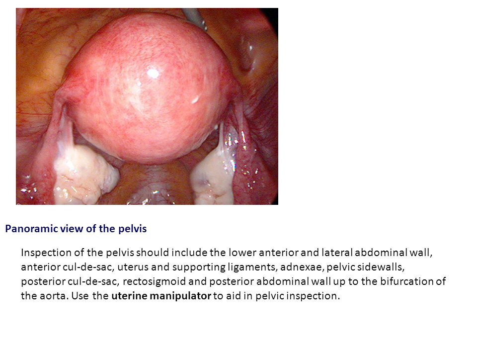
Panoramic view of the pelvis Inspection of the pelvis should include the lower anterior and lateral abdominal wall, anterior cul-de-sac, uterus and supporting ligaments, adnexae, pelvic sidewalls, posterior cul-de-sac, rectosigmoid and posterior abdominal wall up to the bifurcation of the aorta. Use the uterine manipulator to aid in pelvic inspection.

10
מומים מולדים של הרחם והנרתיק

12
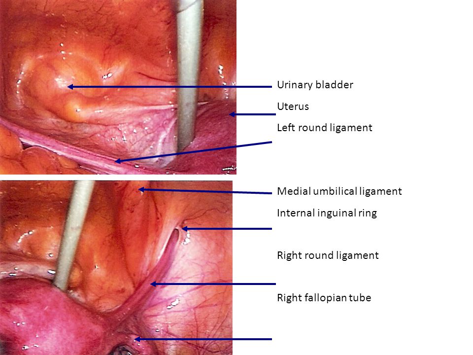
Urinary bladder Uterus Left round ligament Medial umbilical ligament Internal inguinal ring Right round ligament Right fallopian tube

13
Note the relative positions of the three structures: The right fallopian tube, round ligament and ovary. Fallopian tube Ovary Hypogastric Ureter Round ligament

14
מומים מולדים של הרחם והנרתיק

18
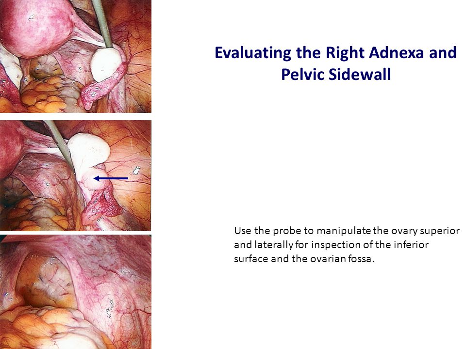
Use the probe to manipulate the ovary superior and laterally for inspection of the inferior surface and the ovarian fossa. Evaluating the Right Adnexa and Pelvic Sidewall

19
Right Pelvic Sidewall Uterus Fallopian tube I-P ligament Uterosacral ligament Ureter Hypogastric artery Sigmoid colon

20
Cannula Uterosacral ligaments Posterior cul-de-sac Sigmoid colon Posterior cul-de-sac

21
Use the suction/irrigation cannula as a probe and to maintain a clear field.

22
Posterior cul-de-sac - Endometriosis The posterior cul-de-sac appears normal. Inspection cannot be considered to be complete until all hidden areas are clearly visualized with the aid of the cannula.

23
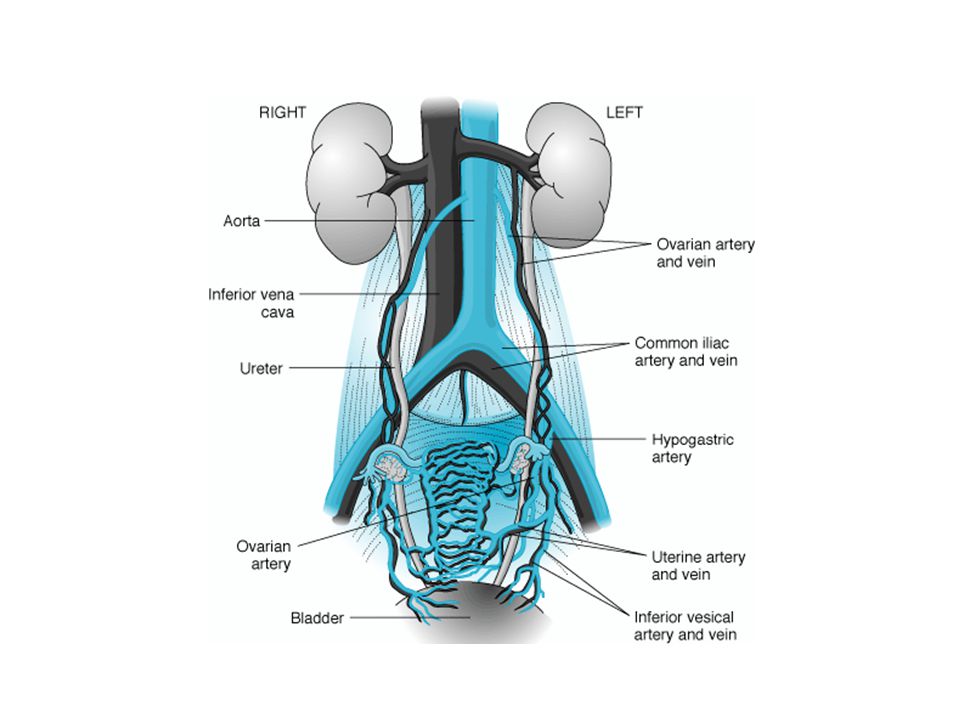
Uterus – Blood Supply Uterine Vessels Ovarian Vessels

25
Where is the uterus here?

27
אספקת דם, ניקוז ורידי, לימפתי ועצבוב

28
לאחר שנתיים... נקראת לחדר לידה להעריך יולדת בשל חוסר התקדמות היולדת מזהה אותך מהתור לבנק לפני שנתיים בבדיקה – פתיחה גמורה הראש SP+1 ללא התקדמות 3 שעות.

32
אגן גרמי

34
כעבור 24 שעות... נקראת למחלקת יולדות להעריך יולדת בשל כאבים במותן ימין לאחר ניתוח קיסרי להפתעתך, שוב את / ה מזהה את עמיתתך לתור בבנק.

36
אספקת דם, ניקוז ורידי, לימפתי ועצבוב

37
מומים מולדים של הרחם והנרתיק

38
Note the relative positions of the three structures: The right fallopian tube, round ligament and ovary. Fallopian tube Ovary Hypogastric Ureter Round ligament

39
כעבור 30 שנה... מגיעה למרפאה אישה בת 54 בתלונות של צניחת איברי האגן וירידה בתפקוד המיני האישה מוכרת לך, אך שכחת ליטול את התרופות לאלצהיימר היום

42
Pelvic Floor Levator Ani Inferior View

43
VIEW OF LEVATOR ANI MUSCLES FROM ABOVE WITH PELVIC ORGANS REMOVED. NOTE ATTACHMENTS TO PUBIC SYMPHISIS, ARCUS TENDINEUS, AND ISCHIAL SPINES Superior view

Similar presentations