Download presentation
Presentation is loading. Please wait.

1
Proteins

2
Proteins Consists of C, H, O, and N
Uses: develop muscle, hair, skin, vital organs, enzymes, some hormones. Last resort energy. Basic unit of structure is an Amino Acid: R is different for each There are 20 different types of Amino Acids Eight essential amino acids Cannot be made by the body, must be obtained through diet.

3
Amino acids

4
Structure of proteins Primary structure: order of the amino acids

5
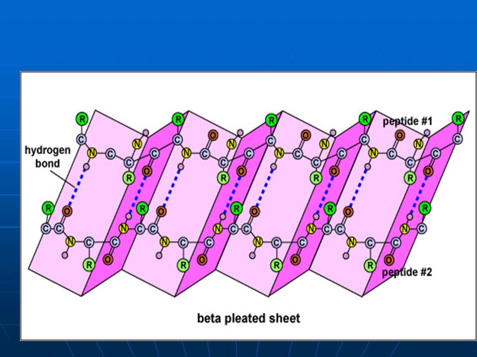
Secondary structure- first level of folding
Pleated sheet. This type of folding makes smooth proteins such as smooth muscle and silk. And puppies. -Tim

6
Diagram of a pleated sheet

8
Pleated sheet

9
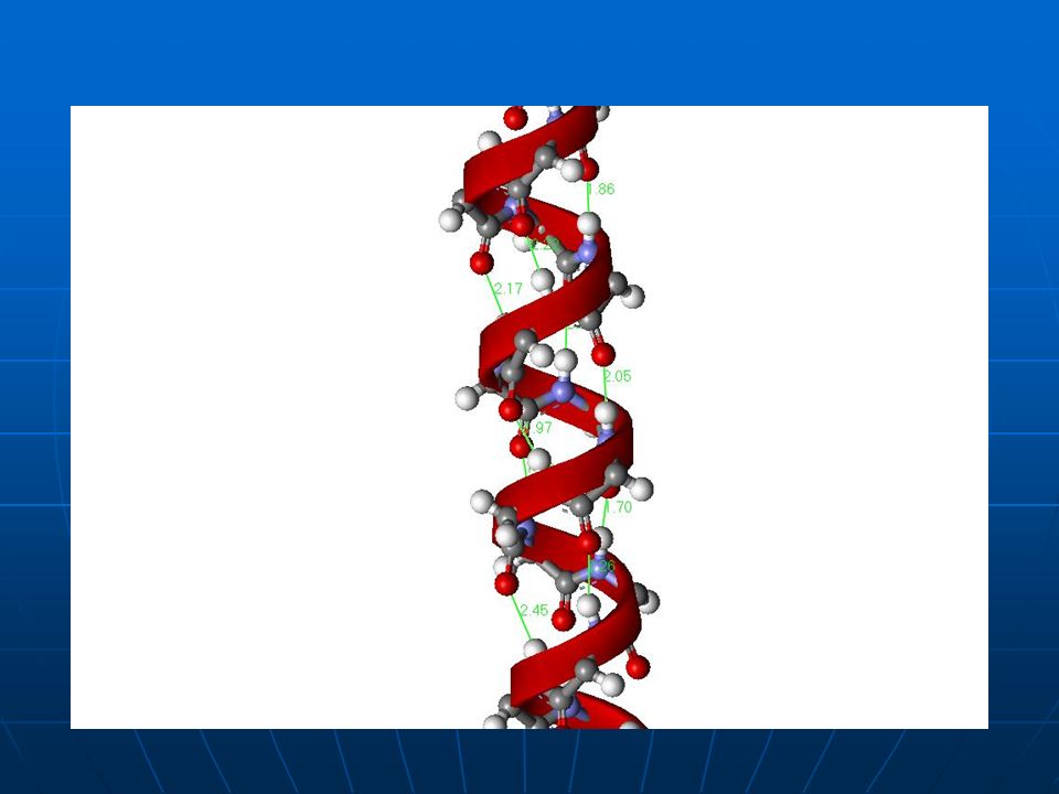
-Alpha helix This type folding creates proteins that are very springy such as hair, tendons, and ligaments. All secondary structure is held together with hydrogen bonding that exists between the C=O and NH groups on the amino acids.

10
Alpha helix

11
Alpha helix

13
Tertiary folding This is the final layer of folding for most proteins. This type of folding is made through the “R” groups on the amino acids. Examples on the appearance of tertiary proteins

14
Tertiary folding cont. Held together by these attraction between R groups: Ionic attraction Covalent bonds Non polar attraction (Van der Waal) Dipole attraction Hydrogen bonding Also Between C=O and H-O- attractions on the amino acids
 Dipole attraction. Hydrogen bonding. Also Between C=O and H-O- attractions on the amino acids.")
15
Here are some examples of the tertiary folding of a complete protein

16
Quadinary folding Has more than one poly-peptide chain
Held together by same attractions as tertiary folding. These proteins are often enzymes and hormones Tend to be globular in shape example:Hemoglobin - Globular to fit Fe+3

17
Denaturing proteins Denaturing is when a change is made to the tertiary and quadinary structure of the proteins. Denaturing can be achieved through changes in: Temperature pH – Metal ions—interfere with ions and attractions between amino acids

18
Identifying Amino Acids
There are two tests commonly used in identifying amino acids: chromatography and electrophoresis. In both tests, proteins are broken down into fragments or into individual Amino Acids first through the use of restriction enzymes. Once the protein is in A.A. or small fragment form, the following two tests can be used.

19
Chromatography Separates A.A. on differences of molecular weight and solubility in solvent. Chromatography strip is placed in a solvent. A.A. are placed on the strip above the solvent level The strip absorbs the solvent. As the solvent travels up the strip the A. A. are picked up and travel with it. The smaller or more soluble A.A. will move faster.

20
Chromatography cont. When the solvent reaches the top of the strip, the strip is removed from the solvent and identification begins. The strip is sprayed with ninhydrin to make the A.A. show up. The distance each A.A. moves is measured. The distances are calculated as ratio: Distance traveled by A.A./Distance traveled by solvent This ratio value, Called Rf, is then compared to a known Rf that is listed on a table for each A.A. Thus, an A.A. can be identified by its Rf value.

21
Electrophoresis Separates A.A. on their differences in molecular weight or their charge, or pH. Each R group has a different charge. This difference affects both the solubility and its migration rate in an electric field. (How fast it moves when voltage is applied to it)
")
22
General electrophoresis
A.A.s are placed in wells in a buffered gel w/ a specific pH, and then voltage is run across the gel for a period of time. The A.A.s will migrate at different rates Stop the voltage (note migration rates) A.A.s are identified based upon these rates by matching the migration rate up with a known migration rate for an amino acid.
 A.A.s are identified based upon these rates by matching the migration rate up with a known migration rate for an amino acid.")
23
Problems with electrophoresis
The pH of the gel is crucial. Every A.A. has pH in which it will not migrate. This is called its iso-electric point or pI. At this point the A.A. has both (+) and (-) in charge so no migration will occur. Methods of avoiding this problem: avoid using a gel with a pH close to the pI point of an A.A. or run several tests with gels of different pHs. Use a gradient pH gel. That is, the pH gradually changes through out the gel. Each A.A. will migrate until it reaches its pI. This method is often used.
 and (-) in charge so no migration will occur. Methods of avoiding this problem: avoid using a gel with a pH close to the pI point of an A.A. or run several tests with gels of different pHs. Use a gradient pH gel. That is, the pH gradually changes through out the gel. Each A.A. will migrate until it reaches its pI. This method is often used.")
24
pH of some Isoelectric points
Amino acid Glutamic acid Phenylalanine Serine Histidine arginine pH of isoelectric point 3.2 5.5 5.7 7.6 10.8

25
Another method Chemically treat the amino acids so that each one has the same charge. This makes the amino acids migrate on the basis or their size, that is, molecular weight. Lighter acids migrate faster and so travel further in a given time frame.

26
Electrophoresis chamber

27
Diagram of an electrophoresis gel

28
A gel with an electric field applied to it. The A.A. migrate across.
Wells w/ A.A.

29
Photograph of an electrophoresis gel. The test was run 4 times.

Similar presentations
 PROTEIN.>")


 Monomer is Amino Acids Growth Repair Hormones Enzymes Antibodies Energy Source.>")
>")


![7.5: PROTEINS Proteins Function Structure. Function 7.5.4: State four functions of proteins, giving a named example of each. [Obj. 1] Proteins are the.](/23/6900992/big_thumb.jpg "7.5: PROTEINS Proteins Function Structure. Function 7.5.4: State four functions of proteins, giving a named example of each. [Obj. 1] Proteins are the.>")
