Download presentation
Presentation is loading. Please wait.

1
Non-Invasive Conference: Aortic Dissection
Ali R. Rahimi, MD MPH September 24, 2008

2
Anatomy Tunica intima, media and adventitia

3
Aortic Dissection “a splitting of the layers of the aortic wall (within the media), permitting longitudinal propagation of a blood-filled space within the aortic wall” Most common cause of death related to the human aorta Incidence 2.6 to 3.5 per 100,000 person-years 65% men with mean age of 63 years
, permitting longitudinal propagation of a blood-filled space within the aortic wall Most common cause of death related to the human aorta. Incidence 2.6 to 3.5 per 100,000 person-years. 65% men with mean age of 63 years.")
4
Pathophysiology Degeneration of aortic media, or cystic medial necrosis, is prerequisite for nontraumatic dissection Primary rupture of intima with secondary dissection of the media vs. hemorrhage within the media and subsequent rupture of overlying intima Propagation can occur both distal and proximal

6
Risk Factors Systemic hypertension (72%) Atherosclerosis (31%)
Pre-existing aortic aneurysm (13% known) more common < 40 yo (19%) Vasculitis (giant cell arteritis, Takayasu arteritis, rheumatoid arthritis, syphilitic aortitis)
 more common < 40 yo (19%) Vasculitis (giant cell arteritis, Takayasu arteritis, rheumatoid arthritis, syphilitic aortitis)")
7
Risk Factors Disorders of collagen (eg, *Marfan syndrome, Ehlers-Danlos syndrome, annuloaortic ectasia) *Present in 50% of those < 40 yo vs. 2% of older patients Bicuspid aortic valve Present in 9% < 40 yo vs. 1% over age 40 Enlargement of aortic root and/or ascending aorta Aortic coarctation, Turner Syndrome, CABG (0.04%), Prior AVR, Cardiac Cath (2%), Trauma (deceleration injury), Weight lifting, Crack cocaine
, Prior AVR, Cardiac Cath (2%), Trauma (deceleration injury), Weight lifting, Crack cocaine.")
8
Ascending aortic dissections ~ 2x as common
It classifies dissections that involve the ascending aorta as type A, regardless of the site of the primary intimal tear, and all other dissections as type B. In comparison, the DeBakey system is based upon the site of origin with type 1 originating in the ascending aorta and propagating to at least the aortic arch, type 2 originating in and confined to the ascending aorta, and type 3 originating in the descending aorta and extending distally or proximally Ascending aortic dissections ~ 2x as common Right lateral wall of ascending aorta most common site Aortic arch involvement ~ 30%

9
Variants Class I - classic separation of intima/media and dual lumens; flap between true and false aneurysm and clot in false lumen; Class II - intramural hematoma with separation of intima/media but no tear or flap; Class III - limited intimal tear without hematoma and eccentric bulge at tear site; Class IV - atherosclerotic ulcer penetrating to adventitia with surrounding hematoma; Class V- iatrogenic or traumatic dissection

10
Clinical Presentation
Severe, sharp or "tearing" posterior chest or back pain (distal to L subclavian) or anterior CP (ascending dissection) Isolation or with syncope, CVA, MI, CHF, … HTN more common in type B dissection (70% vs. 36%) Pulse deficit in 19-30% with an acute type A dissection vs. 9-21% with a type B dissection
 or anterior CP (ascending dissection) Isolation or with syncope, CVA, MI, CHF, … HTN more common in type B dissection (70% vs. 36%) Pulse deficit in 19-30% with an acute type A dissection vs. 9-21% with a type B dissection.")
11
Clinical Finding Artery or Structure
Aortic insufficiency or CHF Aortic valve AMI Coronary artery (right) Tamponade Pericardium Hemothorax Thorax CVA or Syncope Brachiocephalic, CC, Left subclavian UE pulselesness, low BP, pain Subclavian Paraplegia Intercostal LE pain, pulselessness, weakness Common iliac Abdominal pain, mesenteric ischemia Celiac or mesenteric Back or flank pain; ARF Renal artery Horner syndrome Superior cervical sympathetic ganglion
 Tamponade. Pericardium. Hemothorax. Thorax. CVA or Syncope. Brachiocephalic, CC, Left subclavian. UE pulselesness, low BP, pain. Subclavian. Paraplegia. Intercostal. LE pain, pulselessness, weakness. Common iliac. Abdominal pain, mesenteric ischemia. Celiac or mesenteric. Back or flank pain; ARF. Renal artery. Horner syndrome. Superior cervical sympathetic ganglion.")
12
Differential Diagnosis
Acute Coronary Syndrome Pericarditis Pulmonary embolus Aortic regurgitation without dissection Aortic aneurysm without dissection Musculoskeletal pain Mediastinal tumors Pleuritis Cholecystitis Atherosclerotic or cholesterol embolism Peptic ulcer disease or perforating ulcer Acute pancreatitis

13
Diagnosis Clinical Prediction of Aortic Dissection
Study of 250 pts with acute chest and/or back pain (128 with AD) found 96% with acute AD identified using 3 clinical features: Abrupt onset of thoracic or abdominal pain with a sharp, tearing and/or ripping character Mediastinal and/or aortic widening on chest radiograph Variation in pulse (absence of a proximal extremity or carotid pulse) and/or blood pressure (>20 mmHg difference in the right and left arm) Any 2 out of 3 variables (77% of dissections): ≥83% Additional imaging studies obtained in 98% of pts due to limited sensitivity of CXR, especially in type B dissections
 found 96% with acute AD identified using 3 clinical features: Abrupt onset of thoracic or abdominal pain with a sharp, tearing and/or ripping character. Mediastinal and/or aortic widening on chest radiograph. Variation in pulse (absence of a proximal extremity or carotid pulse) and/or blood pressure (>20 mmHg difference in the right and left arm) Any 2 out of 3 variables (77% of dissections): ≥83% Additional imaging studies obtained in 98% of pts due to limited sensitivity of CXR, especially in type B dissections.")
14
Chest X-Ray Type A Type B
63% with mediastinal widening, 11% with no abnormality Type B 56% with mediastinal widening, 16% with no abnormality

15
Electrocardiogram 31% normal 42% non-specific ST and T wave changes
15% ischemic changes 5% changes c/w AMI

16
Advanced Imaging Modalities
Aortic Dissection Involvement of the ascending aorta The extent of dissection and the sites of entry and reentry Thrombus in the false lumen Branch vessel or coronary artery involvement Aortic insufficiency Pericardial effusion

17
Advanced Imaging Modalities
Based on year 2000 IRAD: mean 1.83 studies per patient 61% CT 33% Echocardiography 4% Aortography 2% MRI

18
ESC Guidelines for Diagnostic Imaging of Acute Aortic Dissection
Class I - TTE followed by TEE Class II – CT, CTA to define anatomy Class IIa – CTA in stable pts, MRI in stable pts, Intravascular US Class IIb - CT if detection of tears is crucial, CTA in unstable pts, intravascular US to guide intervention Class III – MRI in hemodynamically unstable patients, routine pre-op CTA

19
ESC Guidelines for Diagnostic Imaging of Chronic Aortic Dissection
Class I – MRI Class IIa – TTE, TEE, CTA

20
CT Scan

21
CT Scan

22
CT Scan

23
CT Scan Sensitivity 83-98% Specificity 87-100% Advantages:
availability at most hospitals ID of intraluminal thrombus and pericardial effusion Disadvantages: Intimal flap seen < 75% of cases and site of entry is rarely identified Potentially nephrotoxic iodinated contrast Unable to assess for aortic insufficiency Accuracy of CT is improved with spiral CT and electron beam or multidetector CT Spiral CT may be more accurate than MRI or TEE in the detection of aortic arch vessel involvement Limitation: Without ECG gating simulate artifact ~ aortic dissection

24
TTE

25
TTE

26
TTE

27
TTE

28
TTE

29
TTE

30
TTE Limited utility for evaluation of the thoracic aorta for dissection unable to adequately visualize distal ascending, transverse, and descending aorta in a substantial majority of patients Intimal flap in the proximal aorta may be seen, though the sensitivity and specificity of TTE are inferior compared to CT, MRI, and TEE Most useful for assessment of cardiac complications of dissection, including AI, pericardial effusion/tamponade, and regional LV dysfunction

31
TEE

32
TEE

33
TEE

34
TEE

35
TEE

36
TEE

37
TEE

38
TEE Advantages sensitivity 97 – 99%
close proximity of esophagus to thoracic aorta and absence of an intervening lung or chest wall portable procedure diagnosis within minutes useful in patients too unstable for CT or MRI intimal dissection flaps can be identified with high spatial resolution true and false lumens can be identified thrombosis in the false lumen, pericardial effusion, aortic regurgitation, and the proximal coronary arteries can be visualized 135º long axis view can define severity and mechanism of aortic regurgitation that complicates acute type A dissections

39
TEE Disadvantages Specificity as low as 77-85%
false-positive findings in the ascending aorta artifacts can be detected by addition of M-mode imaging, increasing specificity to almost 100% Requires sedation and esophageal intubation TEE requires availability of experienced operators (both physicians and technicians) to assure accurate results
 to assure accurate results.")
40
30 Day Mortality Rates for Acute Aortic Dissection
Data from the international registry of acute aortic dissection – published in JAMA in 2000 Acute Type A dissection treated medically - 20% mortality rate at 24 hours 30% at 48 hours 40% at one week 50% by one month Hagan, Etc, JAMA :897

41
Mechanical Composite Root Repair with and w/o Hemiarch Repair

43
Homograft Repair

44
Porcine BioRoot Replacement

45
Valve Sparing Replacement

46
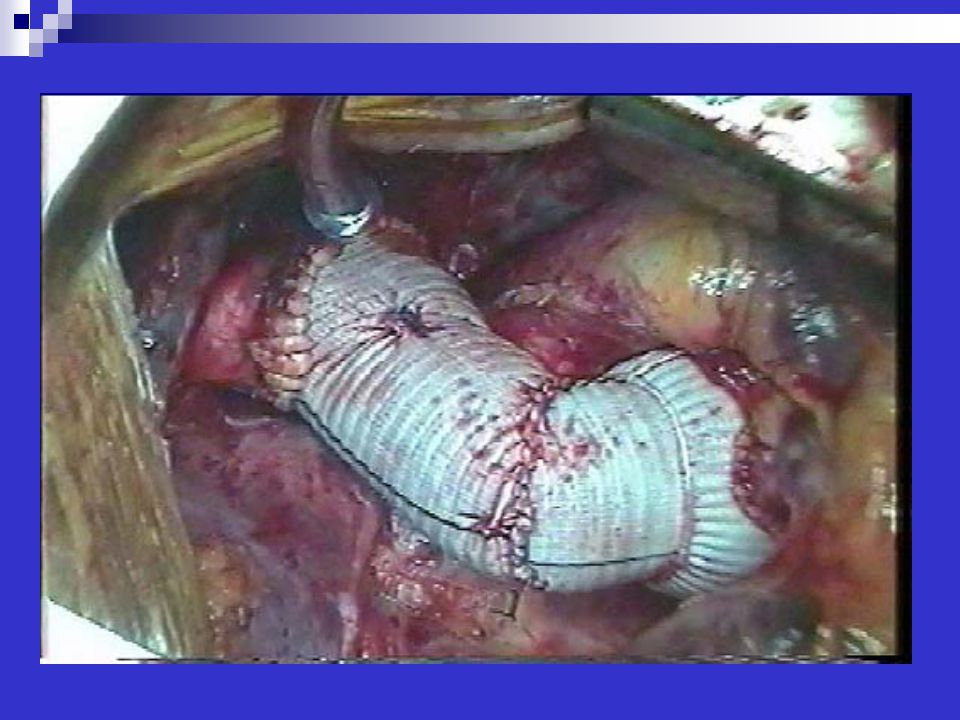
Aortic Dissection Repair: Teflon Felt as Neomedia

47
Endovascular Aortic Stent Grafts

48
Endovascular Aortic Stents

49
References Hurst the Heart, 11th Edition
Manning, W. UpToDate Online Clinical manifestations and diagnosis of aortic dissection.

Similar presentations

 Practice Group Logo here.>")


 Aortic surgery: Update & Decision making วันเสาร์ที่ 17 กันยายน 2554 ห้องประชุมสมาคมศิษย์เก่าแพทย์ศิริราช โรงพยาบาลศิริราช.>")


