Download presentation
Presentation is loading. Please wait.

1
Utilization of Cardiac Serum Marker Measurements to Identify and Exclude Acute Myocardial Infarction Francis M. Fesmire, MD, FACEP Assistant Professor, UT College of Medicine Director, Heart-Stroke Center Erlanger Medical Center, Chattanooga, Tn ffesmire@comcast.net

2
Do You Want A Piece of Me?

3
Ready, Aim…..

4
Fire!!!!

5
Overview Which is the best marker of AMI? – CK-MB activity – CK-MB mass – CK-MB subform ratio – Myoglobin – cTnT – cTnI – Newer assays?????

6
2000 Clinical Policy of the American College of Emergency Physicians reviewed 50 articles comparing serum markers: – CK-MB activity: 7 cutoff values (5-23 IU/L) – CK-MB mass: 14 (4-20 ng/ml) – CK-MB subform ratio: 2 (1.5 & 2.3) – Myoglogin: 9 (35-110 ng/ml) – cTnT: 5 (0.06-0.2 ng/ml) – cTnI: 5 (0.1-2.5 ng/ml)
 – CK-MB mass: 14 (4-20 ng/ml) – CK-MB subform ratio: 2 (1.5 & 2.3) – Myoglogin: 9 ( ng/ml) – cTnT: 5 ( ng/ml) – cTnI: 5 ( ng/ml)")
7
Bias Multitude of Experimental Bias – Positive value of assay also defines AMI – Use the ROC curve optimum value of newer assay to compare against “gold standard” for older assay – Differing patient populations ICU vs general ED Early symptom onset versus late symptom onset

8
Valid Comparison? Conditions for a valid study: – The diagnosis of AMI needs to be independent of positive value of marker under investigation – Statistical Analysis of ROC curve area – Sensitivity and specificity comparison should be performed at a point on the individual ROC curves where likelihood ratio’s are equivalent and clinically meaningful

9
Likelihood Ratios Bayes’ Theorem – Pretest odds of the disease X likelihood ratio = Posttest odds of the disease – Positive LR = sensitivity/(1-specificity) – Negative LR = (1-sensitivity)/specificity In general, a +LR > 10 or < 0.1 should influence clinical decision making The ideal marker of AMI should both identify and exclude AMI
 – Negative LR = (1-sensitivity)/specificity In general, a +LR > 10 or < 0.1 should influence clinical decision making The ideal marker of AMI should both identify and exclude AMI")
10
Definition Reliably Identifies: – sensitivity > 90% with +LR > 10 Reliably Excludes: – specificity > 90% with -LR < 0.1 ACEP Clinical Policy: Suspected AMI or Unstable Angina; Annals of Emergency Medicine 2000; Ann Emerg Med 2000;35:521-544.

11
Diagnostic Marker Cooperative Study Prospective double-blind study comparing CK-MB activity, CK-MB mass, CK-MB subforms, myoglobin, cTnT, and cTnI 955 patients, 119 with AMI Conclude that CK-MB subforms and myoglobin are the most sensitive for early diagnosis of AMI Zimmerman et al: Circulation; 1999;99:1671-1677

12
AMI Definition “The diagnostic standard for myocardial infarction was a CK-MB mass > 7 ng/ml and CK-MB index > 2.5% in greater than 2 samples or in one sample if only one sample was available for analysis” – CK-MB mass > 7 ng/ml both defines AMI and a positive value of CK-MB – No WHO criteria for AMI utilized

13
ROC Curve Area Data 6 Hours: CK-MB subform (0.95) = cTnT (0.95) > CK-MB activity (0.94) > myoglobin (0.92) > cTnI (0.89) 14 Hours: CK-MB activity (0.99) > cTnI (0.97) > CK-MB subform (0.94) > cTnT (0.91) > myoglobin (0.84) – Area of CK-MB mass not given??? – No statistical analysis of ROC curves – No comparison at equal likelihood ratio’s

14
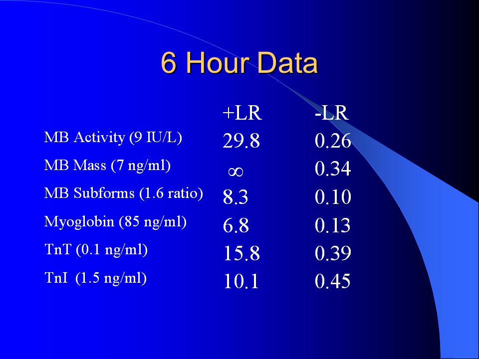
6 Hour Data

16
14 Hour Data

17
*Reliably identifies and reliably excludes

18
Ideal Marker ?? The ideal marker should reliably identify (sensitivity >90%; +LR > 10) and reliably exclude (specificity > 90% and -LR < 0.1): – No marker fulfills this criteria at 2, 4, 6 hours – CK-MB activity: 10, 14, 18 hours – CK-MB mass: 10, 14, 18, 22 hours – cTnI:10, 18 hours – CK-MB subform, myoglobin, cTnT: never
 and reliably exclude (specificity > 90% and -LR < 0.1): – No marker fulfills this criteria at 2, 4, 6 hours – CK-MB activity: 10, 14, 18 hours – CK-MB mass: 10, 14, 18, 22 hours – cTnI:10, 18 hours – CK-MB subform, myoglobin, cTnT: never.")
19
ACEP Evidence-Based Standards “No single determination of one serum biochemical marker of myocardial necrosis reliably identifies or reliably excludes AMI less than 6 hours of symptom onset.” “No serum biochemical marker identifies or excludes unstable angina at any time after symptom onset.” ACEP Clinical Policy: Suspected AMI or Unstable Angina; Annals of Emergency Medicine 2000; 35:521- 544.

20
ACEP Guidelines “In patients presenting with acute chest pain and a negative baseline serum marker level, consider repeat testing at the following time intervals from symptom onset prior to making an exclusionary diagnosis of AMI:” ACEP Clinical Policy: Suspected AMI or Unstable Angina; Annals of Emergency Medicine 2000; In Press

21
ACEP Guidelines

22
“The exact timing of the repeat serum marker should take into account the sensitivity, precision, and institutional norms of the assay being utilized, as well as the release kinetics of the marker being measured.” “cTnT and cTnI are the preferred serum markers in patients presenting greater than 24 hours after symptom onset.” “Myoglobin does not reliably identify or exclude AMI at any time after symptom onset.”

23
Footnote “If time of symptom onset is unknown, unreliable, or more consistent with preinfarctional angina, then time of symptom onset should be referenced to the time of ED presentation.” ACEP Clinical Policy: Suspected AMI or Unstable Angina; Annals of Emergency Medicine 2000; 35:521- 544.

24
WHO Diagnostic Criteria for AMI WHO Criteria: Two of three characteristics: – Typical symptoms – Typical rise and fall in cardiac markers – New Q waves on ECG

25
ESC/ACC Diagnostic Criteria Typical rise and fall of cardiac markers accompanied by one of the following: – Ischemic symptoms – New Q waves – Ischemic ECG changes – Coronary intervention J Am Col Cardiol 2000;36;959-969

26
ESC/ACC Diagnostic Criteria “An increased value for cardiac troponin should be defined as a measurement exceeding the 99 th percentile of a reference control group…. Acceptable imprecision at the 99 th percentile for each assay should be defined as < 10%” J Am Col Cardiol 2000;36;959-969

27
ESC/ACC Cutoff Values 99% (ng/ml)10% CV (ng/ml) Abbott Axsym0.50.8 Bayer Immuno0.10.35 Beckman-Coulter0.040.06 Biosite0.190.5 Dade RXL0.070.14 Dade Stratus CS0.070.06 Ortho Vitros0.080.12 Roche Elecys0.010.035 Am Heart J 2002;144:981-986.
10% CV (ng/ml) Abbott Axsym Bayer Immuno Beckman-Coulter Biosite Dade RXL Dade Stratus CS Ortho Vitros Roche Elecys Am Heart J 2002;144:")
28
Implications Estimated that number of patients with diagnosis of AMI utilizing new definition will increase by??? Ferguson et al (Heart 2002; 88:343-347) – 80 admitted chest pain patients 29% fulfilled WHO criteria 40% fulfilled ESC/AHA criteria
 – 80 admitted chest pain patients 29% fulfilled WHO criteria 40% fulfilled ESC/AHA criteria.")
29
Implications Global Registry of Acute Coronary Events (GRACE Registry) – 3420 patients Redefining AMI based on new troponin cutoff recommendations: – 25% increase in number of patients classified as AMI Gooman et al: J Am Coll Cardiol 2001;37:358A
 – 3420 patients Redefining AMI based on new troponin cutoff recommendations: – 25% increase in number of patients classified as AMI Gooman et al: J Am Coll Cardiol 2001;37:358A")
30
The Future !!! Utilization of Second Generation cTnI Assays for the Early Identification of Acute Coronary Syndromes

31
Stratus CS: 2-Hour cTnI

32
Stratus CS: Delta cTnI

33
What is the best marker of AMI? Troponins by default become best marker of AMI (incorporation bias) Multiple causes of troponin elevations confusing physicians and researchers New definitions on AMI need to focus on measuring changes in troponin values as opposed to absolute values
 Multiple causes of troponin elevations confusing physicians and researchers New definitions on AMI need to focus on measuring changes in troponin values as opposed to absolute values.")
34
Proud Card Member Since 1981

35
Breakfast of Champions !!

36
No Excuses!

37
Utilization of Cardiac Serum Marker Measurements to Identify and Exclude Acute Myocardial Infarction Francis M. Fesmire, MD, FACEP Director Heart-Stroke Center, Erlanger Medical Center Associate Professor, UT College of Medicine Just Do It!!!

Similar presentations


>")


 J. Teixeira, (2) P. Wotquenne, (2) V. D’Orio, (3) D. Gruson, (1)>")


