Download presentation
Presentation is loading. Please wait.

1
Dental Anomalies in Radiology
Developmental VS. acquired 1

2
Developmental Abnormalities

3
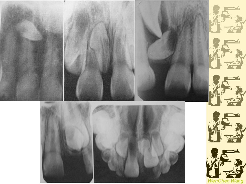
Supernumerary Teeth (hyperdontia, supplemental teeth)
1~4% , familial tendency Mesiodens, paramolar Distodens, distomolar teeth Peridens Single : premaxilla, maxillary molar Multiple : premolar area, mandibular M : F = 2 : 1 Impaction or delay eruption of normal teeth; dentigerous cyst Syndrome: Cleidocranial dysplasia, Gardner’s syn. 2

5
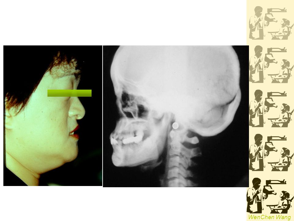
Cleidocranial dysplasia

10
Missing Teeth 8 > 5 > 2 > 1
3~10%, excluding 3rd molars Hypodontia Oligodontia Anodontia 8 > 5 > 2 > 1 Ectodermal dysplasia ; orofaciodigital syndrome 4

11
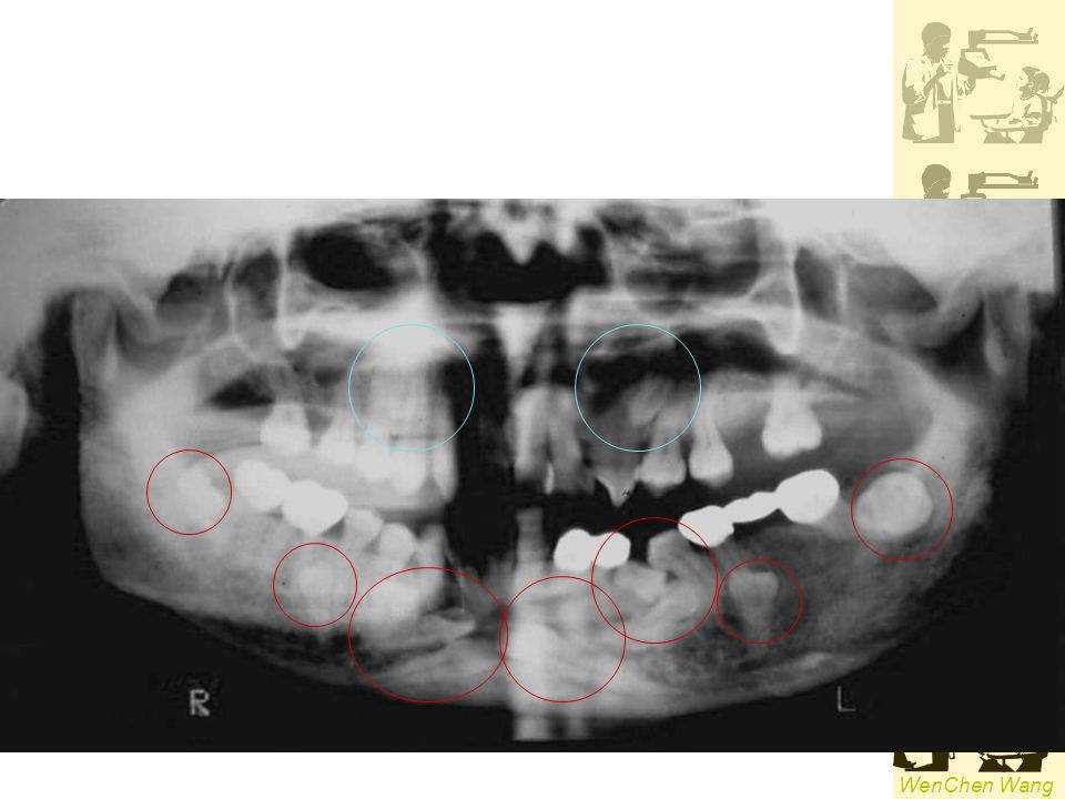
Q: 請就以上同一名患者的根尖X光片,說明有何異常。

12
ectodermal dysplasia

13
SIZE OF TEETH Macrodontia
True generalized type and relative type Macrodontia Hemangioma, hemihypertrophy of the face, pituitary giantism Microdontia pituitary dwarfism supernumerary teeth, 3rd molars, lateral incisors

14
Macrodontia Microdontia

15
Transposition ERUPTION OF TEETH Two teeth exchanged positions
3 & 4 ; 3 & 2, 657

16
Altered Morphology of Teeth

17
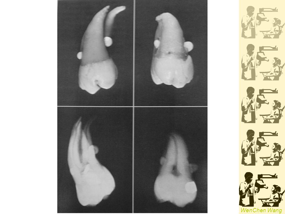
Gemination, Fusion, Concrescence

18
Gemination (twinning)
-Division of a single tooth bud primary dentition , esp. incisor region complete twinning increase tooth number pulp chamber is single & enlarged, maybe partial divided 9

19
Fusion (synodontia) -Adjacent tooth germs combined with dentin or enamel bifid crown or two recognizable teeth, reduced number of teeth more common in the primary dentition, esp. anterior region 7

20
Concresence - Roots of two or more teeth united by cementum
space restriction during develop., local trauma, excessive occlusal force or local infection after development maxillary molars; 3rd molar & a supernumerary tooth 8

21
Fusion / Gemination A tooth with two separated root canals and with one or two roots…Fusion An enlarged tooth with a bifid crown containing an enlarged or possibly partially divided pulp chamber…Gemination 10

22
Taurodontism -Longitudinal enlarged pulp chamber,
increased distance between CEJ to the bifurcation normal crown size & tooth length, shortened roots not recognizable clinically most in molars Trisomy 21 11

23
Dilaceration A sharp bend or curve in the crown or root
maxillary premolars 12

24
Dens in Dente (dens invaginatus)
- Infolding of the outer enamel surface into the interior at the anatomically defined pit caries→pulpal disease 13

25
coronal type: enamel organ infolding into the dental papilla; 2>1>4,5>3
radicular type: invagination of Hertwig’s epithelial root sheath, lined with cementum; 4, 7 14

26
radicular type Dilated odontome

27
Dens Evaginatus - Outfolding of enamel organ
a tubercle on occlusal surface, with enamel surface & dentin core, pulp horn often extends into the evagination premolar or molar pulp infection due to fracture 15

28
Lingual pits Dens Evaginatus

29
Amelogenesis Imperfecta
-Disturbance in enamel development Normal dentin & root autosomal dominant or recessive , X-linked Four general types 16

30
1.Hypoplastic type Thin enamel with pitted, rough or smooth & glossy surface; yellowish to brown undersized, squared crown, lack of contact flat occlusal surface & low cusps, attrition

31
2.Hypomaturation normal thickness of enamel, but mottled surface; cloudy white, yellow or brown, opaque in color softer than normal same density as dentin

32
4.Hypomaturation-hypocalcified with taurodontism
3.Hypocalcified type normal thickness of enamel, density less than dentin normal size & shape when erupt, abrade or fracture away rapidly permeability increase, darkened & stained 4.Hypomaturation-hypocalcified with taurodontism

33
Amelogenesis Imperfecta

34
Dentinogenesis Imperfecta (hereditary opalescent dentin)
autosomal dominant hereditary Type I : D.I. + osteogenesis imperfecta Type II : D.I., no skeletal defects enamel fractures, attrition severely dark brown to black

35
Dentinogenesis Imperfecta Osteogenesis imperfecta

36
Radiographic Features of D.I.
bulbous crown, normal size, constriction of the cervical area short & slender roots occlusal attrition partial or complete obliteration of the pulp chambers, root canals absent or threadlike

37
Dentinogenesis Imperfecta

38
Dentin Dysplasia rare (1:100,000) Type I (radicular)
-autosomal dominant disturbance rare (1:100,000) Type I (radicular) normal color & shaped in both dentition malaligned arch, drifting and exfoliate with little or no trauma short or abnormal root shaped, pulp chamber & root canals completely fill in before eruption 20 % of teeth with type I disease have apical radiolucencies
 Type I (radicular) normal color & shaped in both dentition. malaligned arch, drifting and exfoliate with little or no trauma. short or abnormal root shaped, pulp chamber & root canals completely fill in before eruption. 20 % of teeth with type I disease have apical radiolucencies.")
39
Dentin Dysplasia

40
TypeII (coronal) primary dentition appears as D.I., but permanent dentition is normal obliterated of the pulp chamber & reduced root canals after eruption roots are normal in shape & proportion

41
Dentin Dysplasia

42
Regional Odontodysplasia (odontogenesis imperfecta)
- hypoplastic & hypocalcified of both dentin & enamel only a few adjacent teeth in a quadrant affected either primary or permanent teeth central incisors > lateral incisors >canines (maxillary) delayed eruption ghostlike appearance in image large pulp chamber & wide root canals, roots are short & poorly outlined thin enamel , less dense as usual
 delayed eruption. ghostlike appearance in image. large pulp chamber & wide root canals, roots are short & poorly outlined. thin enamel , less dense as usual.")
43
Regional Odontodysplasia

44
Enamel Pearl (enameloma, enamel drop, enamel nodule)
- small globule of enamel on the roots furcation area of molars prevalence : 3 % mesial or distal aspect in Max. molar and buccal or lingual in Mand. molars

46
Talon Cusp - Anomalous hyperplasia of the cingulum of a Max. or Mand. incisor →a supernumerary cusp T shaped in incisal view Differential diagnosed with supernumerary tooth

47
Turner’s Hypoplasia (Turner’s tooth)
-a local hypoplastic or hypomineralized defect in crown of a permanent tooth extension of a periapical infection or mechanical trauma from deciduous predecessor most common in lower premolars

48
Turner tooth

49
Congenital Syphilis 30 % p’t develop dental hypoplasia
Hutchinson’s incisors & mulberry molars not all p’t with Hutchinson’s teeth or mulberry molars will have congenital syphilis

50
Congenital syphilis Hutchinson’s incisors & mulberry molars

51
Acquired Pathologic Conditions

52
Attrition -Physiologic wearing from occlusal contacts
Incisal, occlusal and interproximal surfaces(contact points) Depends on the abrasiveness of diet, salivary factors, mineralization, emotional tension Bruxism--pathologic condition Crown shorten, reduction of pulp chamber & canals
 Depends on the abrasiveness of diet, salivary factors, mineralization, emotional tension. Bruxism--pathologic condition. Crown shorten, reduction of pulp chamber & canals.")
53
Abrasion -Nonphysiologic wearing by contact with foreign substances
Factitious habits or occupational hazards tooth brushing, flossing, pipe smoking, opening hairpins with teeth

54
Tooth Brushing Injury V-shaped groove in cervical area Sensitive
Maxillary premolars >canines > incisors R-L defect at cervical level, well-defined semilunar shapes

55
Attrition Tooth Brushing Injury

56
Dental Floss Injury Cervical portion of proximal surfaces just above gingiva Narrow semilunar R-L, distal surface often deeper than mesial

57
-Chemical action not involving bacteria
Erosion -Chemical action not involving bacteria Contact acid with teeth: 1. chronic vomiting or acid reflux from GI disorders 2. consumes large amounts of acid foods 3. occupational exposure Regurgitated acids attack lingual surfaces; diet--labial; industrial–all surfaces R-L defect on the crown

58
Resorption -Removal of tooth structure by odontoclast
Chronic infection (inflammation), excessive pressure and function, tumors and cysts
, excessive pressure and function, tumors and cysts.")
59
Internal Resorption - within the pulp chamber or canal, involves resorption of surrounding dentin, results in enlarged pulp space Maybe related to inflammation of pulp tissues--acute trauma to tooth, pulp capping, pulpotomy… 1>6>7; M>F, commonly begins during 30-50y/o Pink spots

60
Radigraphs reveal symptomless early lesions of IR
R-L, round, oval, or elongated within root or crown and continuous with pulp chamber or canal Sharply defined and smooth or slightly scalloped …irregular widening of the pulp chamber or canal

61
Internal Resorption

62
External Resorption -outer surface of tooth resorbed, most commonly in root surface Localized inflammatory lesions, reimplanted teeth, tumor & cyst, excessive mechanical(orthodontic) and occlusal forces, impactions Common sites : apical & cervical (lateral root surface)
 and occlusal forces, impactions. Common sites : apical & cervical (lateral root surface)")
63
Apical ER: -blunting with normal bone and lamina dura -root shortening, except due to periapical inflammatory lesions *canal is visible and abnormal wide at apex Lateral root surface ER: -presence of an unerupted adjacent tooth

64
Apical ER Lateral root surface ER

65
Secondary Dentin - Dentin deposited in pulp chamber after primary dentin formatted completely Normal aging process tertiary dentin: pathologic condition after chronic trauma Reduction in size of pulp chamber and canals Begins in the region adjacent to source of stimuli and alters normal shape of chamber

66
Pulp Stone -- Foci of calcification in the pulp
R-O within pulp chambers or root canals or extending from pulp chamber into root canals, most common in molars No uniform shape or number

67
Pulpal Sclerosis - Diffuse calcification in pulp chamber and canals
Correlation strongly with age Generalized, ill defined collection of fine RO throughout pulp chamber and canals

68
Hypercementosis -Excessive deposition of cementum on roots
1.supraerupated tooth after opposing tooth loss 2.periapical inflammatory lesions 3.hyperocclusion or fractured 4.Paget’s disease 5.hyperpituitarism Smooth or irregular enlargement of root with lamina dura and PDL space

69
Hypercementosis

Similar presentations


>")


