Download presentation
Presentation is loading. Please wait.

1
Mediastinum and pericardium
Prof. Abdulameer Al-Nuaimi E. mail:

2
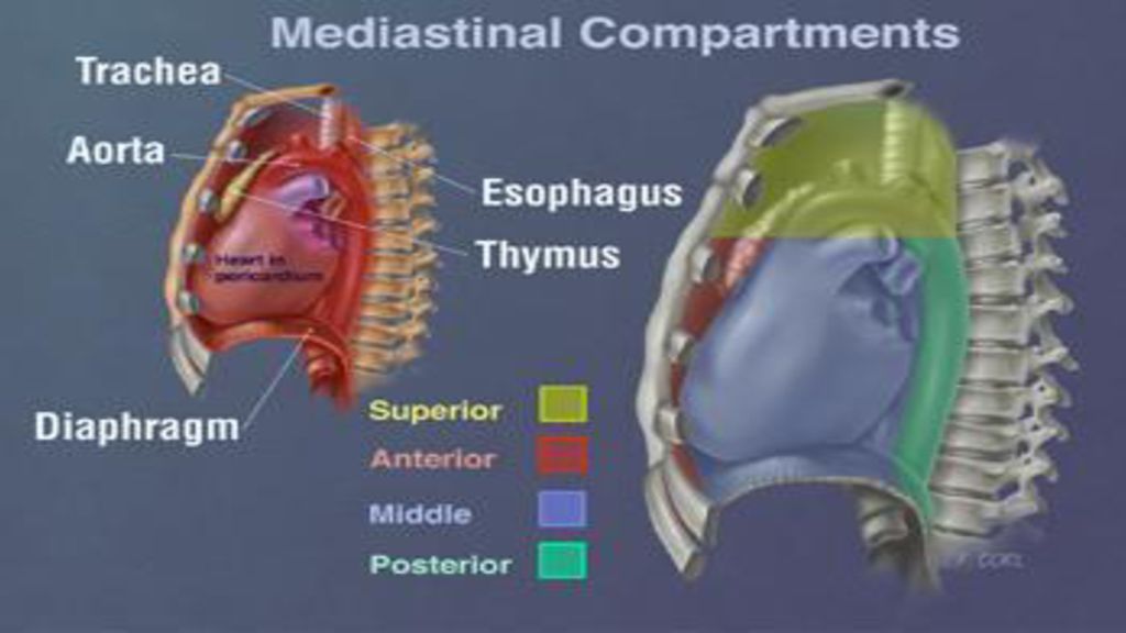
The mediastinum: is the central compartment of the thoracic cavity surrounded by loose connective tissue. The mediastinum contains the heart and its vessels, the esophagus, trachea, phrenic and vagus nerves, the thoracic duct, thymus and lymph nodes of the central chest. The mediastinum lies within the thorax and is enclosed on the right and left by pleurae. It is surrounded by the sternum and chest wall in front, the lungs to the sides and the spine at the back. It contains all the organs of the thorax except the lungs. It is continuous with the loose connective tissue of the neck. The mediastinum can be divided into an upper (superior) and lower (inferior) part: The superior mediastinum starts at the superior thoracic aperture and ends at the thoracic plane. (thoracic plane: is a plane at the level of the sternal angle, and the intervertebral disc of T4–T5). The inferior mediastinum extends from thoracic plane to the diaphragm. It is subdivided into three regions, all relative to the pericardium – the anterior mediastinum being in front of the pericardium, the middle mediastinum contains the pericardium, and the posterior mediastinum behind pericardium
 and lower (inferior) part: The superior mediastinum starts at the superior thoracic aperture and ends at the thoracic plane. (thoracic plane: is a plane at the level of the sternal angle, and the intervertebral disc of T4–T5). The inferior mediastinum extends from thoracic plane to the diaphragm. It is subdivided into three regions, all relative to the pericardium – the anterior mediastinum being in front of the pericardium, the middle mediastinum contains the pericardium, and the posterior mediastinum behind pericardium.")
3
Mediastinum

5
Sternal angle represents several important anatomical features:
1- Beginning and end of the aortic arch 2- Bifurcation of the trachea 3- Bifurcation of the pulmonary trunk 4- Left recurrent laryngeal nerve loops under arch of aorta 5- Ligamentum arteriosum lies at this level 6- Azygos vein drains into superior vena cava 7- Cardiac plexus 8- Thoracic duct empties into left subclavian vein

6
Superior mediastinum Bounderies superiorly by the thoracic inlet; inferiorly by the thoracic plane laterally by the pleurae anteriorly by the manubrium of the sternum posteriorly by the first four thoracic vertebral bodies

7
Contents of the superior mediastinum
muscles Origins of the Sternohyoid, Sternothyroid and lower ends of the Longus colli arteries Aortic arch, brachiocephalic artery, thoracic portions of the left common carotid and the left subclavian Veins brachiocephalic veins, upper half of the superior vena cava and left highest intercostal vein Nerves Vagus nerve, superficial and deep cardiac plexuses, phrenic nerve and left recurrent laryngeal nerve Organs Trachea with paratracheal and tracheobronchial lymph nodes, esophagus, thoracic duct, thymus gland and some lymph glands

8
Lt. Brachiocephalic V Rt. Brachiocephalic V. Lt. Phrenic N. Sup. Vena cava Lt. Vagus N. Ligamentum Art.

11
Inferior mediastinum Inferior mediastinum is divided into 1-Anterior mediastinum Relations laterally is the pleura posteriorly is the pericardium anteriorly is the sternum. Contents A quantity of loose areolar tissue Some lymphatic vessels which ascend from the convex surface of the liver Two or three anterior mediastinal lymph nodes The small mediastinal branches of the internal thoracic artery Thymus (disappears in adults)
")
12
Middle mediastinum It bounds the pericardial sac Pericardium contains the vital organs and is classified into the serous and fibrous pericardium Contents the heart enclosed in the pericardium the ascending aorta the lower half of the superior vena cava with the azygos vein opening into it the bifurcation of the trachea and the two bronchi the pulmonary trunk dividing into its two branches the right and left pulmonary veins the phrenic and vagus nerves some bronchial lymphatic glands Pericardial and phrenic vessels

13
Pericardial and phrenic vessels

14
Posterior mediastinum
Is bounded: Anteriorly by (from above downwards);bifurcation of trachea; pulmonary vessels; fibrous pericardium Inferiorly by the thoracic surface of the diaphragm Superiorly by the thoracic plane Posteriorly by the bodies of the vertebral column from the fifth to the twelfth thoracic vertebra Laterally by the mediastinal pleura Contents Artery: thoracic part of the descending aorta Veins: azygos vein the hemiazygos vein and the accessory hemiazygos vein Nerves: vagus nerve, splanchnic nerves, sympathetic chain Other structures : Esophagus, thoracic duct, some lymph glands
;bifurcation of trachea; pulmonary vessels; fibrous pericardium. Inferiorly by the thoracic surface of the diaphragm. Superiorly by the thoracic plane. Posteriorly by the bodies of the vertebral column from the. fifth to the twelfth thoracic vertebra. Laterally by the mediastinal pleura Contents. Artery: thoracic part of the descending aorta. Veins: azygos vein the hemiazygos vein and the accessory hemiazygos vein. Nerves: vagus nerve, splanchnic nerves, sympathetic chain. Other structures : Esophagus, thoracic duct, some lymph glands.")
15
Contents of the posterior mediastinum

16
The Pericardium Pericardium: Is a membranous flask shaped sac that surrounds and protects the heart. It is formed by two principal portions: An outer fibrous pericardium: Is tough, inelastic, dense irregular connective tissue. It blends inferiorly with the central tendon of the diaphragm and superiorly with the adventitia of the great vessels An inner serous pericardium: is a thin, more delicate membrane that forms a double layer around the heart. The outer parietal layer of the serous pericardium is fused with the fibrous pericardium. The inner visceral layer of the serous pericardium, called the epicardium. It is combined with the underlying delicate areolar tissue and adipose tissue and adheres tightly to the surface of the heart.

17
Between the parietal and visceral layers of the serous pericardium is a thin film of lubricating fluid known as pericardial fluid. The space that contains the few milliliters of pericardial fluid is called the pericardial cavity. The heart is suspended freely inside the pericardial cavity. It is only fixed by the junction between fibrous pericardium and the adventitia of the aorta, pulmonary trunk and superior vena cava superiorly and adventitia of pulmonary veins posteriorly

18
Mediastinum

19
Layers of the Heart Wall
The wall of the heart consists of three layers: epicardium, the myocardium, and the endocardium. Epicardium (external layer): is composed of two tissue layers. The outermost is the visceral layer of the serous pericardium (mesothelium). Beneath the mesosthelium is a variable amount of delicate areolar tissue and adipose tissue. It contains blood vessels, lymphatics, and nerves that supply the myocardium. The middle myocardium (muscle 95% of the heart) is responsible for the pumping action of the heart and is composed of cardiac muscle tissue. Endocardium (innermost layer): is a thin layer of endothelium overlying a thin layer of connective tissue. Provides a smooth lining for the chambers of the heart and covers the valves of the heart. The smooth endothelial lining minimizes the surface friction.
: is composed of two tissue layers. The outermost is the visceral layer of the serous pericardium (mesothelium). Beneath the mesosthelium is a variable amount of delicate areolar tissue and adipose tissue. It contains blood vessels, lymphatics, and nerves that supply the myocardium. The middle myocardium (muscle 95% of the heart) is responsible for the pumping action of the heart and is composed of cardiac muscle tissue. Endocardium (innermost layer): is a thin layer of endothelium overlying a thin layer of connective tissue. Provides a smooth lining for the chambers of the heart and covers the valves of the heart. The smooth endothelial lining minimizes the surface friction.")
21
There are two pericardial sinuses: Transverse sinus and oblique Sinus
Transverse sinus: located between the aorta and pulmonary artery anteriorly and the superior vena cava posteriorly Oblique sinus: is a cul-de-sac sinus, enclosed between the limbs of an inverted U shaped pericardial reflection; it lies posterior to the left atrium. Pericardial reflection is a double layer of visceral mesoderm supporting the heart in the pericardial cavity. Oblique sinus Transverse sinus Inf vena cava Oblique sinus

22
Boundaries of the oblique sinus
right (in ascending order): inferior vena cava, right inferior pulmonary vein and right superior pulmonary vein Superior: it is separated from the transverse pericardial sinus above by a double reflection of serous pericardium that extends transversely between the left and right superior pulmonary veins Left (in ascending order): left inferior pulmonary vein and left superior pulmonary vein
: inferior vena cava, right inferior pulmonary vein and right superior pulmonary vein. Superior: it is separated from the transverse pericardial sinus above by a double reflection of serous pericardium that extends transversely between the left and right superior pulmonary veins. Left (in ascending order): left inferior pulmonary vein and left superior pulmonary vein.")
24
Thank You

Similar presentations