Download presentation
Presentation is loading. Please wait.

1
Managing EVAR Graft Complications
Michael J. Reardon, M.D. Professor of Cardiothoracic Surgery Methodist DeBakey Heart & Vascular Center

2
Conflict of Interest Consultant to Medtronic CoreValve Trial
Steering committee member SurTAVI Trial National PI

3
Signs and Symptoms Low grade pyrexia Sepsis Weight loss Anorexia
Fatigue Sepsis Fever Rigors Shock

4
Presentation Imaging signs of graft infection Increase in sac diameter
Air in sac Loss of tissue planes Increase in sac diameter Abscess formation Destruction of spinal vertebral bodies Anaemia Occult gastrointestinal bleeding

5
Presentation Rupture Fistula Aortoenteric Aorto-oesophageal
Aortobronchial Aortocutaneous

7
Culture Blood cultures x3 before antimicrobials given Culture fluid, bone, thrombus, device if removed Pre-operative antibiotic course improves outcome If rupture no opportunity

8
Antimicrobials Positive culture: sensitivities to drugs
Negative culture: broad spectrum When to stop? When indices return to normal: White blood cells, erythrocyte sedimentation rate, c-reactive protein For ever When patient refuses to take any more PICC lines, portacath with central line

9
Organisms Culture negative 25% Staphylococcus aureus 30%
Salmonella % Streptococcus 10% Staphylococcus albus Escherichia Coli

10
CT guided aspiration - propionibacterium acnes

11
Other Reported Organisms
Proteus Serratia Enterobacter Neisseria Mycobacterium Propionibacterium Clostridium Enterococcus Bacteriodes Candida Klebsiella Actinobacter

12
Treatment Options Intravenous antimicrobials Drainage and irrigation
Better outcome if further from procedure Drainage and irrigation Further endovascular repair Useful to control haemorrhage Inevitably will become infected Bridge to definitive repair Timing of definitive repair very important

13
Treatment Options Removal of the device and extensive debridement
In situ reconstruction: antibiotic soaked grafts, silver impregnated grafts, autologous grafts, homografts Extra-anatomic reconstruction Omentum to cover aorta and sac Drainage

14
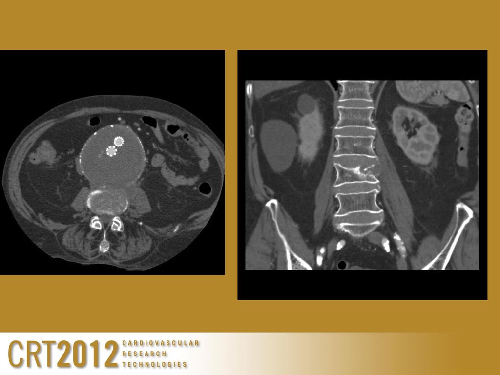
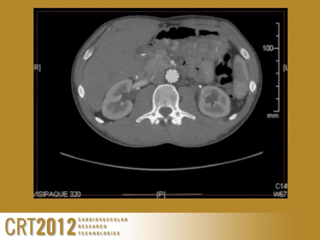
Large abdominal germ cell tumor
25 year old male Large abdominal germ cell tumor Duodenal aorto fistula with 25 unit bleed Endograft abdominal aorta – stops bleed Chemotherapy – no evidence of disease Intermittent fever and chills

17
Allograft
18
47 year old female had bleeding into her left chest after a previous spinal fusion with instrumentation Receives a thoracic endograft that controls bleeding

19
5 months after endograft she develops fever and hemoptysis
Evidence of contrast outside of endograft on CT scan Open repair
20
Infected Endografts 62 cases
49 (79%) removed with either in-situ or extra-anatomic reconstruction Mortality rate 16% 11 (18%) were treated with antibiotics with or without drainage Mortality rate 36% at 3 months Fiorani P et al J Endovasc Ther 2003; 10:
 removed with either in-situ or extra-anatomic reconstruction. Mortality rate 16% 11 (18%) were treated with antibiotics with or without drainage. Mortality rate 36% at 3 months. Fiorani P et al J Endovasc Ther 2003; 10:")
21
Belfast 409 patients EVAR AAA 6 (1.5%) infected 2 psoas abscess:
graft removed with extra-anatomic bypass OK 2 infected grafts: one removed with extra-anatomic bypass OK one treated conservatively died 1 died suddenly: Post Mortem aortoenteric fistula 1 died untreated as inoperable cancer Sharif MA et al J Vasc Surg 2007; 46: 442-8

22
Chicago 2000-7 Infections Mean time from implantation 243 days
5/389 EVAR (0.26%) 5/106 TEVAR (4.77%) Mean time from implantation 243 days 2 had contained rupture Rest infections and/or abscess on imaging Propionibacterium 3, Staph 3, Strep 2, Enterobacter 1
 5/106 TEVAR (4.77%) Mean time from implantation 243 days. 2 had contained rupture. Rest infections and/or abscess on imaging. Propionibacterium 3, Staph 3, Strep 2, Enterobacter 1.")
23
Chicago All EVAR removed with extra-anatomic bypass in 3 and in situ in 2 TEVAR 1 removal 4 treated medically with 1 survivor, 2 died of rupture and of MSOF from sepsis Heyer et al J Vasc Interv Radio 2009; 20: 173-9

24
University of Michigan
9 patients Mean time 33 months post implant Investigations: CT, MR, white cell scan Rifampicin soaked in situ grafts 4 Extra-anatomic bypass 5 E coli, Bacteroides, Staph, Strep, Candida 1 died of aortoenteric fistula 3 others developed an aortoenteric fistula 2 died Laser A et al J Vasc Surg 2011; Feb Epub

25
Guy’s & St Thomas’ 10 infected aortic grafts:
3 TEVAR drainage and long term antibiotics All alive and well; portacath, PICC line, oral 7 EVAR all removed with axillobifemoral grafts 2 deaths: iliac haemorrhage, respiratory failure 5 alive and well 1 concurrent spinal reconstruction 1 spinal brace

26
World Literature 2010 EVAR for AAA 102 reported infections since 1991
Options: Antibiotics and percutaneous drainage Explantation and in situ or extra-anatomic bypass Outcome best for explantation and in situ replacement Setacci C et al J Cardiovasc Surg (Torino) 2010; 51: 33-41
 2010; 51:")
27
Summary Infected endografts likely to be an increasing problem
Culture and antibiotic therapy Drainage Removal in-situ autologous reconstruction Silver impregnated, antibiotic soaked grafts Extra-anatomic reconstruction

28
Summary Antibiotic therapy Long term intravenous PICC, portacath
Long term oral; reduced dose Stop when no evidence of infection Pragmatically when patient stops taking them

29
Thank You

Similar presentations

 ・ In 1998, we developed a modified elephant trunk (ET) technique using a single four-branched arch graft with a sewing “collar” and “long.>")


 LECT7 ALI B ALHAILIY.>")


