Download presentation
Presentation is loading. Please wait.

1
Dr. Mustafa Fadil Alhammami University of Mustansyria College of medicine Department of medicine Neuromedicine Tue.6/10/2015

3
Sagital gross anatomy 1-The spinal cord has 31 segments: 8 cervical, 12 thoracic, 5 lumbar, 5 sacral, 1 coccygeal. 2- Cervical spinal nerves C1 to C7 exit spinal column at neural foramina above corresponding vertebral body.Herniation of intervertebral disk between C4 and C5 can affect exiting C5 nerve root. Cervical spinal nerve C8 and all spinal nerves below this level exit spinal column at neural foramina at the corresponding vertebral body.

5
Transverse Section anatomy

6
A-absence of reflexes, lower motor neuron paresis, atrophy and fasciculations at level of the lesion. B- Spastic paraparesis or quadriparesis (depending on level of the lesion) C- Sensory level: panmodality sensory loss at and below level of the lesion.
 C- Sensory level: panmodality sensory loss at and below level of the lesion..")
7
A- Early sphincter dysfunction. B- Urinary retention with absence of urinary sensation. C- Saddle anesthesia. D-paraplagia. E- Variable presence of reflexes.

8
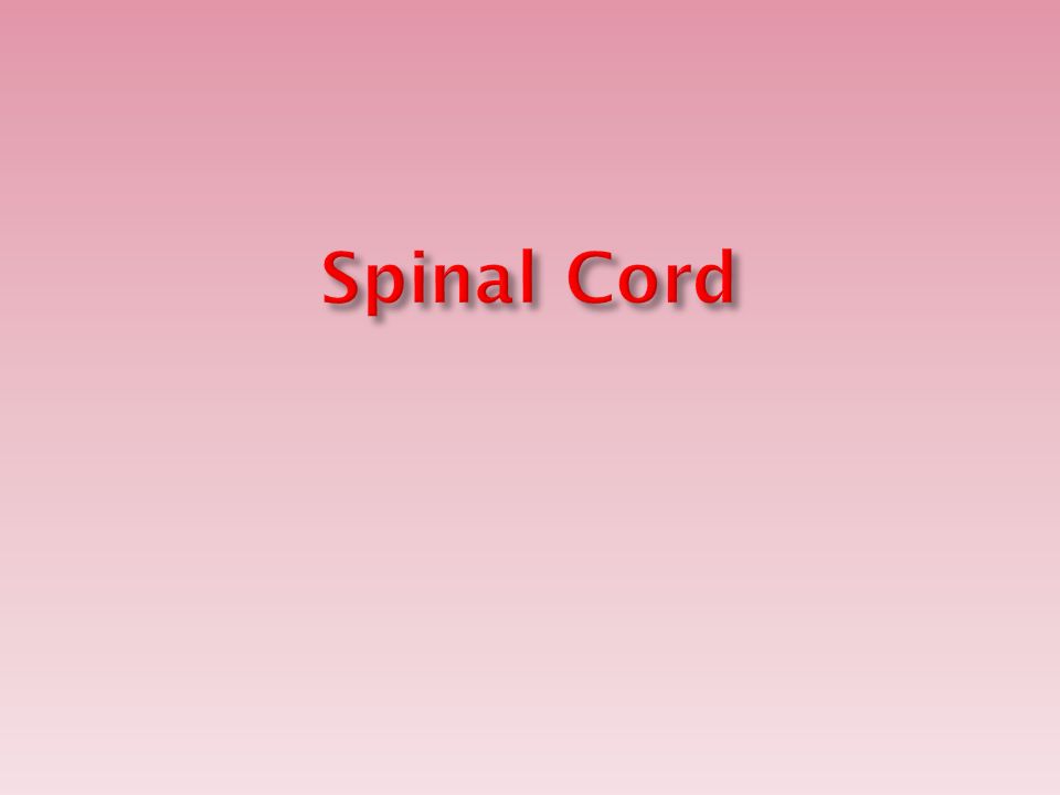
A- Contralateral loss of pain and temperature sensation. B- Ipsilateral loss of vibration and proprioceptive sensation below level of the lesion. C- Ipsilateral pyramidal weakness (spastic weakness). D- Ipsilateral lower motor neuron paresis with atrophy, fasciculations at the level of the lesion.
. D- Ipsilateral lower motor neuron paresis with atrophy, fasciculations at the level of the lesion..")
10
A- “dissociated” selective sensory loss of pain and temperature (capelike distribution if it occurs in cervical spinal cord). B- With further expansion, lateral corticospinal tracts may be involved, producing upper motor neuron signs below level of the lesion. C- Posterior columns may be relatively preserved.

11
1- Hereditary : spastic paraplegia,leukodystrophy etc… 2-vascular :anterior spinal artery infarction. 3-compressive :tumor,abscess,disc herniation, etc… 4-truma :sharp,blunt. 5- Neutritional : vit. B12 defeciency, copper defeciency etc… 6-Toxin and drugs :chemotherapy,heavy metals. 7-Infection : viral, bacterial,fungal. 8-Inflimmation : sarcoidosis, sjogren. 9- Demyelination :multiple sclerosis, Devic.

12
The clinical presentation: 1-patient is usually young age develop pain in the cervical area that may radiate to the arm. 2-if the herniation is lateral so lead to radicular symptoms. 3- But if the herniatin is central so lead to compression on the spinal cord itself causing spastic quadriplegia with sensory level and incontinence.

14
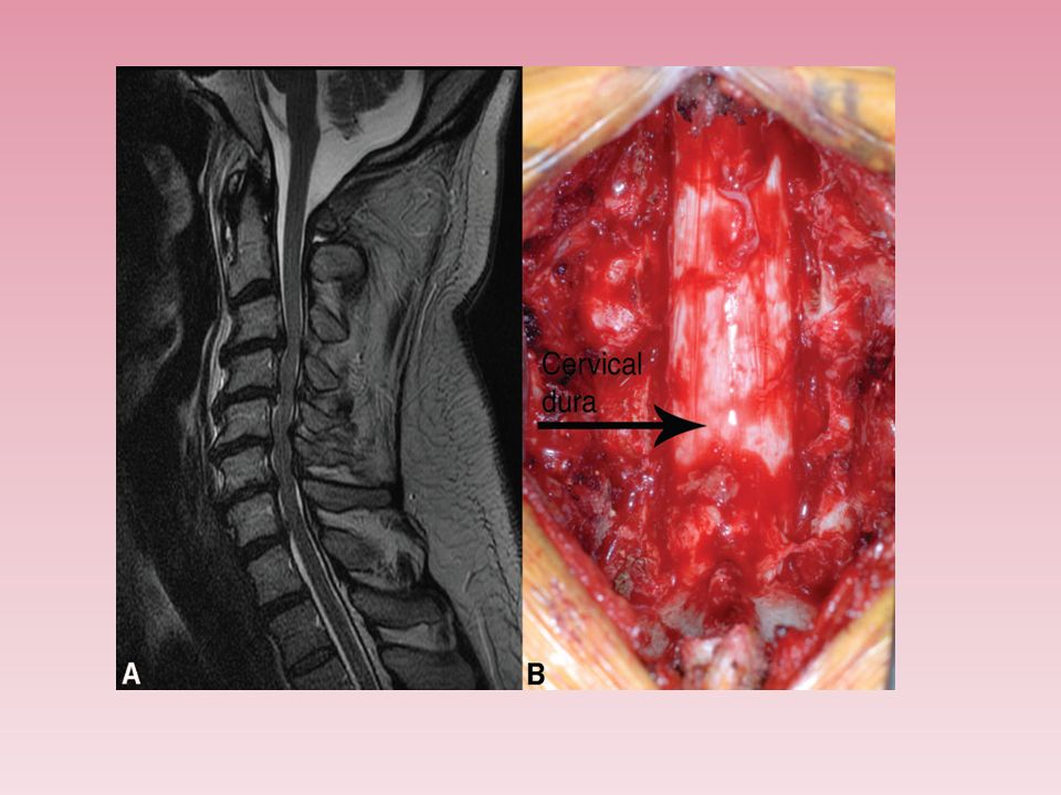
The investigation: 1- MRI of spine :to see the direction and extent of the compression. Treatment : 1-rest for few days. 2- analgesia. 3-surgery :is indicated in: A-sever pain not respond to treatment. B-neurological symptoms like incontinence or weakness(SC compression).
..")
15
The clinical presentation: 1-Acute lumbar disc herniation is often precipitated by trauma, usually by lifting heavy weights while the spine is flexed. 2-The onset may be sudden or gradual. Alternatively, repeated episodes of low back pain may precede sciatica by months or years. 3- lower motor neurons weakness of the lower limbs. 4- incontinence. 5-Root pressure is suggested by limitation of flexion of the hip on the affected side if the straight leg is raised (Lasègue's sign). Investigations and treatment : The same as cervical disc prolapsed.
. Investigations and treatment : The same as cervical disc prolapsed..")
16
Blood supply of the spinal cord : 1-Blood supply to the spinal cord emerges from 1 anterior and 2 posterior spinal arteries (branches of vertebral arteries). 2-The anterior spinal artery supply the anterior 2/3 of the spinal cord(sparing the dorsal column).
..")
17
Causes 1-Aortic dissection 2- Aortic surgery 3-Aortic atherosclerosis 4-Severe hypotension 5-Spinal trauma 6- Cardiogenic embolism 7- Vertebral artery dissection 8-Vasculitis

19
Clinical pictures : 1-Flaccid weakness below the lesion (paraplegia or quadriplegia) due to spinal shock. 2-This is followed by spastic paraplegia. 3-Anesthesia or hypoesthesia to pain and temperature below the lesion. 4-Urinary incontinence. 5-Constipation (paralytic ileus). 6- Preservation of proprioception and vibration. 7- Back or radicular pain at the level of the lesion 8- Respiratory failure (with rare mid cervical and upper cervical infarctions).
. 6- Preservation of proprioception and vibration. 7- Back or radicular pain at the level of the lesion 8- Respiratory failure (with rare mid cervical and upper cervical infarctions)..")
20
Investigations: 1-CT may be useful to exclude alternative diagnoses. 2-MRI of the spine may show cord swelling or T2 hyperintensity at the affected area. 3-CT angiogram or MRA of the spine can also detect the lesion. Treatment: 1-Vasopressors are used to increase perfusion pressure. 2-CSF is removed to reduce the resistance to microcirculatory flow. 3-treat the risk factors for ischemia like hypertension,DM, etc… 4-Treat the complication like bed sore, DVT,aspiration pneumonia.

22
Syringomyelia is a cystic dilation or fluid-filled cavity within the spinal cord that typically involves the cervical region, but may extend up into the brainstem or down to the conus.

23
Causes: 1-congenital. 2-trauma. 3-tuomr. 4-spinal cord infarction. 5-infection.

25
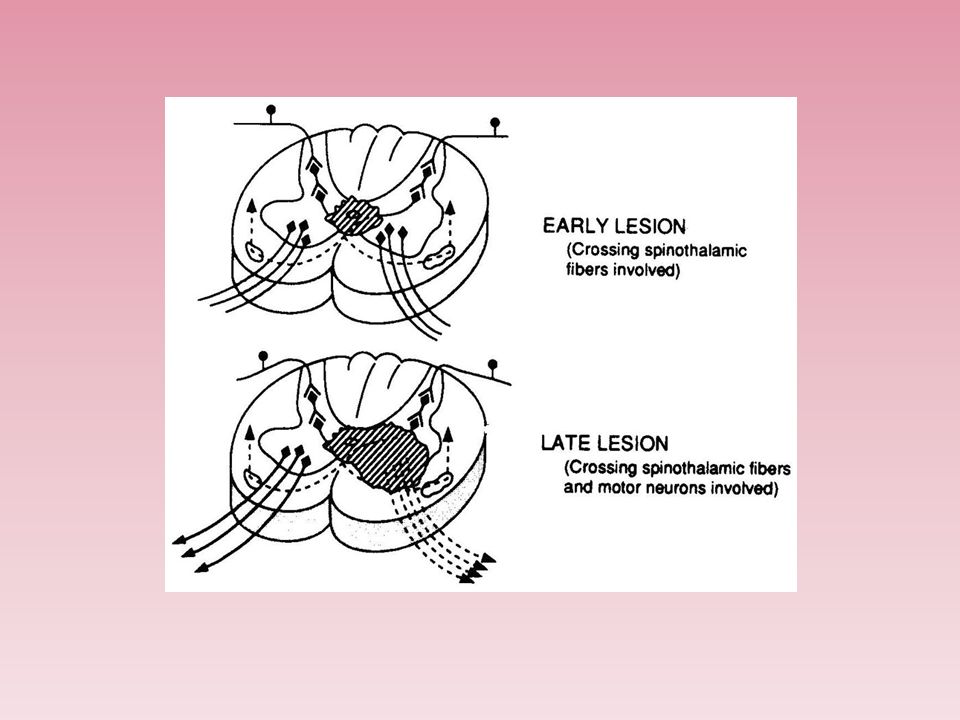
Clinical Features: 1-Clinical symptoms follow the destructive path of the widening syrinx, which begins in the center and expands outward and longitudinally(central cord syndrome). 2-flaccid weakness in lower cervical segments and corticospinal tract changes in the legs. 3-loss of pain and temperature sensation occurs in a capelike distribution due to injury of the decussating spinothalamic tract fibers. 4-the dorsal column still spared till late stage.

26
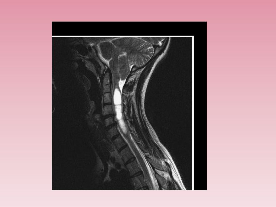
Investigations: 1-MRI of cervicothoracic spine with contrast is the investigation of choice which show cystic area filled with CSF. 2- MRI of brain and lumbosacral area should be done also to exclude chiari malformation and spina bifida. 3-Investigations for the cause of syrinx. Treatment: consists of surgical drainage with restoration of CSF flow in patients with progressive symptoms.

Similar presentations