Download presentation
Presentation is loading. Please wait.

1
Lab 9 The Nervous System: Histology and The Brain

2
Objective 1 Neuron Structure

3
Multipolar Neuron model Dendrites (receive) Cell Body (process) Axon (send) Axon Terminals (transfer) Axon Terminals Main parts of a neuron
 Cell Body (process) Axon (send) Axon Terminals (transfer) Axon Terminals Main parts of a neuron")
4
Dendrites (receptive regions) Cell body (biosynthetic center and receptive region) Nucleus Nucleolus Axon (impulse generating and conducting region) Impulse direction Dendrite Neuron cell body Nissl bodies (rough ER) Neurofibrils
 Cell body (biosynthetic center and receptive region) Nucleus Nucleolus Axon (impulse generating and conducting region) Impulse direction Dendrite Neuron cell body Nissl bodies (rough ER) Neurofibrils")
5
Impulse direction Neurilemma (sheath of Schwann) Schwann cell (one internode) Node of Ranvier Axon Axon hillock
 Schwann cell (one internode) Node of Ranvier Axon Axon hillock")
6
Terminal branches (Telodendria) Axon terminals (secretory component) Impulse direction
 Axon terminals (secretory component) Impulse direction")
7
Cell Body Spinal Cord Smear – Motor Neuron What you need to draw and label

8
Schwann cells - supporting cells of the PNS that myelinate axons. myelin sheath Schwann cell nucleus axon neurilemma Node of Ranvier Myelin sheath – whitish lipoprotein that surrounds and insulates the axon (nerve fiber) Neurilemma - external layer containing bulk of cytoplasm with nucleus and organelles
 Neurilemma - external layer containing bulk of cytoplasm with nucleus and organelles.")
9
Nodes of Ranvier - Gaps between successive Schwann cells along the length of the axon

10
What you need to draw and label Node of Ranvier Neurilemma Axon

11
Objective 2 Neuron Classification

12
Pseudounipolar: -a short process emerges from the cell body and divides into proximal and distal branches -most sensory neurons soma Distal process (toward periphery)Proximal process (toward CNS )
Proximal process (toward CNS )")
13
Pseudounipolar Cell Bodies in the Dorsal Root ganglion of a Spinal Nerve

14
Spinal Cord Dorsal Root Ganglion Centrally located nuclei Pseudounipolar cell bodies

15
Bipolar neuron -a single axon and a single dendrite attached to opposite sides of the cell body -found in special sense organs (eye, ear, etc.) Bipolar neurons Human retina
 Bipolar neurons Human retina")
16
Bipolar neurons Bipolar neurons in the human retina

17
Purkinje cell of the Cerebellum Pyramidal cell of the hippocampus Neuron from the cerebral cortex Multipolar: -multiple dendrites and a single axon -includes most neurons; all motor neurons and most CNS neurons

18
Multipolar neurons have diverse morphologies

19
Silver Stained Neuron In Gray Matter

20
Multipolar neurons Spinal Cord - Anterior Horn

21
Multipolar neurons you will be drawing Purkinje cellPyramidal cell

22
(Low Power – Cerebrum) Pyramidal cell
 Pyramidal cell")
23
Purkinje cell (Low Power - Cerebellum)
")
24
Objective 3Nerves Nerves are structures of the PNS that consist of axons and dendrites bundled together by connective tissues Fascicle:a bundle of axons or dendrites Epineurium: tough, fibrous connective tissue sheath surrounding a nerve Perineurium: loose, areolar connective tissue sheath surrounding fascicles Endoneurium: delicate connective tissue wrapping around each nerve fiber

25
a = epineurium b= perineurium

26
perineurium endoneurium axon

27
LAB ACTIVITY Draw and label the components of the nerve cross section

28
Nervous System The Brain Sheep HumanHuman BrainBrain Brain

29
OBJECTIVES Identify the components of the human brain using models and diagrams Identify the structures of the sheep brain

30
Objective 4:Anatomy of the human brain Cerebrum (gold area) Diencephalon (Violet area) Brain stem (green area) Cerebellum (pink area)
 Diencephalon (Violet area) Brain stem (green area) Cerebellum (pink area)")
31
Cerebrum

32
Fissure Sulcus Gyrus Ventricles Structures of the Cerebrum Cerebral Hemispheres Left HemisphereRight Hemisphere Gray Matter – Cerebral cortex & Basal nuclei White Matter – Myelinated fiber tracts (axons) Frontal cut
 Frontal cut")
33
Structures of the Cerebrum Human Brain, Lateral ViewOccipital lobe lobe Frontal Temporal lobe Parietal lobe Gyrus Lateral sulcus Central sulcus Transverse Fissure Parieto-occipital Sulcus

34
Structures of the Cerebrum Human Brain, Superior View Longitudinal fissure Central sulcus Postcentral gyrus Precentral gyrus Frontal pole Occipital pole

35
Midsagittal Section of Human Brain Corpus Callosum Fornix Septum pellucidum Parietal lobe Occipital lobe Frontal lobe

36
Internal Structures of the Cerebrum Frontal Section Corpus callosum Fornix Lateral Ventricles Septum pellucidum Longitudinal fissure

37
Diencephalon

38
Functions of the Diencephalon Endocrine System Relay Stations Thalamus

39
Structures of & related to the Diencephalon Midsagittal Section Corpus callosum Fornix Intermediate mass HypothalamusMammillary body Pineal body/ gland Thalamus

40
Pituitary gland Thalamus Hypothalamus Infundibulum Sella Turcica

41
Brainstem

42
Pons Medulla oblongata Midbrain: Corpora quadrigemina Superior colliculi Inferior colliculi Cerebral peduncle Spinal cord

43
Corpora quadrigemina Superior colliculus Inferior colliculus Brainstem Posterior Pineal body/ gland

44
Structures of the Brainstem Inferior View Midbrain (Cerebral peduncle) Pons Medulla oblongata Spinal cord Optic chiasma Infundibulum Mammilary Bodies
 Pons Medulla oblongata Spinal cord Optic chiasma Infundibulum Mammilary Bodies")
45
Cerebellum “Little Brain”

46
Arbor Vitae Cerebellar Peduncles Fourth ventricle Structures of the Cerebellum Midsagittal Section

47
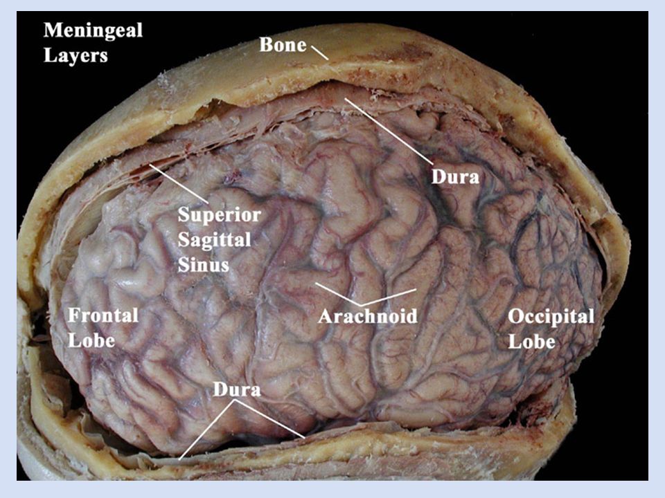
Meninges of the Brain

48
A mnemonic PAD from deep to superficial the layers of the meninges are pia, arachnoid, dura (they pad the brain)
")
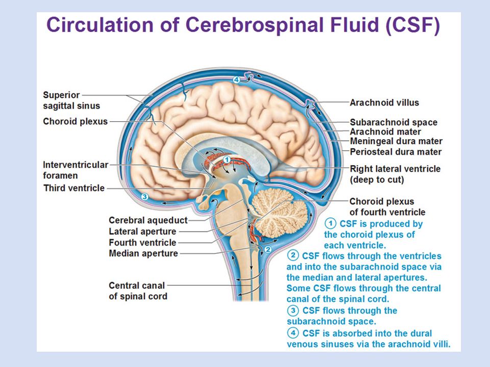
50
Cerebral Aqueduct Third Ventricle Fourth Ventricle Ventricles of the Brain The spaces within the brain through which cerebrospinal fluid flows

51
Cerebrospinal fluid is continually produced by the choroid plexus found in the ventricles Die-cast model

53
Objective 5: Sheep Brain Dissection

54
Identify the external structures Inferior Sheep brain structures are the same as the human brain, only slightly different sizes

55
Remove Dura mater (w/ pituitary gland) Identify the external structures Superior
 Identify the external structures Superior")
56
Identify the external structures Inferior

57
Pull back the cerebellum to reveal Corpora Quadrigemina & Pineal Gland

58
Make a midsagittal cut through the brain

59
Identify the internal structures Midsagittal

60
Identify the internal structures Midsagittal Lateral Ventricle Third VentricleCerebral AqueductFourth Ventricle

Similar presentations