Download presentation
Presentation is loading. Please wait.

1
ABDOMINAL CAVITY

2
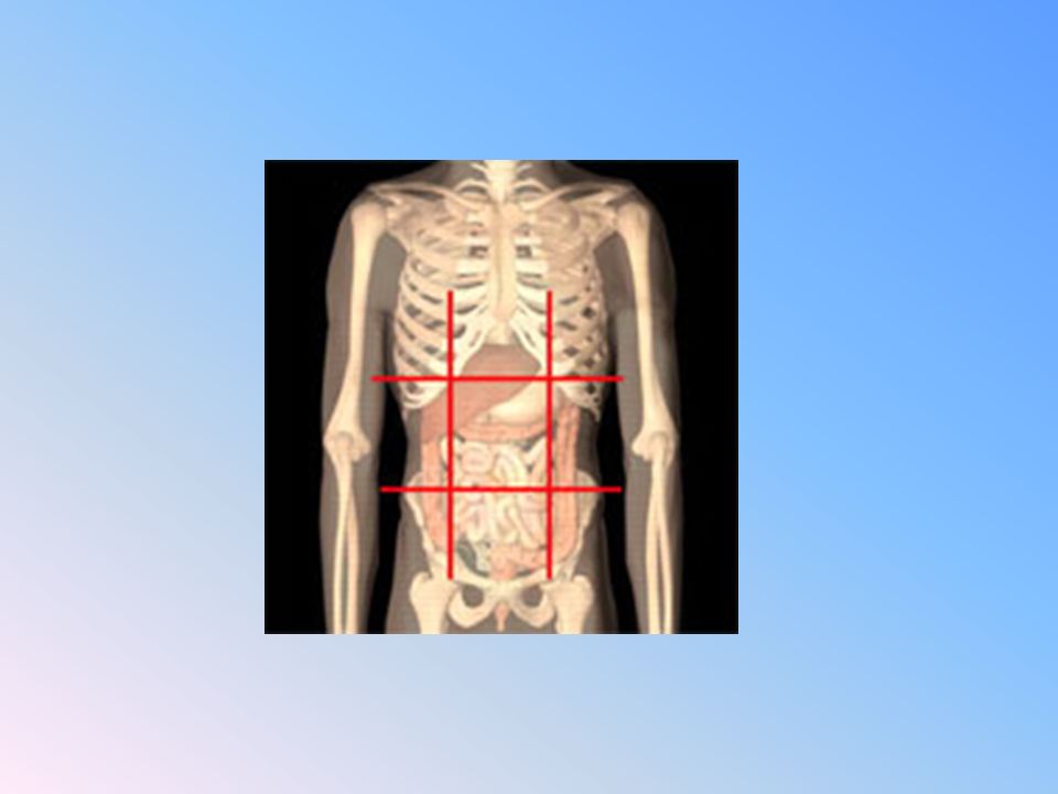
The abdominal cavity is a large cavity so it is subdivided descriptively into 9 regions.
This division is made by 2 horizontal planes and two vertical planes. The vertical planes are: They are one on each side. Each extends from the mid-clavicular point down to the mid-inguinal point. The horizontal planes are: Subcostal plane. Inter-tubercular plane.

3
Subcostal plane: It lies at the lowest limit of the costal margin (level of L3). Inter-tubercular plane: It passes between the tubercles of the two iliac crests (level of L5).
.")
4
The divisions of the abdominal cavity are:
Right hypo-chondrium Epigastrium Left hypo-chondrium Right lumbar Umbilical Left lumbar Right iliac fossa Hypogastric (suprapubic) region Left iliac fossa
 region. Left iliac fossa.")
6
Transpyloric plane: It cuts the body of the first lumbar vertebra.
It lies midway between the xiphoid process of sternum and umbilicus. Structures at this level: Pylorus of stomach. Hilum of left kidney. Fundus of gall bladder. Body of L1

7
Peritoneum

8
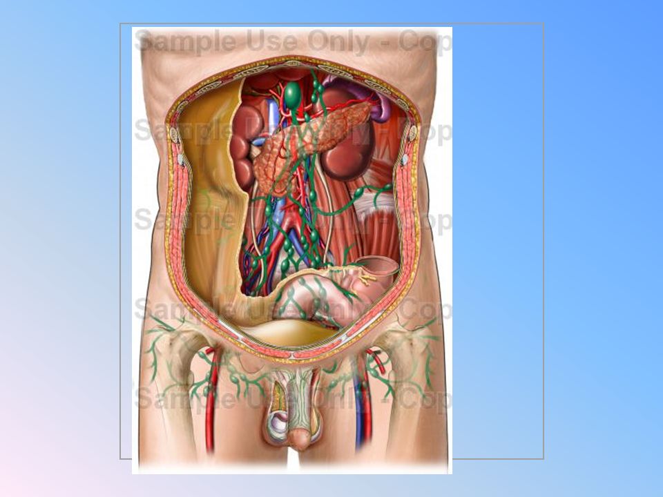
Peritoneum: It is a closed serous sac present inside the abdominal cavity. The peritoneum looks like a balloon. The intra-abdominal organs have different relations with the peritoneum: - Enclosed within a fold of peritoneum (intraperitoneal) and so freely mobile. - Present behind the peritoneum and so they are fixed; retroperitoneal.
 and so freely mobile. - Present behind the peritoneum and so they are fixed; retroperitoneal.")
9
Parietal peritoneum: The part of peritoneum that lines the abdominal wall. Visceral peritoneum: The part of peritoneum that surrounds the abdominal organs (viscera). Peritoneal cavity: Is a potential space between the parietal and visceral peritoneum that contains a very thin film of fluid. Increase in the amount of this fluid is called ascites.
. Peritoneal cavity: Is a potential space between the parietal and visceral peritoneum that contains a very thin film of fluid. Increase in the amount of this fluid is called ascites.")
10
Peritoneal Reflections

11
The folds of peritoneum are called:
1- Omentum: if related to the stomach.

12
2- Mesentry: if related to the small intestine.
3- mesocolon: if related to the large intestine.

13
4- ligaments: eg. Ligaments of the liver, falciform, right and left triangular. Ligaments of the spleen; gastrosplenic and lienorenal.

14
The main contents of the peritoneal folds are:
1- blood vessels supplying the related organ. 2- autonomic nerve plexus. 3- lymphatics and lymph nodes.. 4- extraperitoneal connective tissue. 5- the organs themselves ( for freely mobile organs).
.")
15
The peritoneum (peritoneal sac) is a closed sac in male however in females it is not completely closed as the two lateral ends of the Fallopian tubes open in it.
 is a closed sac in male however in females it is not completely closed as the two lateral ends of the Fallopian tubes open in it.")
16
The peritoneal cavity is divided into two main sacs:
Greater sac. Lesser sac. The two sacs are connected together through the epiploic foramen (foramen of the lesser sac)
")
17
Greater sac: It is the part of peritoneum that is present just behind the anterior abdominal wall.

18
Lesser sac (omental bursa):
It is a small diverticulum of the peritoneal sac that lies behind the stomach.

19
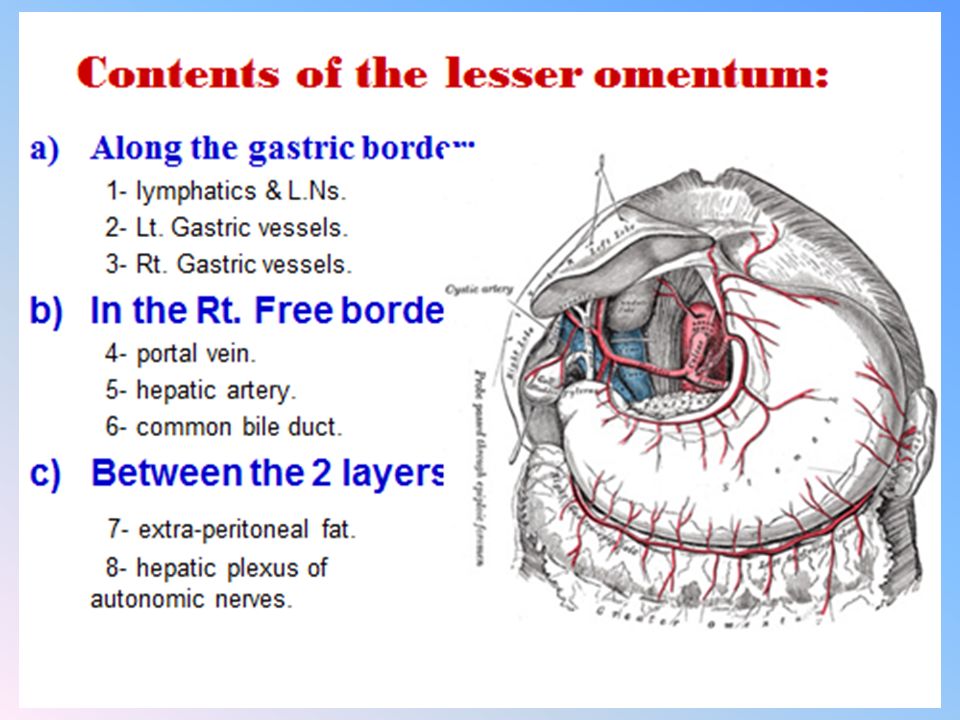
Boundaries of the lesser sac:
Anterior wall: From above downwards; 1- lesser omentum. 2- peritoneum covering the posterior surface of the stomach and 1st inch of duodenum. 3- anterior 2 layers of the greater omentum.

20
Posterior wall: The lower part formed by the posterior 2 layers of the greater omentum. The upper part is formed by the peritoneum covering the following structures:

21
Body of pancreas. Upper part of abdominal aorta. Left crus of diaphragm. Left kidney and left suprarenal gland.

23
Boundaries of the lesser sac:
Left border: formed by the gastrosplenic & lienorenal ligaments. Right border: the epiploic foramen (foramen of Winslow).
.")
24
Epiploic foramen: It is a slit-like opening connecting the greater with the lesser sac.

25
Boundaries of the epiploic foramen:
Anteriorly: Free margin of lesser omentum containing bile duct, portal vein & hepatic artery. Posteriorly: IVC

26
Superiorly: Caudate lobe of the Liver. Inferiorly: 1st part of duodenum

27
Lesser Sac (omental bursa) – Clinical Correlations
The lesser sac has important clinical significance. As it is located posterior to the stomach, the posterior wall of the stomach is in contact with the sac. Thus any posterior ulceration of the stomach will cause fluid to pass into this sac. Also an injured pancreas or inflamed pancreas will cause pancreatic secretions to enter the lesser sac. This can cause a pancreatic pseudocyst. Also sometimes the loop of the small intestine will enter the epiploic foramen and go into the lesser sac. The boundaries of the epiploic foramen contain important vessels, hence surgical removal of the intestine is deemed dangerous.

28
Nerve supply of the peritoneum:
Parietal peritoneum: Supplied by somatic nerves that supply the abdominal wall (lower 5 intercostal & subcostal nerves) & diaphragm (phrenic nerve). The parietal peritoneum is senstive to pain, temperature and touch.
 & diaphragm (phrenic nerve). The parietal peritoneum is senstive to pain, temperature and touch.")
29
The visceral peritoneum:
Supplied by autonomic nerves that supplies the viscera. It is only sensitive to traction and distension of the viscera.

Similar presentations