Download presentation
Presentation is loading. Please wait.

1
Digestive system

2
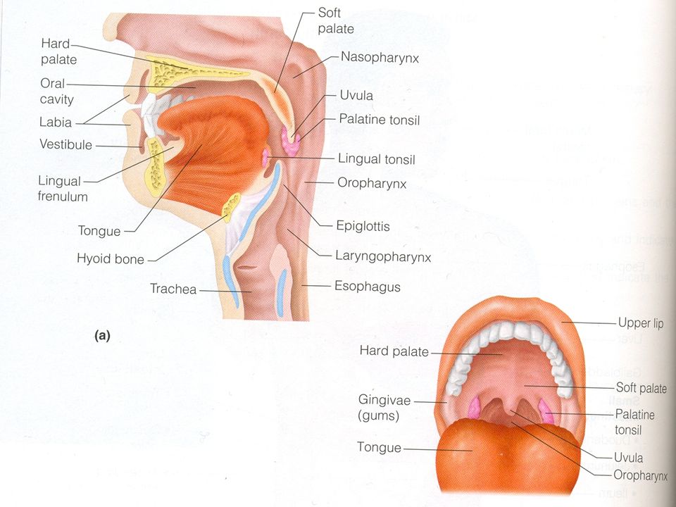
Mouth Oral cavity, lips, lateral wall is the cheek, roof is hard and soft palate, Uvula is fleshy fingerlike projection of soft palate. Vestibule is the space between cheek, and teeth. Lingual frenulum a fold of mucous membrane prevent the tongue from falling back Tongue tie short frenulum

5
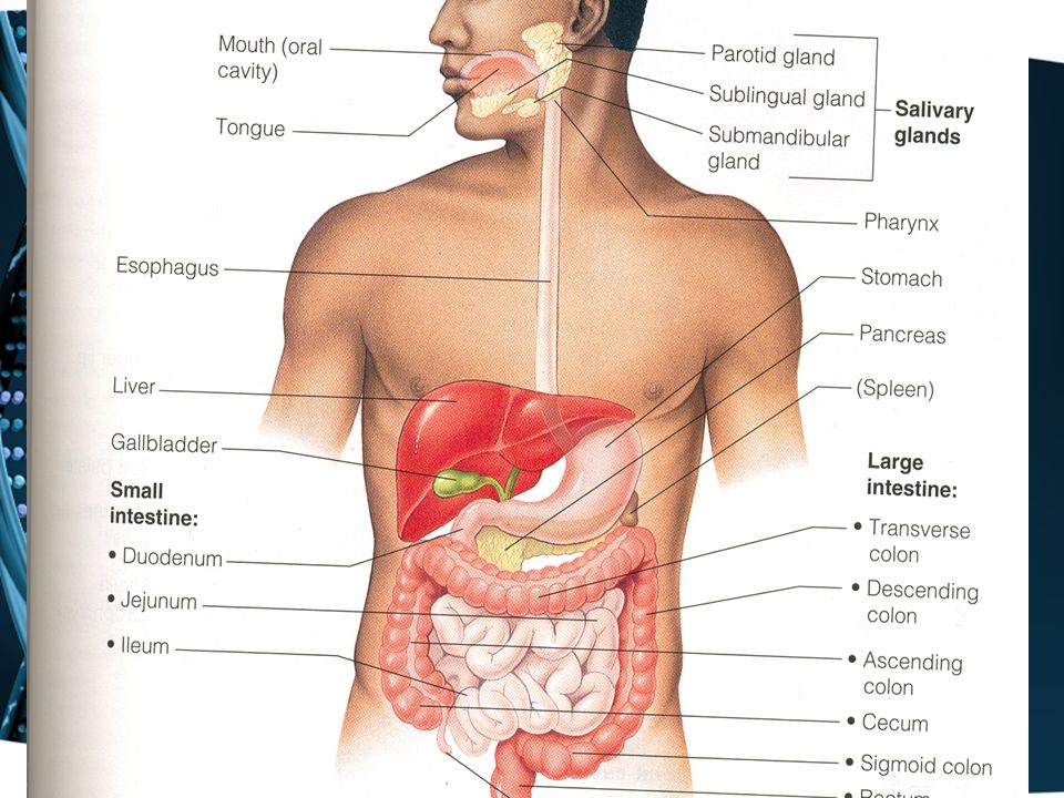
Esophagus Run from the pharynx to stomach Is 25cm in length? Enter abdomen through esophageal opening of the diaphragm at level of T10.

7
Stomach Is C shaped, lie in epigastric, and left hypochondrium. 25 cm long It has greater curvature, and lesser curvature The mucous membrane has folds called rugae Lesser omentum is a double layer of peritoneum from liver to lesser curvature Greater omentum another double layer drapes down and cover the abdominal organs, it is riddled with fat which insulate, cushion and protect abdominal organs Has large collection of lymph nodes.

9
Cardia: contain sphincter through which food enter stomach Fundus: the part to the left and above the cardia Body: between fundus and pylorus Pylorus: terminal stomach ends in pyloric sphincter.

10
Small intestine Is muscular tube from pyloric sphincter to ileocecal valve? It is 4 meter in length in living person. Attached to posterior abdominal wall by mesentery

11
Small intestine is divided into Duodenum Jejunum Ileum Duodenum: is 25 cm in length, C shaped surround head of pancreas It is divided into four parts Bile duct join pancreatic duct to open in major duodenal papilla in posterio-medial par of 2 nd part of D just below middle Jejunum: is 2.5 meter Ileum: is3.5 meter ends in ileocecal valve Peyer’s patches local collection of lymphatic in SM.

12
Duodenum

13
Gallbladder Thin wall sac; lie in inferior surface of liver Concentrate, and store 50cm of bile. Cystic duct open in to common hepatic duct to form bile duct Biliary colic Acute cholecystitis

14
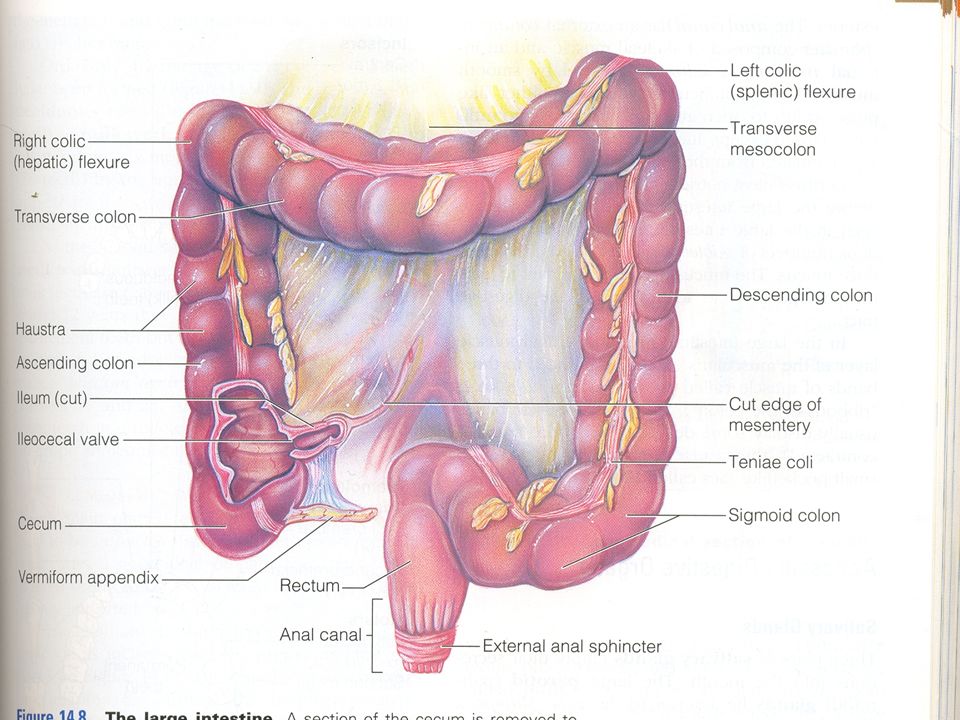
Large intestine Is 1.5 meter in length? Extend from ileocecal valve to anus Much larger in diameter Divided in to Caecum,Appendix,Colon,Rectum Anal canal.

15
Colon is divided into Ascending, transverse,descending, Sigmoid colon. Appendix: Narrowest part of the alimentary canal, worm like tube. Arise from caecum 2 cm below ileo- caecal junction. Length is variable from 1.5 cm to 22cm. Position is extremely variable, so symptoms of appendicitis may vary.

16
Colon Teniae coli represent outer longitudinal muscle layer of colon,3 in number. TC start at base of appendix and end at rectosigmiod junction Hustration because teniae is shorter than colon by one foot Appendices epiplocie condensation of fat, hanging from bowel wall.

18
Rectum: The last 15 cm of the colon Has three lateral flexions LT,RT,LT. Rectum is divided into upper third, middle third and lower third.

19
Anal canal: length is 4cm. Have two sphincters Internal is involuntary, formed of smooth muscle fibers External is voluntary formed of striated muscle fibers

20
Piles: is dilatation of veins may be either internal or external Fissure: tear in anal mucosa severely painful Abscess may lead to fistula

21
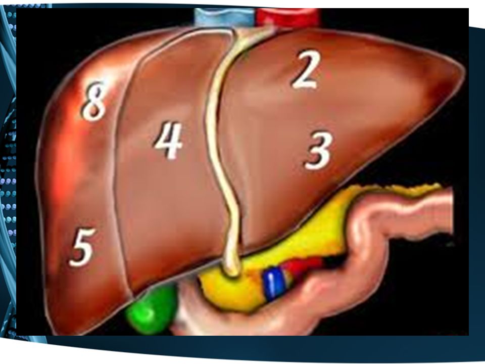
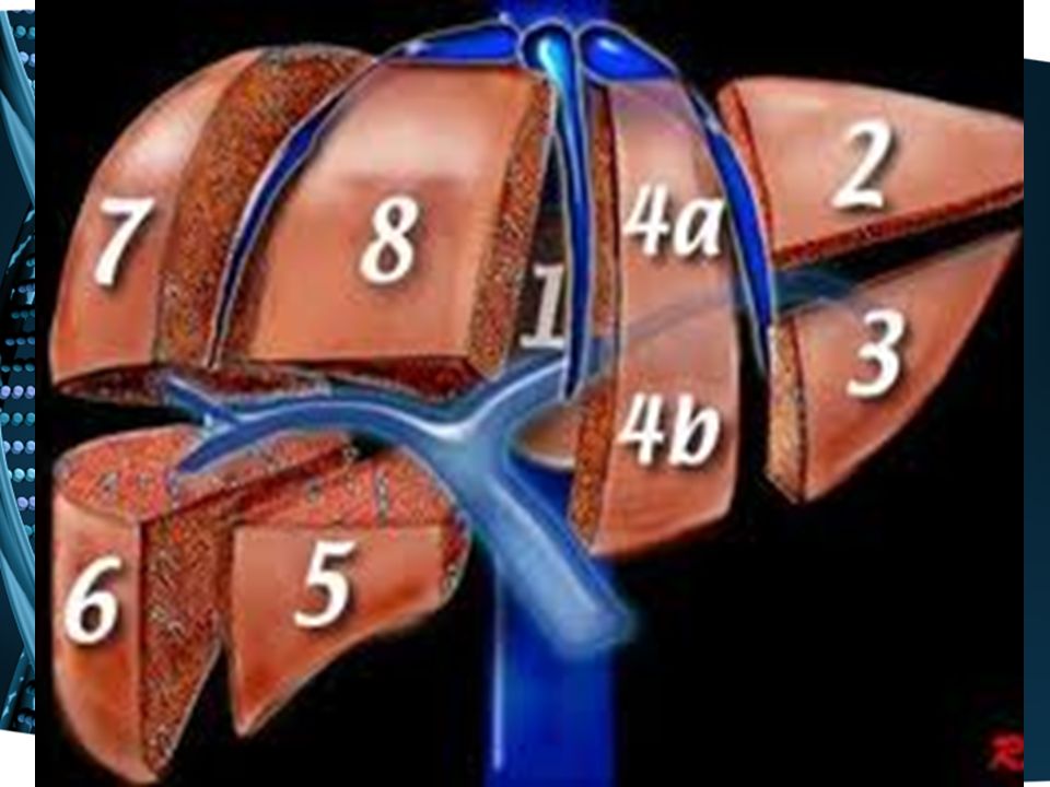
Liver and gallbladder Largest organ in the body, weight is 1.5kilo. Liver has three surfaces,superior,posterior,and inferior The umbilical fissure and falciform ligament divide antro- superior surface into RT and left lobes

22
RT lobe of liver surgically is divided into segment V, VI,VII, VIII. LT lobe is divided into II, III, IV Segment I is caudate lobe of liver Lie in RT hypochondrium Produce bile, leave liver through RT, LT hepatic ducts, join cystic duct to form bile duct

25
Blood supply Portal vein70%, hepatic artery 30% Bile duct, join pancreatic duct to open in major duodenal papilla in 2 nd part of duodenum.

26
Pancreas Triangular in shape Head, neck, body, and tail. Extend from duodenum to spleen Lie retro peritoneal Lie in epigastric region Mixed gland with endocrine and exocrine function Have two ducts which join bile duct and open in 2 nd part of duodenum Pancreatitis

27
Salivary glands Three pairs of salivary glands Parotid gland Lie behind the lobule of the ear The largest salivary gland Two lobes,one large the superficial, one small, the deep lobe Has duct open in the mouth, opposite upper 2 nd molar tooth

29
Submandibular gland Lie below angle of the mandible Duct opens in floor of the mouth, opposite to frenulum of tongue Small sublingual gland Under the tongue

30
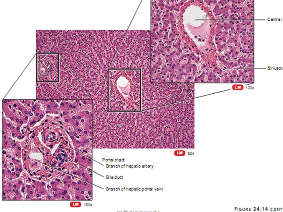
Mucosa,,submucosa,,,musculosa,,serosa Structure of digestive tube

33
Epithelial lining of digestive tube Non-keratinized st.sq.epithelium Oral cavity Esophagus Anal canal Simple columnar epithelium Stomach,,,,,rugae Small intestine….. folds,villi,microvilli(the combination of the folds of Kerckring(valvulae conniventes), the villi, and the microvilli increases the total absorptive area of the mucosa perhaps 1000-fold, making a tremendous total area of 250 or more square meters for the entire small intestine. Large intestine…..no villi
, the villi, and the microvilli increases the total absorptive area of the mucosa perhaps 1000-fold, making a tremendous total area of 250 or more square meters for the entire small intestine. Large intestine…..no villi.")
Similar presentations


![Anatomy Practical [PHL 212]](/14/4428258/big_thumb.jpg "Anatomy Practical [PHL 212]>")


>")


 Salivary Oral cavity glands (mouth) Esophagus Stomach>")
 What Tooth? Bicuspid Incisor Crown Neck (Found Below Gum Line) Root 15 6.>")


