Download presentation
Presentation is loading. Please wait.

1
CARDIOVASCULAR ASSESSMENT AND PHYSICAL EXAMINATION

2
1. Review anatomy & physiology of the cardiovascular system. 2. Discuss relevant aspects of the patient history. 3. Describe physical assessment of cardiovascular status. 4. Review diagnostic procedures, tests and medications relative to the cardiovascular system.

3
Anatomy & Physiology Functions of the heart & CV system Pumps blood to tissues to supply O 2 & nutrients Pumps blood to tissues to supply O 2 & nutrients Remove CO2 & metabolic wastes Remove CO2 & metabolic wastes

4
PERICARDIUM / PERICARDIAL SAC Protects heart from trauma Protects heart from trauma Serous fluid lubricates and Serous fluid lubricates and prevents friction prevents friction Prevents heart from over filling Prevents heart from over filling Anatomy & Physiology

5
CORONARY ARTERIES Right & Left arteries encircle the heart and supply blood to the myocardium during ventricular relaxation( diastole) LEFT MAIN CORONARY ARTERY L ANTERIOR DESCENDING (LAD) L CIRCUMFLEX (LCX) RIGHT CORONARY ARTERY POSTERIORMARGINAL
 LEFT MAIN CORONARY ARTERY L ANTERIOR DESCENDING (LAD) L CIRCUMFLEX (LCX) RIGHT CORONARY ARTERY POSTERIORMARGINAL")
6
CORONARY ARTERIES ( R) ARTERY ( L) ARTERY LAD CIRCUMFLEX
 ARTERY ( L) ARTERY LAD CIRCUMFLEX")
7
CARDIAC LOAD Preload = degree of myocardial fiber stretch at the end of diastole and just before contraction Afterload = pressure against which ventricles must eject blood. This pressure is affected by systemic vascular resistance (SVR)
.")
8
8

9
OTHER ELEMENTS OF CARDIAC ASSESSMENT Previous cardiac hx Previous cardiac hx Other medical conditions that may affect heart function Other medical conditions that may affect heart function Chest injury Chest injury Previous heart surgery Previous heart surgery Past medical hx Past medical hx Medications: prescribed, OTC, herbals Medications: prescribed, OTC, herbals Activity tolerance Activity tolerance Health habits Health habits Family hx Family hx

10
CARDIOVASCULAR EXAMINATION HISTORY PHYSICAL EXAM LAB TEST ECG CARDIAC IMAGING

11
CARDIOVASCULAR SYMPTOM CHEST PAIN SHORTNESS OF BREATH DOE ( DYSPNEA ON EXERTION) PND ( PAROXYSMAL NOCTURNAL DYSPNEA) WHEEZING
 PND ( PAROXYSMAL NOCTURNAL DYSPNEA) WHEEZING")
12
CONTINUED DIZZINESS SYNCOPE PALPITATION FATIGUE EDEMA INTERMITTENT CALAUDICATION CYANOSIS

13
CONTINUED AGGRAVATING FACTORS ALLEVIATING FACTORS PREVIOUS LABORATORY TESTS RISK FACTORS

14
EXAMINATION Inspection Inspection Palpation Palpation Percussion Percussion Auscultation Auscultation

15
GENERAL APPEARANCE VITAL SIGNS JUGULAR VEINS HEART PRIPHERAL PULSES

16
VITAL SIGNS BP HEART RATE RHYTHM RESPIRATORY RATE TEMPERATURE

17
17 INSPECTION

18
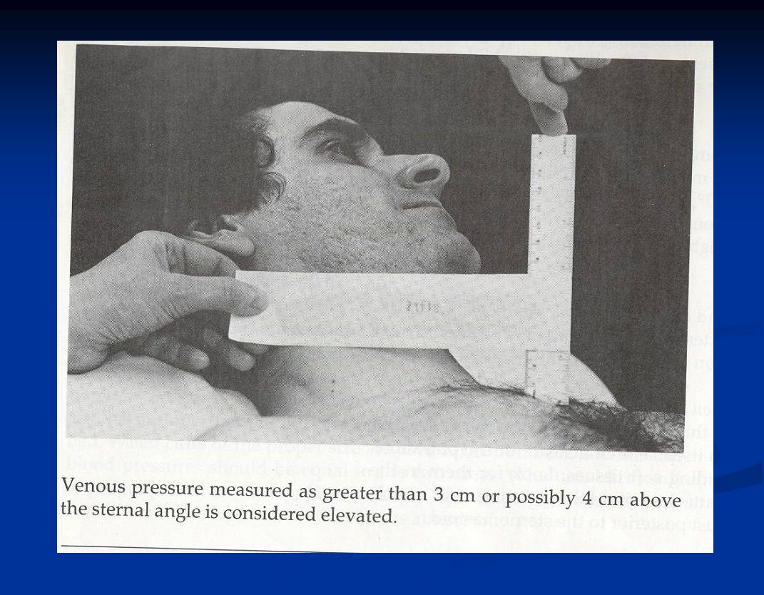
18 JUGULAR VEINS/ JUGULAR VENOUS PRESSURE RIGHT SIDE, HEAD TILTED TO L ADJUST ANGLE OF BED TO SEE PULSATION AT MIDNECK RECORD DISTANCE FROM R ATRIUM TO TOP OF PULSATION

21
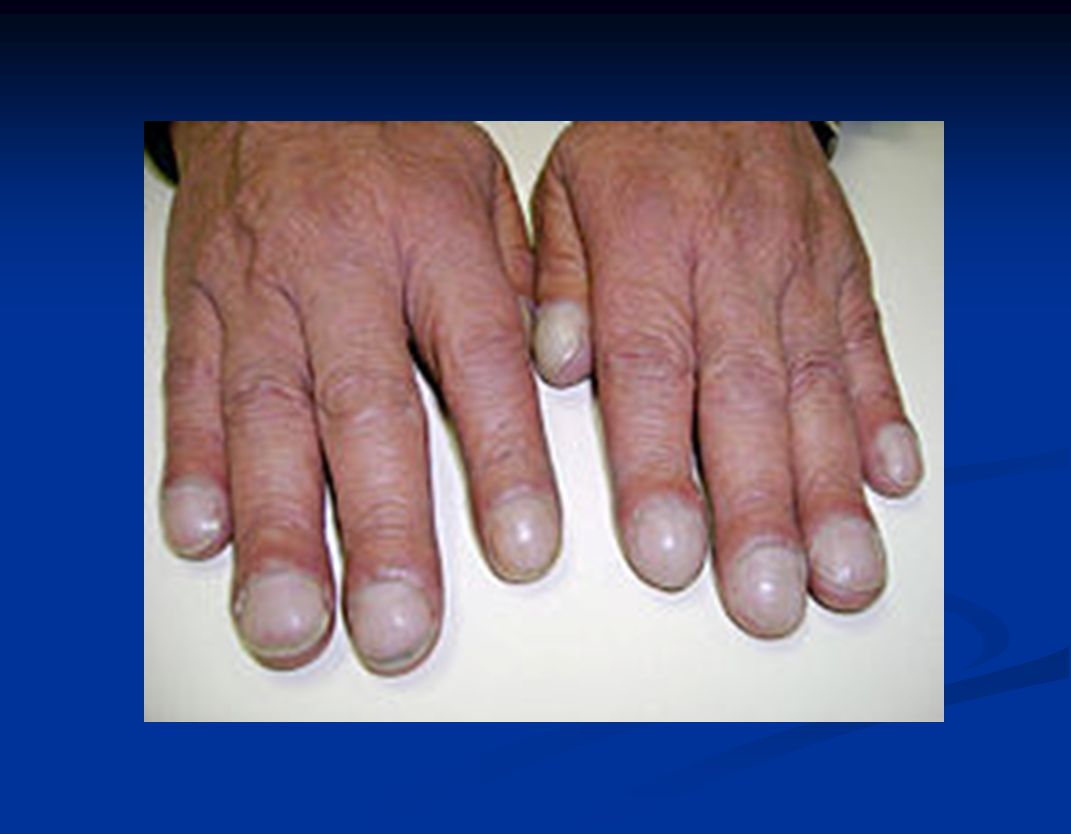
INSPECTION (continued) Lips, nail beds Heart: apical impulse point of maximal impulse Extremities: (edema, venous or arterial insufficiency)
 Lips, nail beds Heart: apical impulse point of maximal impulse Extremities: (edema, venous or arterial insufficiency)")
22
22

24
IMPULSES – FINGER PADS THRILLS ( vibration palpated secondrary to a murmur – turbulant blood flow through a heart valve) APICAL IMPULSE ( normally 5 th ICS and medial to mid- clavicular line) Point of max impulse Left lateral decubitus : position apical impulse more easily palpable
 APICAL IMPULSE ( normally 5 th ICS and medial to mid- clavicular line) Point of max impulse Left lateral decubitus : position apical impulse more easily palpable")
25
AUSCULTATION Diaphragm – medium and high frequency sounds Diaphragm – medium and high frequency sounds Bell – low frequency sounds Bell – low frequency sounds Normally hear closure of valve Normally hear closure of valve Sounds from left side of heart louder than equivalent sounds from right side of heart Sounds from left side of heart louder than equivalent sounds from right side of heart

26
AUSCULTATION S 1 – closure of mitral and tricuspid valves S 1 – closure of mitral and tricuspid valves S 2 – closure of aortic and pulmonic valves S 2 – closure of aortic and pulmonic valves S 1 systole S 2 diastole S 1 S 1 systole S 2 diastole S 1 Simultaneous palpation of carotid pulse can help in differentiating S 1 and S 2 Simultaneous palpation of carotid pulse can help in differentiating S 1 and S 2

27
FIRST AND SECOND HEART SOUNDS Aortic component (A 2 ) normally louder than pulmonic component (P 2 ) Aortic component (A 2 ) normally louder than pulmonic component (P 2 ) Mitral component (M 1 ) normally louder than tricuspid component (T 1 ) Mitral component (M 1 ) normally louder than tricuspid component (T 1 )
 normally louder than pulmonic component (P 2 ) Aortic component (A 2 ) normally louder than pulmonic component (P 2 ) Mitral component (M 1 ) normally louder than tricuspid component (T 1 ) Mitral component (M 1 ) normally louder than tricuspid component (T 1 )")
28
28

29
DIAPHRAGM Right 2 nd intercostal space Right 2 nd intercostal space Aortic Area Left 2 nd intercostal space Left 2 nd intercostal space Pulmonic Area Third intercostal space Third intercostal space Erb’s point Left lower sternal border Left lower sternal border Tricuspid area Apex – over apical impulse Apex – over apical impulse Mitral area

30
BELL Left lower sternal border Left lower sternal border Apex Apex Apex with patient in left lateral decubitus position Apex with patient in left lateral decubitus position Light pressure only! Light pressure only!

31
31 Abnormal heart sound and murmur S3,s4,click …… Systolic murmur: AS,MR,PS,TR,VSD Diastolic murmur: AR,MS,PR,TS

Similar presentations


Chapter (8) Assessment of Cardiovascular SystemAssessment of Cardiovascular System.>")


 Transport O 2, nutrients, hormones, cell wastes, etc…>")

