Download presentation
Presentation is loading. Please wait.

2
SynapsesOfC.n.sSynapsesOfC.n.s By Dr. Khaled Ibrahim By

3
SYNAPSESSYNAPSES Definition: It is the Communication (area of contact) between 2 neurons in the central nervous system. Definition: It is the Communication (area of contact) between 2 neurons in the central nervous system. Types of synapses ELECTRICAL SYNAPSES CHEMICAL SYNAPSES
 between 2 neurons in the central nervous system. Types of synapses ELECTRICAL SYNAPSES CHEMICAL SYNAPSES.")
4
Extremely rare. Structure: -The membranes of the two neurons come close together and are held together by connecting protein channels called “connexons”, which contain pores that permit the passage of ions between cells easily. -This allows rapid direct conduction of electrical potentials from one neuron to the next. 1- ELECTRICAL SYNAPSES :

5
2- CHEMICAL SYNAPSES: Much more common (almost all synapses in CNS). Presynaptic neuron: is the neuron which conduct the impulse toward the synapse & release the chemical transmitter. Postsynaptic neuron: is the neuron which conduct impulse away from the synapse. Presynaptic neuron Postsynaptic neuron

6
Physiological Anatomy: Formed of: 1) a presynaptic terminal. 2) postsynaptic membrane. 3) a synaptic cleft. a) Presynaptic Terminal: - It is a terminal branch of the axon of the presynaptic neuron. - This terminal can make contact with any part of the postsynaptic neuron. So, chemical synapses can be classified into: Axodendritic ----> junction the terminal & postsynaptic dendrites. Axosomatic -----> junction the terminal & postsynaptic soma. Axoaxonic -----> junction the terminal & postsynaptic axon. According to predominance: Axodendritic > Axosomatic > Axoaxonic. But according to excitability: Axoaxonic > Axosomatic > Axodendritic.
 postsynaptic membrane. 3) a synaptic cleft. a) Presynaptic Terminal: - It is a terminal branch of the axon of the presynaptic neuron. - This terminal can make contact with any part of the postsynaptic neuron. So, chemical synapses can be classified into: Axodendritic ----> junction the terminal & postsynaptic dendrites. Axosomatic -----> junction the terminal & postsynaptic soma. Axoaxonic -----> junction the terminal & postsynaptic axon. According to predominance: Axodendritic > Axosomatic > Axoaxonic. But according to excitability: Axoaxonic > Axosomatic > Axodendritic..")
8
Structure: * Most of the terminals resemble small round or oval knobs. So, they are called terminal buttons or synaptic knobs. * Synaptic knob contains: - large number of vesicles which contain the chemical transmitter released in the synaptic cleft. - large number of mitochondria which supply energy (ATP) for synthesis of the transmitter substance either in the endoplasmic reticulum or the cytoplasm of the synaptic knob. - Then the transmitter is transported into the vesicles by specific transporters in the vesicle membrane
 for synthesis of the transmitter substance either in the endoplasmic reticulum or the cytoplasm of the synaptic knob. - Then the transmitter is transported into the vesicles by specific transporters in the vesicle membrane.")
9
c) Postsynaptic Membrane: - It shows thickening directly opposite the synaptic knob called "active zone". - This active zone is the site of receptors which can bind chemical transmitters released from the synaptic knob. b) Synaptic Cleft: - It is the narrow space separating the membrane of the synaptic knob from the postsynaptic membrane. - 20 to 30 nm in width. - It is filled with thin filaments that help to bind the pre-and postsynaptic membranes together.
 Synaptic Cleft: - It is the narrow space separating the membrane of the synaptic knob from the postsynaptic membrane to 30 nm in width. - It is filled with thin filaments that help to bind the pre-and postsynaptic membranes together..")
10
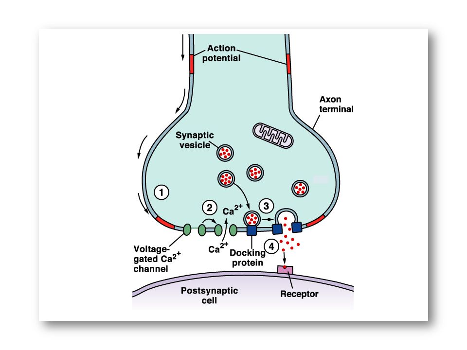
Mechanism of Synaptic Transmission 1 Release of the transmitter into the synaptic cleft. 2 Action of the transmitter on the postsynaptic membrane. 3 Termination of synaptic transmission.

11
1) Release of the transmitter into the synaptic cleft: (Role of Ca 2+ ions) The membrane of synaptic knob contains voltage-activated Ca 2+ channels near the release sites of the presynaptic membrane.
 Release of the transmitter into the synaptic cleft: (Role of Ca 2+ ions) The membrane of synaptic knob contains voltage-activated Ca 2+ channels near the release sites of the presynaptic membrane.")
13
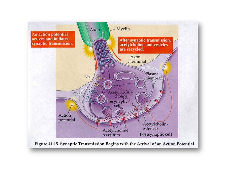
Mechanism: Arrival of action potential to the synaptic knob -----> depolarization of the membrane -----> transient opening of Ca 2+ channels ------> Ca 2+ influx near the release sites ------> fusion of the vesicles with the knob membrane ------> rupture to release its chemical transmitter content by "exocytosis". Then vesicle membrane separates from the knob membrane -----> mobilized back to fuse with a cistern of the smooth endoplasmic reticulum (where the chemical transmitter is synthesized) ------> inserted into packets in the cistern membrane ------> pinched off forming new synaptic vesicles.
 > inserted into packets in the cistern membrane > pinched off forming new synaptic vesicles..")
14
The number of vesicles ruptured (& so the amount of the transmitter released) is proportional to the intracellular Ca 2+ concentration in the synaptic knob. i.e., ↑ intracellular Ca 2+ concentration -----> ↑released transmitter & vice versa. Intracellular Ca 2+ concentration is increased by: a) ↑frequency of impulses reaching the presynaptic terminal. So, successive action potential will produce a greater response which is called "post-tetanic potentiation". b) ↓rate of removal of Ca 2+ from the synaptic knob.
 ↑frequency of impulses reaching the presynaptic terminal. So, successive action potential will produce a greater response which is called post-tetanic potentiation . b) ↓rate of removal of Ca 2+ from the synaptic knob..")
15
2) Action of the transmitter on the postsynaptic membrane: The released chemical transmitter binds a SPECIFIC RECEPTOR on the postsynaptic neuron producing a change in the postsynaptic membrane potential known as POSTSYNAPTIC POTENTIALS which will be SUMMATED to generate an ACTION POTENTIAL propagated along the postsynaptic neuron.
 Action of the transmitter on the postsynaptic membrane: The released chemical transmitter binds a SPECIFIC RECEPTOR on the postsynaptic neuron producing a change in the postsynaptic membrane potential known as POSTSYNAPTIC POTENTIALS which will be SUMMATED to generate an ACTION POTENTIAL propagated along the postsynaptic neuron.")
16
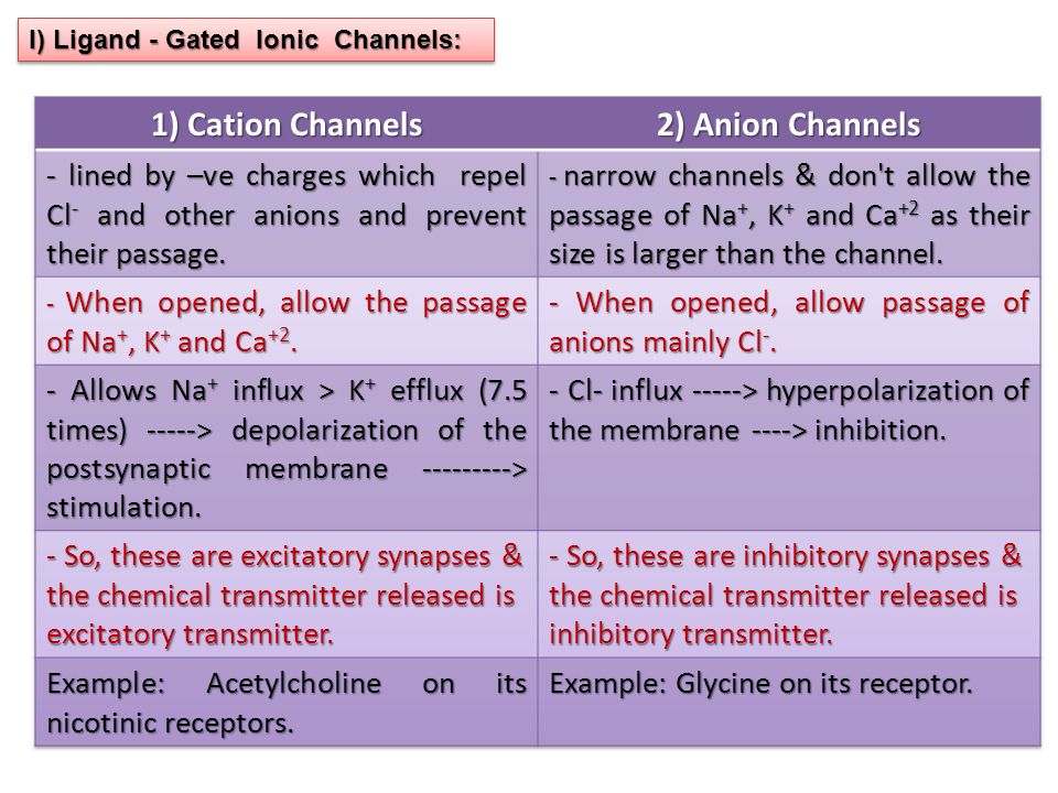
Postsynaptic Receptors Ligand - Gated Ionic Channels. G-Protein Coupled Receptors.

17
I) Ligand - Gated Ionic Channels:
 Ligand - Gated Ionic Channels:")
19
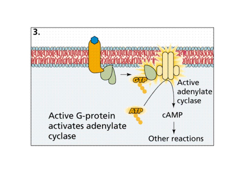
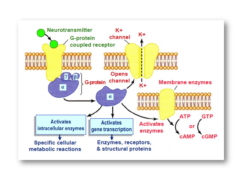
GDP Inactive adenyl cyclase II) G-Protein Coupled Receptors: Chemical transmitter
 G-Protein Coupled Receptors: Chemical transmitter")
20
The Transmitter binds the receptor

22
1 3 2 II) G-Protein Coupled Receptors:
 G-Protein Coupled Receptors:")
23
These receptors are provided by specific membrane proteins which are not ionic channels. These receptors use a group of proteins known as “G-proteins” which are bound to the receptor proteins. G- proteins are the key molecule of the system. G-protein molecule consists of three components; alpha( ), beta ( ), and gamma ( ) components. They are called G-proteins as they are bound to GTP in their active form & to GDP in their inactive form.
, beta ( ), and gamma ( ) components. They are called G-proteins as they are bound to GTP in their active form & to GDP in their inactive form..")
25
The separated active “ component” may: 1) Open specific second - messenger gated ion channels in the post synaptic membrane: e.g. K + channels which when opened ------> ↑ K + efflux -----> hyperpolarization of the postsynaptic membrane. - Mechanism: A neurotransmitter binds to its specific membrane receptor -----> altered receptor binds G-proteins ------> replacement of GDP bound to them by GTP ( the G- protein is activated) -------> Separation of the “ component” from the remainder of the G-protein molecule & becomes free to move within the postsynaptic cell.
 > Separation of the component from the remainder of the G-protein molecule & becomes free to move within the postsynaptic cell..")
26
2) Activate particular enzyme systems ------> formation of the second messengers, such as : c AMP, c GMP, inositol - 3 - phosphate (IP3), diacylglycerol (DAG), and arachidonic acid. These systems control many highly specific metabolic pathways in the neuron. 3) Regulate gene transcription by controlling the binding of RNA polymerase to the gene ------> control of synthesis and production of certain proteins within the neuron, including enzymes, receptor molecules, and cellular structural proteins.
 Regulate gene transcription by controlling the binding of RNA polymerase to the gene > control of synthesis and production of certain proteins within the neuron, including enzymes, receptor molecules, and cellular structural proteins..")
27
Postsynaptic Potentials: They are changes of the potential of the postsynaptic membrane as a result of binding of a neurotransmitter to its specific receptor. The “postsynaptic potentials” are of two types: (i) Excitatory postsynaptic potentials (EPSPs). (ii) Inhibitory postsynaptic potentials (IPSPs).
 Excitatory postsynaptic potentials (EPSPs). (ii) Inhibitory postsynaptic potentials (IPSPs)..")
28
(i) Excitatory postsynaptic potentials (EPSPs): Definition: A state of partial depolarization of the postsynaptic membrane as a result of binding of an excitatory transmitter to its specific receptor. During depolarization, membrane potential became closer to the threshold ------> ↑ excitability of the neuron. So, this change is called “excitatory postsynaptic potential”: (EPSP). Mechanism: 1)opening of ligand-gated cation channels -----> more Na + influx than K + efflux -----> depolarization. 2)Closure of K + channels -----> ↓K + efflux -----> depolarization.
. Mechanism: 1)opening of ligand-gated cation channels -----> more Na + influx than K + efflux -----> depolarization. 2)Closure of K + channels -----> ↓K + efflux -----> depolarization..")
29
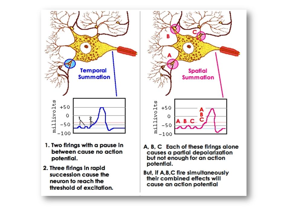
Characters: 1- Short duration (15 m.s.) then rapidly declines as +ve charges diffuse out of the postsynaptic membrane to restore normal resting membrane potential. 2- Can be summated: - EPSP produced by a single discharge from one synaptic knob is quite small, and produces a depolarization of about 0.5 mV. - A depolarization of at least 10 to 20 mV is required to reach the usual threshold for excitation of the postsynaptic neuron. - So, EPSPs should summate to ↑the amplitude of depolarization & reach the threshold.

30
- There are two types of summation: spatial summation & temporal summation. a) Spatial summation: Summation of several EPSPs produced at the same time (when more than one presynaptic is active at the same time). b) Temporal Summation: Summation of rapid successive EPSPs at the same site with time interval less than 15 ms. (when one presynaptic terminal is stimulated rapidly & repeatedly) - In both types of summation, if the summated EPSPs reach the threshold, an action potential is produced.
 Spatial summation: Summation of several EPSPs produced at the same time (when more than one presynaptic is active at the same time). b) Temporal Summation: Summation of rapid successive EPSPs at the same site with time interval less than 15 ms. (when one presynaptic terminal is stimulated rapidly & repeatedly) - In both types of summation, if the summated EPSPs reach the threshold, an action potential is produced..")
32
(ii) Inhibitory postsynaptic potentials (IPSPs). Definition: A state of hyperpolarization caused by binding of inhibitory neurotransmitter to its specific receptor. During hyperpolarization, membrane potential became away from the threshold ---> ↓ excitability of the neuron. So, this change is called “inhibitory postsynaptic potential”: (IPSP).
..")
33
Mechanism: 1) opening of ligand - gated anion channels (Cl - channels) -----> Cl - influx - ----> hyperpolarization. 2) Opening of specific K + channels -----> ↑ K + efflux ------> hyperpolarization. Characters: 1- Short duration (15 m.s.). 2- Can be summated: Both by spatial & temporal summation (see before)
 Opening of specific K + channels -----> ↑ K + efflux > hyperpolarization. Characters: 1- Short duration (15 m.s.). 2- Can be summated: Both by spatial & temporal summation (see before).")
34
- - + + + - - Postsynaptic inhibition Presynaptic inhibition

35
Types of inhibition: 1- Postsynaptic inhibition: - It is a type of inhibition in which the inhibitory knob of a presynapic neuron is applied directly to a postsynaptic membrane. - The inhibitory presynaptic terminal may be applied to any part of the postsynaptic neuron: Axodendritic, Axosomatic, Axoaxonic junctions. Mechanism: see before. Extent: affect the entire postsynaptic neuron.

36
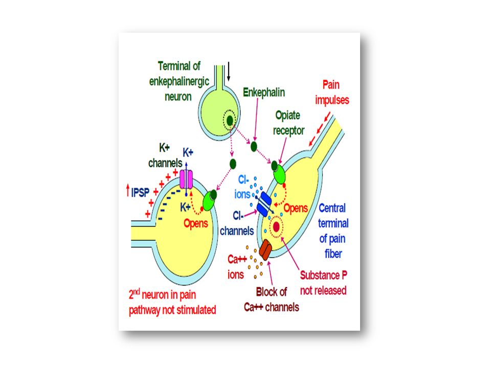
2- Presynaptic Inhibition: - It is a type of inhibition in which the inhibitory knobs of an interneuron is applied to an excitatory presynaptic terminal. - The inhibitory interneuron forms an axo-axonic synapse with the presynaptic neuron. - The inhibitory interneuron acts to modify the rate of release of the neurotransmitter from the presynaptic neuron. - Extent: It affects only a localized area on the postsynaptic neuron.

38
Mechanism: * Most of the inhibitory interneurons which inhibit the presynaptic terminals release the inhibitory neurotransmitter “gamma amino butyric acid: GABA”. * Release of GABA ----> opening Cl - channels in the presynaptic terminal ------> ↑Cl - influx -----> neutralization of the excitatory effect of Na + influx occurred when an action potential arrives the presynaptic terminal - ----> ↓ amplitude of the action potential ------> ↓opening of voltage - gated Ca ++ channels -----> ↓ Ca ++ influx ----> ↓ release of the transmitter -----> ↓postsynaptic response.

39
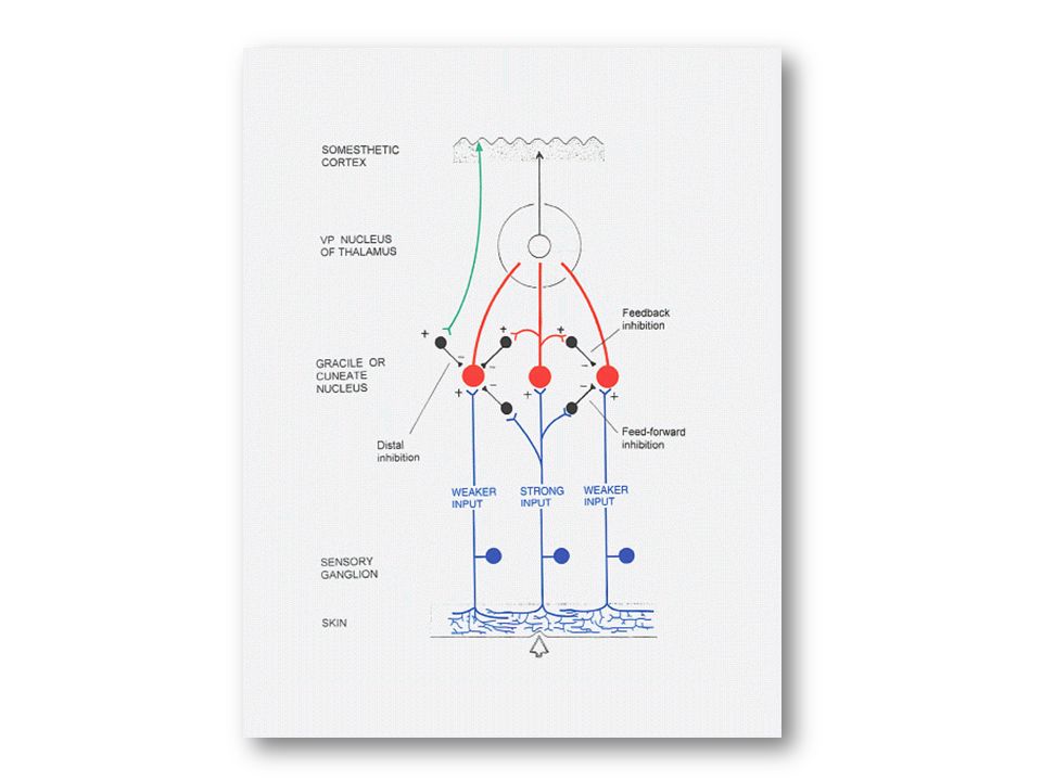
Significance: * Presynaptic inhibition occurs in many sensory pathways in the nervous system. * It is involved in: 1) The process of lateral inhibition in which the more strongly stimulated neurons inhibit the less stimulated surrounding neurons. This minimizes the sideways spread of signals in the sensory tracts and focuses on the most important ones. 2) The pain control system in which presynaptic inhibition blocks transmission of pain signals along the central pathways of pain.
 The process of lateral inhibition in which the more strongly stimulated neurons inhibit the less stimulated surrounding neurons. This minimizes the sideways spread of signals in the sensory tracts and focuses on the most important ones. 2) The pain control system in which presynaptic inhibition blocks transmission of pain signals along the central pathways of pain..")
41
Termination of synaptic transmission: - This occurs when the neurotransmitter is removed from the synaptic cleft. - This is done by one or more of the following ways:- i) Re-uptake of the transmitter into the presynaptic terminal by an active transport mechanism, specific for each transmitter. ii) By enzymatic degradation within the cleft itself. Some of the breakdown products are then transported back into the presynaptic terminal. iii) By diffusion of the transmitter out of the synaptic cleft into the surrounding interstitial fluid.
 Re-uptake of the transmitter into the presynaptic terminal by an active transport mechanism, specific for each transmitter. ii) By enzymatic degradation within the cleft itself. Some of the breakdown products are then transported back into the presynaptic terminal. iii) By diffusion of the transmitter out of the synaptic cleft into the surrounding interstitial fluid..")
42
General Properties of Chemical Synaptic Transmission 1- One – Way Conduction: At synapses, conduction of impulses occurs in one direction only; from the presynaptic to the postsynaptic neurons & not in the opposite direction. This is known as “Bell- Magendie law”. Mechanism: The backward impulse produces no effect because there are no transmitter vesicles in the presynaptic neuron.

43
2- Synaptic Delay: Definition: It is time passed between arrival of an action potential to the synaptic knob and the occurrence of response in the postsynaptic neuron. This represents the time required for: a) Release and diffusion of the neurotransmitter b) Binding of the transmitter to the postsynaptic receptors. c) Generation of PSPs & its summation to generate action potential. The minimum time for synaptic delay is about 0.5 millisecond. Significance: can be used to determine the number of synapses present in a polysynaptic reflex.
 Release and diffusion of the neurotransmitter b) Binding of the transmitter to the postsynaptic receptors. c) Generation of PSPs & its summation to generate action potential. The minimum time for synaptic delay is about 0.5 millisecond. Significance: can be used to determine the number of synapses present in a polysynaptic reflex..")
44
3- Synaptic Fatigue: Definition: It is progressive decline in rate of discharge of the postsynaptic neuron, following rapid strong stimulation of the presynaptic neuron. Mechanism: Depletion of the stores of the neurotransmitter in the synaptic knobs, because there is no enough time resynthesis and reuptake mechanisms that refill the stores under intensive stimulation. Synaptic fatigue disappears when the transmitter has been replenished. Hypoxia accelerate synaptic fatigue while strychnine delays it. In sever conditions, synaptic transmission completely stops. This is called "synaptic block". Significance: Synaptic fatigue when occurs protects CNS from overexcitability (a protective mechanism).
..")
45
4- POST-TETANIC FACILITATION (POTENTIATION) When a presynaptic terminal is stimulated rapidly for a period of time, the synapse becomes more responsive to subsequent stimuli than normal for a period of seconds or minutes. The physiological significance of the post-tetanic facilitation may have a role in the memory process in C.N.S. This is called “Post-Tetanic Facilitation or Potentiation”. It is related to the excess rise of Ca 2+ inside the synaptic knob (due to delay in Ca 2+ extruding mechanism outside the knob). The elevated Ca 2+ inside the knob causes more and more vesicles to release their transmitter, producing a greater response of the post synaptic neuron.
. The elevated Ca 2+ inside the knob causes more and more vesicles to release their transmitter, producing a greater response of the post synaptic neuron..")
46
5- Long - Term Potentiation: Brief rapid repeated stimulation of presynaptic terminal -----> prolonged ↑in rate of postsynaptic discharge. Mechanism: It is initiated mainly by an increase of the intracellular Ca 2+ concentration in the postsynaptic neuron rather than the presynaptic knob through opening of certain Ca 2+ channels in the postsynaptic membrane. Significance: It occurs in some parts of the nervous system, but it has a special significance in the hippocampus; which plays an important role in learning and memory.

47
6- Effect of Hypoxia on Synaptic Transmission: Hypoxia ↓neuronal excitability & synaptic transmission. This is due to the need of the neurons for adequate oxygen supply. interruption of cerebral circulation for about 7 seconds causes loss of consciousness (coma) & for about 2-3 minutes causes irreversible damage of the brain. 7- Effects of pH on Synaptic Transmission: In brain synapses, alkalosis ↑ excitability and synaptic transmission while acidosis ↓it. So, ↑pH to 7.8 to 8.0, produces severe convulsions whereas, ↓pH around 7.0 produces coma. This latter condition is always seen in severe uremic or diabetic acidosis.
 & for about 2-3 minutes causes irreversible damage of the brain. 7- Effects of pH on Synaptic Transmission: In brain synapses, alkalosis ↑ excitability and synaptic transmission while acidosis ↓it. So, ↑pH to 7.8 to 8.0, produces severe convulsions whereas, ↓pH around 7.0 produces coma. This latter condition is always seen in severe uremic or diabetic acidosis..")
48
7- Effect of Drugs on Synaptic Transmission:

49
NEUROTRANSMITTERSNEUROTRANSMITTERS Neurotransmitters of the central nervous system can be broadly classified into two groups: (i) Small- molecule neurotransmitters. (ii) Neuropeptides Small - Molecule Neurotransmitters rapidly acting cause most of the fast responses of the nervous system, such as transmission of sensory impulses to the brain and motor signals to the muscles. synthesized in the presynaptic terminals stored there in the transmitter vesicles untill released during the process of synaptic transmission to act on the postsynaptic membrane receptors Class I Acetylcholine Class II Biogenic Amines Dopamine Norepinephrine Serotonin Histamine Class III Amino Acids Glutamate Aspartate γ- Aminobutyric acid (GABA) Glycine Class IV Nitric oxide (NO) Carbon monoxide (CO)
 Neuropeptides Small - Molecule Neurotransmitters rapidly acting cause most of the fast responses of the nervous system, such as transmission of sensory impulses to the brain and motor signals to the muscles. synthesized in the presynaptic terminals stored there in the transmitter vesicles untill released during the process of synaptic transmission to act on the postsynaptic membrane receptors Class I Acetylcholine Class II Biogenic Amines Dopamine Norepinephrine Serotonin Histamine Class III Amino Acids Glutamate Aspartate γ- Aminobutyric acid (GABA) Glycine Class IV Nitric oxide (NO) Carbon monoxide (CO).")
50
Neuropeptides several families of peptides that posses relatively high molecular weight whose actions are usually slow and prolonged synthesized in the soma of neurons where they are stored within secretory vesicles that are then transported to the nerve terminals by the mechanism of “axonal streaming” of the axon cytoplasm. Act through G-protein coupled receptors -----> long-term effects in the post- synaptic neurons as: 1.metabolic changes in the cell. 2.activation or deactivation of specific genes. 3.Prolonged alterations in the numbers of excitatory or inhibitory receptors in the postsynaptic membrane. Some of these effects can last for days or perhaps even months or years. Examples: Angiotensin, substance-P, neuropeptide-Y, calcitonin-gene related peptide, soamtostatin, opioid peptides.

Similar presentations


 Nervous system functions Structure of a neuron Sensory, motor, inter- neurons Membrane potential Sodium.>")


