Download presentation
Presentation is loading. Please wait.

1
Hassan Bukhari 12/10

2
Objective Answer few questions Can we quantify hemothoraces? Should we drain all hemothoraces? Should we administer antibiotic prior to thoracostomy insertion? Retained hemothorax: VAT vs. 2 nd chest tube? Come up with an algorithm for hemothorax management.

3
Content Occult hemothorax Prophylactic antibiotics prior to thoracostomy insertion. Retained hemothorax.

4
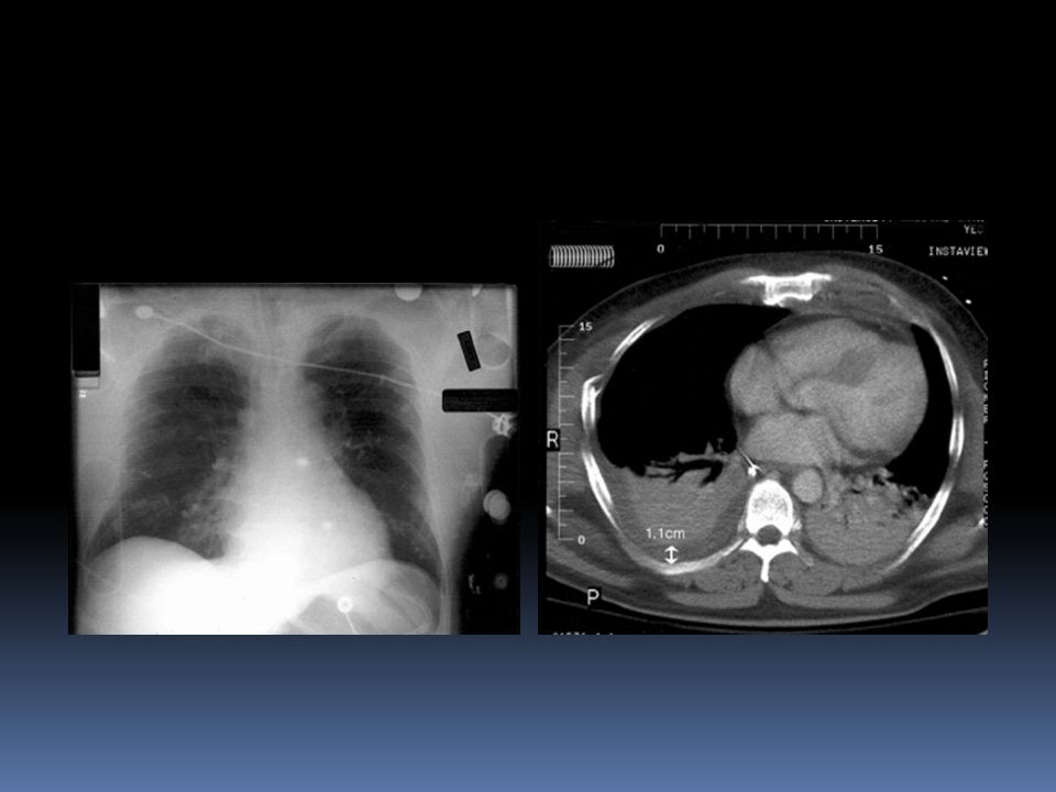
Occult hemothorax Definition Seen on chest CT but not on supine CXR Incidence 20-30% Management Only 2 retrospective studies addressed this topic

6
Quantifying hemothorax CXR, U/S and Chest CT CT is the most reliable test Decubitus CXR can be used Measurements (on CT) small: < 1.5 cm 260 ml Moderate: 1.5-4.5 cm 260-1000 ml Large: > 4.5 cm > 1000 ml
 small: < 1.5 cm 260 ml Moderate: cm ml Large: > 4.5 cm > 1000 ml")
8
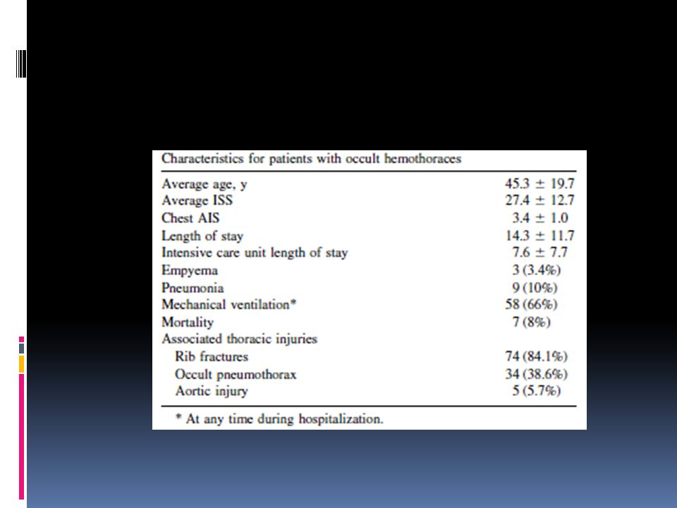
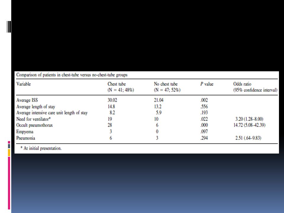
Methods: retrospective

10
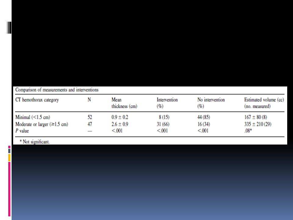
Results Minimal hemothorax (52 patients) 8 (15%) needed drainage 48 (85%) treated conservatively 44 (92%) succeeded in avoiding drainage Mod/large hemothorax (47 patients) 31 (66%) needed drainage 28 (34%) treated conservatively 16 (57%) succeeded in avoiding drainage
 8 (15%) needed drainage 48 (85%) treated conservatively 44 (92%) succeeded in avoiding drainage Mod/large hemothorax (47 patients) 31 (66%) needed drainage 28 (34%) treated conservatively 16 (57%) succeeded in avoiding drainage")
11
Conclusion Hemothorax ≥ 1.5 cm 4 times more likely to undergo drainage. Recommendation < 1.5 cm hemothorax can be safely observation

13
Quantifying hemothorax CT +/- 3D reconstruction Occult hemothorax Not seen on CXR < 2 cm is considered minimal

14
Methods: retrospective

17
Conclusion Occult hemothorax Seen only on CT and size < 2cm can be safely observed. Occult hemopneumothorax unknown

18
Algorithm Blunt trauma stable Occult hemothorax or < 2cm Observe Hemothorax seen on CXR or > 2cm on CT Chest Tube insertion

19
Prophylactic antibiotics

20
Method: systemic review

21
Result Antibiotic given before chest tube insertion and continued for < 24 hrs Reduces risk of empyema from 7.6% to 1.1% Reduces risk of pneumonia from 16% to 6.6% RR of 0.19 (95% CI 0.07-0.5) for development of empyema RR of 0.44 (95% CI 0.27-0.73) for development of pneumonia
 for development of empyema RR of 0.44 (95% CI ) for development of pneumonia")
22
Conclusion Prophylactic antibiotics prior chest tube insertion for isolated chest injury (blunt or penetrating) has Protective effective against the development of posttraumatic empyema and pneumonia
 has Protective effective against the development of posttraumatic empyema and pneumonia")
23
Algorithm Blunt trauma stable Occult hemothorax or < 2cm Observe Hemothorax on CXR or > 2cm Prophylactic antibiotic Drain

24
Retained hemothorax

25
Definition Persistent hemothorax after chest tube infection, which is diagnosed within 72 hrs. Incidence 1-20%

26
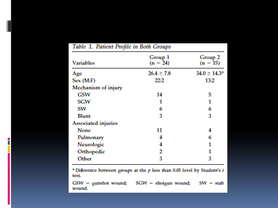
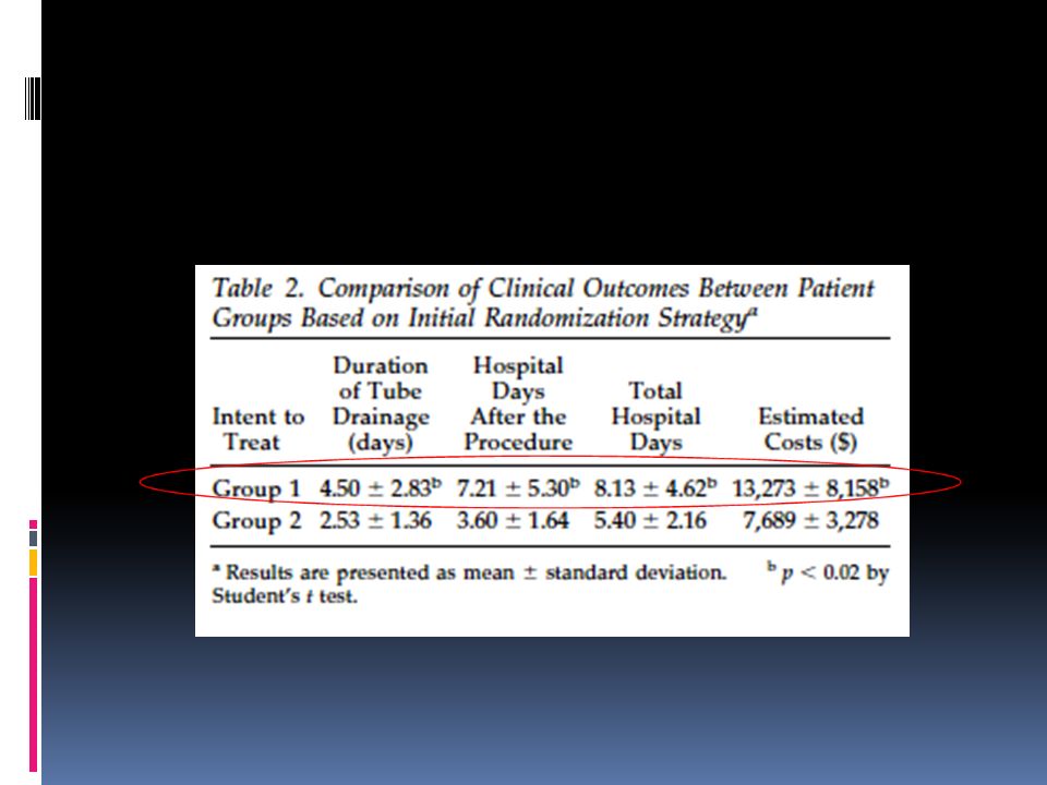
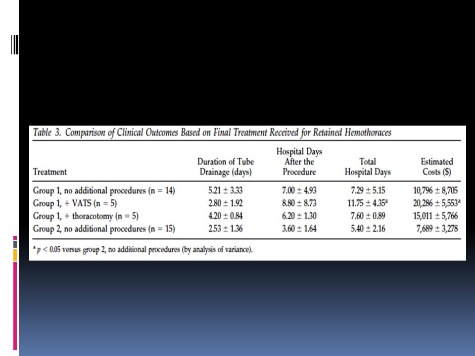
Method: prospective randomized trial Retained hemothorax on CXR after 72 hrs from chest tube insertion 2 groups Group 1 second chest tube insertion If failed randomized to VATS vs. thoracotomy Group 2 VAT Duration: 4 years

27
Patient and port position

31
Conclusion Retained hemothorax Second chest tube (G1) vs. VATS (G2) VATS decreases duration of CT, hospital stay and lowered hospital cost. No mortality VATS vs. thoracotomy for failed G1 No difference
 VATS decreases duration of CT, hospital stay and lowered hospital cost. No mortality VATS vs. thoracotomy for failed G1 No difference.")
32
AAST proposed study Prospective, observational, multicenter Management of posttraumatic retained hemothorax.

33
Chest Trauma Occult hemothorax or < 2cm Observe + Follow up CXR Worsen Hemothorax seen on CXR or >2cm on CT Prophylactic antibiotic Chest tube insertion Retained hemothorax on CXR or CT within <5 days* VATSThoracotomy *Surg Endosc. 2008 Jan;22(1):91-5
:91-5.")
Similar presentations


, FCCP>")
 Admitted D6 with 1 week H/O: SOBE, Cough, minimal sputum SOBE, Cough, minimal sputum ? Fever &>")


 in elderly and critically ill patients as a safe and effective.>")


