Download presentation
Presentation is loading. Please wait.

1
Exercise-Induced Pulmonary Arterial Hypertension James J. Tolle, MD; Aaron B. Waxman, MD, PhD; Teresa L. Van Horn, BA Paul P. Pappagianopoulos, MEd; David M. Systrom, MD Circulation;118;2183-2189; Nov 18,2008

2
Background The clinical relevance of exercise-induced pulmonary arterial hypertension (PAH) is uncertain Its existence has never been well studied by direct measurements of central hemodynamics Using invasive cardiopulmonary exercise testing Exercise-induced PAH A symptomatic stage of PAH Physiologically intermediate between resting pulmonary arterial hypertension and normal.
 is uncertain Its existence has never been well studied by direct measurements of central hemodynamics Using invasive cardiopulmonary exercise testing Exercise-induced PAH A symptomatic stage of PAH Physiologically intermediate between resting pulmonary arterial hypertension and normal.")
3
Methods Patients Four-hundred six complete CPETs performed over a 3-year period in the Massachusetts General Hospital Cardiopulmonary Exercise Laboratory with radial and pulmonary arterial catheters in place radionuclide ventriculographic scanning The CPETs Evaluation of dyspnea or fatigue of unclear etiology Evaluation for cardiac or pulmonary transplantation.

4
Cardiopulmonary Exercise Testing Pulmonary gas exchange and minute ventilation (VE) were measured breath by breath Radial and pulmonary artery catheters were placed with the use of standard techniques, the latter by the internal jugular approach
 were measured breath by breath Radial and pulmonary artery catheters were placed with the use of standard techniques, the latter by the internal jugular approach")
5
Mean end-expiratory values were obtained for right atrial pressure (RAP), mPAP, and mean systemic arterial pressure PO2, PCO2, pH, hemoglobin concentration([Hb]), and O2 saturation Right ventricular (RV) and left ventricular (LV) ejection fractions (RVEF,LVEF) and LV end-diastolic volume were measured at rest and near peak exercise by a first- pass cardiac radionuclide scan
![Mean end-expiratory values were obtained for right atrial pressure (RAP), mPAP, and mean systemic arterial pressure PO2, PCO2, pH, hemoglobin concentration([Hb]), and O2 saturation Right ventricular (RV) and left ventricular (LV) ejection fractions (RVEF,LVEF) and LV end-diastolic volume were measured at rest and near peak exercise by a first- pass cardiac radionuclide scan](http://images.slideplayer.com/39/10859456/slides/slide_5.jpg "Mean end-expiratory values were obtained for right atrial pressure (RAP), mPAP, and mean systemic arterial pressure PO2, PCO2, pH, hemoglobin concentration([Hb]), and O2 saturation Right ventricular (RV) and left ventricular (LV) ejection fractions (RVEF,LVEF) and LV end-diastolic volume were measured at rest and near peak exercise by a first- pass cardiac radionuclide scan")
6
A single bout of incremental cycling exercise to exhaustion Two minutes of rest → 2 minutes of unloaded cycling Mean systemic arterial pressure and end-expiratory RAP and mPAP Continuously End-expiratory pulmonary capillary wedge pressure (PCWP) At rest and during each minute of exercise At cessation of exercise shortness of breath, leg fatigue or pain, or chest pain, alone or in combination.
 At rest and during each minute of exercise At cessation of exercise shortness of breath, leg fatigue or pain, or chest pain, alone or in combination.")
7
Data Analysis Ventilatory and pulmonary gas exchange data final 30 seconds of the 2-minute rest period over contiguous 30-second intervals during exercise Predicted values for VO2max utilizing age, gender, and height were those of Hansen and colleagues V E /V CO2 was measured at the ventilatory threshold

8
Cardiac output (Qt) Fick principle: Qt= VO2/(Ca-VO2) Predicted maximal Qt predicted VO2max assumed arterial-venous O2 content difference([Hb]10) PVR = (mPAP-PCWP)/Qt Maximum effort Peak heart rate≥ 80% of predicted Peak respiratory exchange ratio ≥ 1.00
![Cardiac output (Qt) Fick principle: Qt= VO2/(Ca-VO2) Predicted maximal Qt predicted VO2max assumed arterial-venous O2 content difference([Hb]10) PVR = (mPAP-PCWP)/Qt Maximum effort Peak heart rate≥ 80% of predicted Peak respiratory exchange ratio ≥ 1.00](http://images.slideplayer.com/39/10859456/slides/slide_8.jpg "Cardiac output (Qt) Fick principle: Qt= VO2/(Ca-VO2) Predicted maximal Qt predicted VO2max assumed arterial-venous O2 content difference([Hb]10) PVR = (mPAP-PCWP)/Qt Maximum effort Peak heart rate≥ 80% of predicted Peak respiratory exchange ratio ≥ 1.00")
9
PAH mPAP≥30 mm Hg, PCWP< 20 mm Hg, and PVR ≥ 80 dyne · s · cm-5 Resting PAH mPAP ≥ 25 mm Hg, PCWP < 15 mm Hg at rest Exercise induced PAH mPAP <25 mm Hg at rest PVH was defined as PCWP ≥ 20 mm Hg, at maximum exercise LV systolic dysfunction : PVH with LVEF<0.55 LV diastolic dysfunction : PVH with LVEF≥ 0.55 Peripheral limitation VO2max 80% of predicted Ca-vO2<[Hb]
![PAH mPAP≥30 mm Hg, PCWP< 20 mm Hg, and PVR ≥ 80 dyne · s · cm-5 Resting PAH mPAP ≥ 25 mm Hg, PCWP < 15 mm Hg at rest Exercise induced PAH mPAP <25 mm Hg at rest PVH was defined as PCWP ≥ 20 mm Hg, at maximum exercise LV systolic dysfunction : PVH with LVEF<0.55 LV diastolic dysfunction : PVH with LVEF≥ 0.55 Peripheral limitation VO2max 80% of predicted Ca-vO2<[Hb]](http://images.slideplayer.com/39/10859456/slides/slide_9.jpg "PAH mPAP≥30 mm Hg, PCWP< 20 mm Hg, and PVR ≥ 80 dyne · s · cm-5 Resting PAH mPAP ≥ 25 mm Hg, PCWP < 15 mm Hg at rest Exercise induced PAH mPAP <25 mm Hg at rest PVH was defined as PCWP ≥ 20 mm Hg, at maximum exercise LV systolic dysfunction : PVH with LVEF<0.55 LV diastolic dysfunction : PVH with LVEF≥ 0.55 Peripheral limitation VO2max 80% of predicted Ca-vO2<[Hb]")
10
Results Patient Demographics Of the 406 patients, the indication for testing was known in 340 255 (75%) for dyspnea of uncertain etiology 22 (6%) for fatigue 28 (8%) to differentiate a cardiac from pulmonary limit to exercise 28 (8%) had known left-ventricular systolic dysfunction and were being considered for cardiac transplantation 7 (2%) had other indications. All exercise tests were symptom limited shortness of breath, leg fatigue, or both, with only 1 patient, in the normal group, additionally experiencing chest pain

13
Plateau Versus Takeoff Patterns of mPAP Versus VO2

14
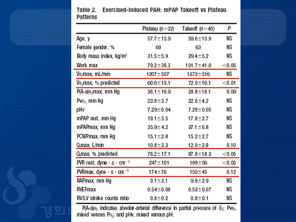
Takeoff Versus Plateau Patterns of mPAP Versus VO2 Normals 15 interpretable mPAP versus VO2 log-log plots 14 a takeoff and 1 a plateau pattern Exercise-induced PAH 40 a takeoff pattern, 32 a plateau pattern, and 6 were uninterpretable Resting PAH group 2 a takeoff pattern, 9 a plateau pattern, and 4 were uninterpretable.

16
Discussion Symptomatic patients The National Institutes of Health registry criteria Do not have an elevated mPAP at rest At maximum exercise, their overall aerobic capacity and central hemodynamics lie between those of the normal subject and the patient with resting PAH

17
Prior Studies Utilized noninvasive techniques, Stress Doppler transthoracic echocardiography a useful screening modality for resting PAH not well validated during exercise RAP normally rises : 5 mm Hg during exercise At rest by transthoracic echocardiography on the basis of inspiratory changes in inferior vena cava caliber Never been validated during exercise, when venous compliance is known to decrease

18
The contribution of the PCWP to an exercise-induced RV systolic pressure rise cannot be assessed directly with transthoracic echocardiography. PCWP has been estimated by echocardiography at rest but not during exercise Suspected cases of PAH based on echocardiography actually have PVH, especially in the elderly

19
Unexplained exertional intolerance Exercise-induced PAH and LV diastolic dysfunction Distinction of the 2 is important Treatment is usually quite different The influence of cardiac output on the RV systolic pressure, and therefore the PVR response to exercise, cannot be directly measured by echocardiography → Estimates of cardiac output and PVR have been made recently by transthoracic echocardiography at rest, but they have not yet been validated during exercise.

20
I Exercise induced PAH in 2 of a total of 16 patients with connective tissue disease or idiopathic PAH Measurements of PCWP during exercise, and PVR is unknown II 3 patients with unexplained exertional dyspnea who had normal resting mPAP and an exaggerated rise at peak cycling exercise The mean PCWP at peak exercise was 20 mm Hg raising the possibility that some of the patients had diastolic heart failure

21
Clinical Significance Symptoms vs asymptomatic or preclinical Exercise-induced PAH mild, intermediate physiological stage of PAH. Highest in resting PAH, Lowest in normals, and intermediate in exercise-induced PAH.

22
The takeoff pattern of mPAP versus VO2 normal and less severe exercise-induced PAH patients The plateau pattern of mPAP versus VO2 more severely affected exercise-induced PAH patients and of those with resting PAH similar plateau of invasively measured RV systolic pressure in resting PAH → a continuum of pulmonary vascular responses to exercise, beginning with the normal takeoff pattern, moving through 2 stages of exercise-induced PAH, and finally reaching the plateau pattern of resting PAH.

23
Screening and early detection - preventing progression to resting PAH Exercise parameters Resting mPAP(21~ 25 mm Hg) : intermediate → Invasive exercise testing Systematic longitudinal follow-up our exercise-induced PAH patients Limited longitudinal data - exercise-induced PAH patients may remain relatively stable from a clinical hemodynamic standpoint over several years → long-term clinical and hemodynamic follow-up
 : intermediate → Invasive exercise testing Systematic longitudinal follow-up our exercise-induced PAH patients Limited longitudinal data - exercise-induced PAH patients may remain relatively stable from a clinical hemodynamic standpoint over several years → long-term clinical and hemodynamic follow-up")
24
Potential Mechanisms In the normal human cardiac output ↑ from rest to maximum exercise → widening of input and outflow pressure difference → PVR falls (as a result of both passive and active pulmonary vascular recruitment and distension) have normal oxygen uptake, central hemodynamics, and fall in PVR at peak exercise.
 have normal oxygen uptake, central hemodynamics, and fall in PVR at peak exercise.")
25
In long-standing pulmonary hypertension, intimal proliferation and fibrosis, medial hypertrophy, and in situ thrombosis characterize the pathological findings in the pulmonary vasculature, although at an earlier stage, changes may be confined to the small pulmonary arteries. These changes, as well as the upstream sequelae such as RV dysfunction, are time dependent and result in progressive symptoms and impairment of exercise tolerance → patients with PAH of varying duration and severity will exhibit very different mPAP responses to exercise.

26
The takeoff pattern of mPAP pulmonary vasoconstriction late during incremental exercise in normals and those with mild exercise- induced PAH During incremental exercise, including those of arterial blood lactate concentration, ventilation, CO2 output,and humoral catecholamines

27
pulmonary arterial vasoconstrictive effects of desaturated and acid mixed-venous blood Catecholamines -> drive skeletal muscle glycolysis and potentiate PVR interleukin-6 humoral cytokine that rises measurably in proportion to exercise intensity, has been related to catecholamines → pulmonary hypertension.

28
Limitations of the Study The principal CPET indication at our institution is unexplained dyspnea, which might make our results generally applicable. 2 mPAP patterns during CPET can only be addressed indirectly in this study to generate several testable hypotheses This study is largely - cross-sectional systematic longitudinal studies

29
Conclusions Exercise-induced PAH is an early, mild, and clinically relevant phase of the PAH spectrum

Similar presentations


 Department of Clinical Pharmacy Salman Bin AbdulAziz University College Of Pharmacy.>")


 吳惠東.>")


