Download presentation
Presentation is loading. Please wait.

1
Chapter 38, (page 734 - ) Locomotion Skeleton + Muscles Csaba Bödör, bodor@korb1.sote.hu
 Locomotion Skeleton + Muscles Csaba Bödör,")
2
Skeletal system Internal skeleton that can grow Bones and cartilage, (Ca impregnated)
")
3
Cartilage and Bones Main components of the skeletal system Cartilage in humans: external ear, tip of the nose, etc. Cells of cartilage: chondrocytes They secrete: matrix and collagen fibers > cartilage Lacuna: small cavity within the matrix Cartilage lacks nerves and blood vessels Bone cells: osteocytes: secrete and maintain the matrix Bone is a highly vascular tissue (rich is blood vessels) Typical bone: outer layer- compact bone, inner layer- spongy bone Compact bone: units called osteons Osteons: osteocytes are arranged (lamella) Central microscopic canal: Haversian canal
 Typical bone: outer layer- compact bone, inner layer- spongy bone Compact bone: units called osteons Osteons: osteocytes are arranged (lamella) Central microscopic canal: Haversian canal.")
5
Structure of a typical bone Typical long bone (example: radius) periosteum: outer connective tissue membrane (+ layers > diameter) endosteum: inner lining outer compact bone,> yellow marrow inner spongy bone > red marrow diaphysis and 2 epiphyses metaphysis (between d and e) disk of cartilage: growth center (length) they disappear at maturity and become epiphyseal lines
 periosteum: outer connective tissue membrane (+ layers > diameter) endosteum: inner lining outer compact bone,> yellow marrow inner spongy bone > red marrow diaphysis and 2 epiphyses metaphysis (between d and e) disk of cartilage: growth center (length) they disappear at maturity and become epiphyseal lines")
6
Process of bone formation Enchondral bone development (ossification) bone is formed from a cartilage template primary ossification in diaphysis secondary in epiphyses
 bone is formed from a cartilage template primary ossification in diaphysis secondary in epiphyses")
7
Process of bone formation Intramembraneous bone development (ossification) bone is formed from other connective tissue (mesenchyme) typical for flat bones
 bone is formed from other connective tissue (mesenchyme) typical for flat bones")
8
Process of bone formation Continuous formation and resorbtion (destruction) Osteoblasts: bone-building cells: matrix, collagen fibers They differentiate (mature) into osteocytes Osteoblasts are rich in RER, Golgi The inorganic part of bones is hydroxyapatite (mainly calcium phosphate)
 Osteoblasts: bone-building cells: matrix, collagen fibers They differentiate (mature) into osteocytes Osteoblasts are rich in RER, Golgi The inorganic part of bones is hydroxyapatite (mainly calcium phosphate)")
9
Process of bone formation Continuous formation and resorbtion (destruction) Osteoclasts: break down bone (resorb) large multinucleated cells (sometimes 50 nuclei) release H + to dissolve crystals and enzymes to digest collagen dynamic process, the whole skeleton is completely replaced every 10 years osteoporosis: disease with increased bone resorbtion bones become too fragile
 Osteoclasts: break down bone (resorb) large multinucleated cells (sometimes 50 nuclei) release H + to dissolve crystals and enzymes to digest collagen dynamic process, the whole skeleton is completely replaced every 10 years osteoporosis: disease with increased bone resorbtion bones become too fragile")
10
Muscle tissue Is specialized to contract Long cells > muscle fibers, in muscle fibers: longitudinal, contractile units: Myofibrils (myosin + actin, …)
")
11
Muscle tissue Types

12
SKELETAL MUSCLE SMOOTH MUSCLE CARDIAC MUSCLE

13
Muscle structure Elongated cells: muscle fibers, wrapped by connective tissue Plasma membrane: sarcolemma Sarcolemma inward extensions: T tubules Cytoplasm of a muscle fiber: sarcoplasm Endoplasmic reticulum: sarcoplasmic reticulum Longitudinal units : myofibrils In myofibrils smaller structures: myofilaments Myofilaments: myosin and actin filaments Myosin filaments: thick, mainly protein myosin Actin filaments: thin, protein actin, tropomyosin, troponin

14
Muscle structure

15
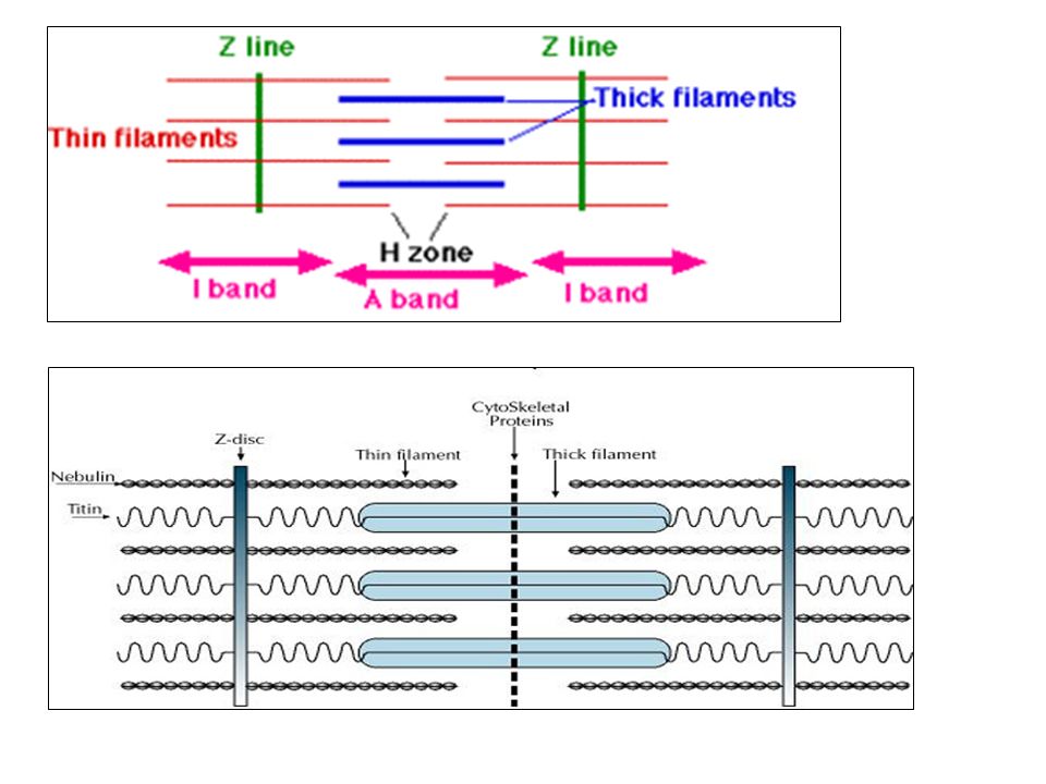
Muscle structure - sarcomere Sarcomere : basic unit of muscle contraction M line I band A band H zone Sarcomere

18
Muscle contraction Contraction: sarcomeres shorten Theory: sliding filament model

19
Muscle contraction Theory: sliding filament model

20
Muscle contraction MOTOR neuron Neuromuscular junction

21
Muscle contraction MOTOR unit Motor neuron + ~150 muscle fibers

22
Muscle contraction

23
Ca channels in SR open > Ca released into the sarcoplasm a.p. travels along the sarcolemma into the T tubules Depolarization, action potential Acetylcholine + receptors Acetylcholine: released into synaptic cleft Ca binds to troponin Muscle contraction (sarcomeres shorten)
.")
24
Muscle contraction Ca binds to troponin Change of the shape of the troponin Troponin pushes the tropomyosin away from active sites on actin filaments Myosin binding sites are exposed

25
Muscle contraction ATP + MYOSIN at rest ATP is split : chemical e. > mechanical e Myosin + ADP+P: energized state „cocked” state Energized myosin binds to actin (cross bridge)
.")
26
Muscle contraction P is released > conf. change in myosin head Power stroke: actin is pulled closer to the center of the sarcomere (ADP is released) Myosin is detached after binding of a new ATP If sufficient Ca present > new cycle with new active sites > shortening cont.
 Myosin is detached after binding of a new ATP If sufficient Ca present > new cycle with new active sites > shortening cont..")
27
Acetylcholin-esterase Ca back to SR Muscles in partially contracted state Muscle tone:

28
Muscle contraction, energy ATP is the immediate energy source, ATP hydrolysis provides the energy to “cock” the myosin Creatine phosphate: used for intermediate energy storage Glycogen: energy is stored in form of glucose

29
Muscle action Agonist muscle contracts Antagonist muscle relaxes

30
Specialized muscle fibers Types of myosin: Type I., IIa, IIx Slow (red fibers): mainly type I. rich in mitochondria and myoglobin Endurance activities (swimming, long distance running, etc.) White (fast fibers): type IIa., IIx few mitochondria Rapid response (sprinting, lift weight) They can sustain activity for only a short time Single stimulus: single quick contraction called simple twitch Series of stimuli close together: single sustained contraction: tetanus Contraction of smooth muscles tends to be sustained !! No sustained contraction in cardiac muscle !!
 White (fast fibers): type IIa., IIx few mitochondria Rapid response (sprinting, lift weight) They can sustain activity for only a short time Single stimulus: single quick contraction called simple twitch Series of stimuli close together: single sustained contraction: tetanus Contraction of smooth muscles tends to be sustained !. No sustained contraction in cardiac muscle !!.")
Similar presentations


 and lengthening (passive). Three types in mammals: 1.Skeletal muscle – used to move.>")
