Download presentation
Presentation is loading. Please wait.

1
Prepared & presented by:
Anatomy of the Ear Prepared & presented by: Prof. Dr. Hassan M. Serry © Anatomy and Embryology department, ASU, Month/Year 12/2012

2
By the end of this lecture, you should be able to:
ILOs By the end of this lecture, you should be able to: List parts of the ear. Describe parts of External ear: auricle & external auditory meatus and give their nerve supply and its applied anatomy. Describe roof, floor, and walls of Middle ear and enumerate its contents. Identify parts and function of auditory tube. Name parts of Internal ear: bony & membranous labyrinth.

3
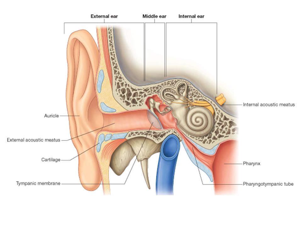
The Ear

4
It is the organ of Hearing and also concerned with Equilibrium

7
External Ear

10
AURICLE

16
EXTERNAL ACOUSTIC (AUDITORY) MEATUS
 MEATUS")
18
1/3 2/3 24 mm in length

20
It describes an S-shaped curve
Lateral Medial

21
TYMPANIC MEMBRANE (EAR DRUM)
")
22
about 10 mm in diameter it is obliquely directed making an angle of 55 with the floor of the meatus

23
posterior anterior

29
Middle Ear

31
Epitympanic recess Tympanic cavity proper

32
Posterior Anterior

33
Anterior (carotid) wall
 wall")
34
Canal for Tensor tympani Bony part of Auditory tube Internal carotid artery

36
Posterior(mastoid) wall
 wall")
37
Aditus to Mastoid antrum Pyramid

39
Roof (tegmental) wall
 wall")
40
Tegmen tympani

41
Floor (Jugular) wall
 wall")
42
Tympanic branch of XI Internal jugular vein

43
Medial (Labyrinthine)
wall

46
Promontory Prominence of Facial n. canal Fenestra vestibuli Fenestra chocleae Sinus tympani Tympanic plexus of ns.

48
Lateral (membranous) wall
 wall")
50
CONTENTS OF TYMPANIC CAVITY

52
Auditory Ossicles

53
Malleus Lateral Incus Anterior Stapes Posterior Medial

56
Muscles of tympanic cavity

58
Action The tensor tympani and stapedius usually contract together in reflex response to sounds of fairly high intensity, exerting a 'protective damping effect before vibrations reach the internal ear The tensor pulls the tympanic membrane inwards to tense it and also pushes the stapes more tightly into the fenestra vestibuli. The stapedius muscle opposes the tensor in the latter action.

59
Pharyngotympanic (Auditory) tube
 tube")
60
Tympanic caviry Pharyngotympanic tube 1/3 2/3 Pharynx 36 mm in length

61
The internal ear (Labyrinth)
")
62
The labyrinth is situated in the petrous part of the temporal bone, medial to the middle ear.
It consists of the bony labyrinth, comprising a series of cavities within the bone. and the membranous labyrinth, comprising a series of membranous sacs and ducts contained within the bony labyrinth. Labyrinth

64
Bony Labyrinth Labyrinth

65
Cochlea Vestibule Semicircular canals

66
Vestibule Labyrinth

67
Right bony labyrinth Anterolateral view
Semicircular canals Vestibule Posterior Anterior Oval window (Fenestra vestibuli) Scala vestibuli
 Scala vestibuli.")
68
Right bony labyrinth Top view
Semicircular canals Aqueduct of vestibule Vestibule Lateral Medial Oval window (Fenestra vestibuli) Scala vestibuli
 Scala vestibuli.")
69
Semicircular canals Labyrinth

70
Right bony labyrinth Anterolateral view
Anterior semicircular canal Lateral semicircular canal Ampullae of semicircular canals Posterior Anterior. Posterior semicircular canal

71
Semicircular canals Anterior Lateral Posterior

72
Cochlea Labyrinth

73
Cochlea (snail shell -shaped)القوقعة
It opens into the anterior part of the vestibule it consists of a central pillar, the modiolus, around which a hollow bony tube makes two and one half spiral turns Each successive turn is of decreasing radius so that the whole structure is conical. The apex faces anterolaterally and the base faces posteromedially The first basal turn of the cochlea is responsible for the promontory seen on the medial wall of the middle ear. Labyrinth

74
Right bony labyrinth Anterolateral view
Cochlea Apex Basal turn Posterior Anterior.

75
Section of cochlea Cochlea Modiolus Basal turn

76
Internal auditory meatus
Section of cochlea Cochlea tube Scala vestibuli Spiral lamina Scala tympani Modiolus Internal auditory meatus

77
Longitudinal section of cochlea
Cochlea tube Scala vestibuli Modiolus Scala tympani Spiral lamina Spiral ganglion

78
Section of cochlea Basilar membrane Vestibular membrane
Scala vestibuli Modiolus Scala tympani Spiral ganglion Spiral lamina

79
Membranous Labyrinth it is contained within the bony labyrinth but much smaller in size. It is filled with endolymph Its walls receive the branches of the vestibulocochlear nerve. Labyrinth

80
Anterior semicircular duct Ampulla
Lateral semicircular duct Anterior semicircular duct Ampulla Endolymphatic duct and sack Dura mater Posterior semicirular duct Cochlear duct Utricle Utricosaccular duct Saccule Scala vestibuli Cochlear canaliculus Scala tympani Labyrinth

81
Labyrinth

82
Thank You

Similar presentations


>")


